
Miryam Frost, Sung-tae Hwang, Roque Lavín Delgado, and Valentina Wang, American School of Madrid, Pozuelo, Madrid, Spain
Reviewed on 9 May 2026; Accepted on 22 June 2026; Published on 26 October 2026
With help from the 2026 BioTreks Production Team.
As the burden of cancer continues to rise despite exponential developments in the medical field, concerns regarding the treatment of cancer have arisen to the surface of discussion altogether. In 2026, cancer accounted for an estimated 2,114,850 new cases and 626,140 deaths in the United States, leaving the issue of cancer still unresolved. While modern therapies for cancer have improved the survival rate of those diagnosed, they simultaneously have been causing daunting side effects on the hematopoietic function of bone marrow producing leukocytes, hence inducing neutropenia. For patients receiving anticancer treatment, neutropenia can progress to febrile neutropenia, an oncologic emergency that requires antibacterial management along with cancer therapy. This condition interrupts and potentially delays the cancer therapy, affecting treatment efficiency and thus the patients after all. Granulocyte-Stimulating Factor (G-CSF) is a key hematopoietic cytokine that stimulates the production of neutrophils by initiating hematopoiesis. G-CSF is currently being used in the clinical field to reduce the potential infection related to chemotherapy-induced febrile neutropenia by systematically increasing neutrophil levels in blood. In our project, we aim to mass-produce Human G-CSF in E. coli (E. coli) BL21(DE3) strain using an engineered pET expression system through the insertion, multiplication, and purification of our designated gene and its product. This project will enhance the accessibility of recombinant G-CSF with microbial expression engineering, improving neutropenia management and hence resolving the risk of febrile neutropenia.
Keywords: Febrile neutropenia, protein engineering, recombinant protein, G-CSF, pET
Authors are listed in alphabetical order. Meghan Perks mentored the group. Please direct all correspondence to mailto:meghanperks@asmadrid.es.
Background
Cancer as a threat to human society
Cancer is a leading cause of morbidity and mortality on a global scale. In 2022, there were approximately 20 million new cancer cases and 9.7 million deaths. Projections estimate 35 million new cases per year by 2050, indicating a 77% increase compared to 2022 (Bray et al., 2024; International Agency for Research on Cancer [IARC], 2024). Since the absolute burden of cancer remains extremely high and modern oncology still relies on intensive treatments, complications related to these treatments have arisen as daunting tasks to be resolved in the medical field.
Side effects of cancer treatments
As of today, anticancer therapies are designed to eliminate rapidly dividing cells, targeting malignant cells that cause the malfunction of tissues and organs. However, some of these therapies can also injure healthy tissues that are needed for rapid cellular replacement, including the bone marrow (van den Boogaard et al., 2022). All therapies that affect the bone marrow may provoke myelosuppression, a condition where bone marrow activity is decreased and leads to the reduced production of blood cells (Daniel & Crawford, 2006). Anticancer therapies such as cytotoxic chemotherapy usually provoke myelosuppression and hinder hematopoiesis, the major process of new blood cell formation through cell differentiation and specialization (Mayo Clinic Staff, 2025).
They do this by rapidly targeting dividing cells in DNA synthesis, DNA damage responses, or mitosis. In the bone marrow, hematopoietic stem and progenitor cells (HSPCs), especially hematopoietic progenitor cells (HPCs), frequently undergo these processes, leaving them vulnerable. (Centers for Disease Control and Prevention [CDC], 2025; Daniel & Crawford, 2006). What makes cytotoxic chemotherapies myelosuppressive are the drugs used as agents that interfere with DNA synthesis, DNA integrity, and mitosis. Most common agents used in chemotherapy include a range of substances: from 5-fluorouracil (5-FU), which disrupts nucleotide metabolism and interferes with DNA/RNA synthesis (Casale & Patel, 2024) to carboplatin, which forms DNA crosslinks that impair DNA replication and transcription (National Institute of Diabetes and Digestive and Kidney Diseases [NIDDK], 2020). These agents lead to cell-cycle arrest and apoptosis in proliferating cell populations, including bone marrow precursors, consequently causing reduced bone marrow activity and reduced production of blood cells: erythrocytes, leukocytes, and platelets (Daniel & Crawford, 2006; van den Boogaard et al., 2022). Particularly, in the case of leukocytes, their deficient production reduces the number of neutrophils, which are the most abundant circulating leukocytes that fight against infection (Rout et al., 2024). When neutrophil production is impaired, patients become more susceptible to severe infections, and the abnormally low level of neutrophils leads to the condition of neutropenia (Rout et al., 2024).
Neutropenia
Neutropenia refers to a condition of an abnormally low level of neutrophils, granulated leukocytes produced from bone marrow, that are crucial for the immune response to inflammation and against infection (Rout et al., 2024). It is considered a severe disease since the number of neutrophils corresponds with the individual’s immune system against bacterial infection and inflammatory response in wound healing. Based on absolute neutrophil count (ANC), severe neutropenia is commonly defined as having < 0.5 × 10⁹/L in the blood circulation, and it poses a higher risk of getting an infection simply with lower numbers of white blood cells fighting pathogens (Rout et al., 2024). It can be congenital or acquired; acquired neutropenia has been arising onto the issue recently, being one of the notorious side effects of cancer therapies, specifically in the case of febrile neutropenia (Punnapuzha et al., 2023). Febrile neutropenia is defined by the condition of fever (> 38°C) along with the decreased level of neutrophils to a severe level (ANC < 0.5 × 10⁹/L). It's one of the common complications of systemic cancer treatments (Borgeaud et al., 2024).
For patients receiving anticancer treatment and experiencing febrile neutropenia, it necessitates the complication of antibacterial management along with cancer therapy. This condition interrupts and potentially delays the cancer therapy, affecting treatment efficiency and thus the patients. Febrile neutropenia is difficult to detect since a fever might be the only early signal of a severe infection, so clinical pathways prioritize rapid evaluation and empiric antimicrobial coverage (Freifeld et al., 2011).
In many cases, neutropenia forces delays of cancer treatments as it raises the risk of corresponding severe infections, leading to the necessary reduction of the relative dose intensity (RDI). Lower RDI has been associated with worse survival outcomes in a systematic review, showing how neutropenia not only increases infection risk but may also delay the cancer treatment and exacerbate the patient's conditions accordingly (Nielson et al., 2021) (Figure 1).
| Figure 1. Impact of reduced chemotherapy relative dose intensity (RDI) on patient survival. Lower relative dose intensity of chemotherapy is associated with decreased overall survival in patients with advanced solid tumors. Source: Adapted from Nielson et al. (2021). | 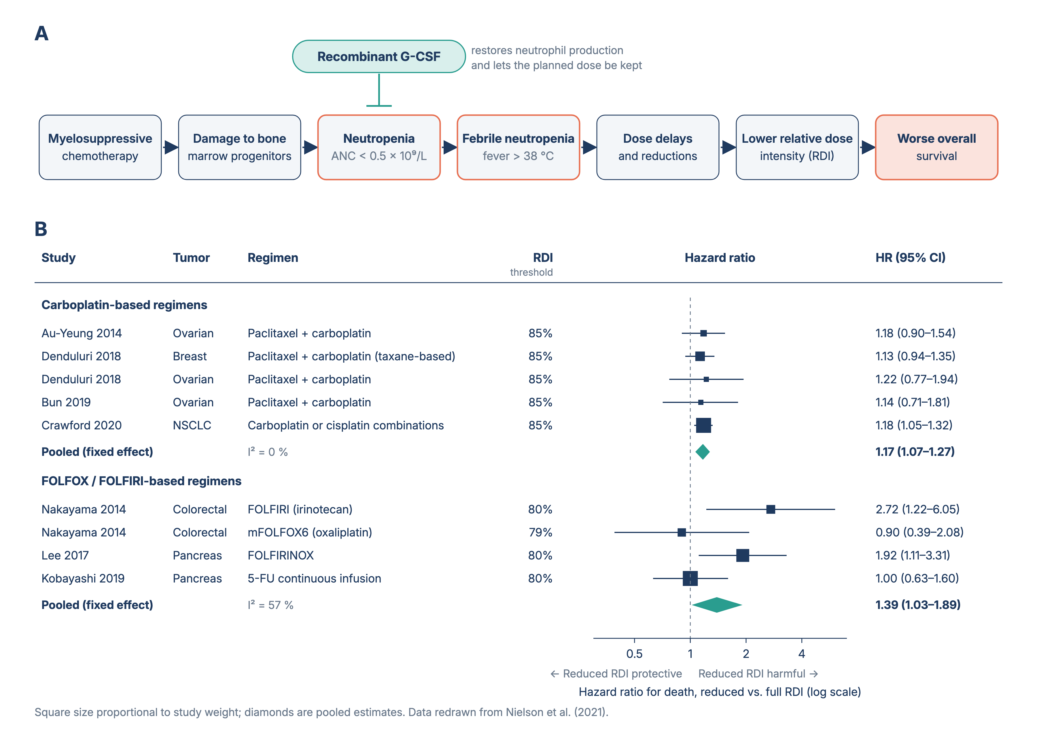 |
|---|
Other causes of neutropenia
Neutropenia can also occur not only due to certain cancers and their treatments, but also from certain infections, autoimmune diseases, genetic conditions and even unknown reasons (called chronic idiopathic neutropenia), signifying low levels of nutrients such as vitamin B-12, copper or folate, or some conditions of the spleen (Mayo Clinic Staff, 2025). Although these etiologies are important at the clinical level, neutropenia is particularly critical and dangerous to a patient in oncology, considering that it occurs frequently during oncological treatment and can interfere with a safe anticancer therapy (Freifeld et al., 2011).
Neutrophils
| Figure 2. Neutrophil functions in host defense and inflammation. Note. Adapted from Mayadas et al. (2014), Annual Review of Pathology: Mechanisms of Disease. | 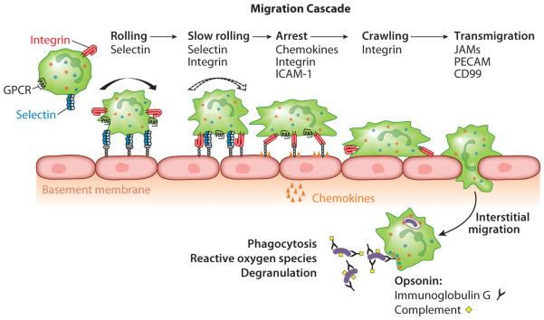 |
|---|
Neutrophils are the most predominant leukocytes in humans, produced in the bone marrow in large numbers on a daily scale. Approximately 100-200 billion neutrophils are produced daily, and certain proportions of them migrate to the bloodstream using signals, where they circulate throughout the body (Rosales, 2018). Through the constant circulation and replacement in the blood, neutrophils play critical roles in the innate immune system against infections and clearing pathogens (Rosales, 2018). The controlled migration of neutrophils from the bloodstream to the site of infection follows the sequential steps of marginalization, rolling, firm adhesion, diapedesis, and chemotaxis. Neutrophils bind to endothelial cells (on the surface of the bloodstream) and emigrate to the damaged tissues by squeezing through the endothelial cells and following the gradients of chemokines (Schultz et al., 2011). Upon the arrival at the site of infection, neutrophils perform phagocytosis through phagosomes, engulfing and eradicating the pathogens, hence, exterminating the infection at certain sites (Hampton & Dickerhof, 2023).
Neutrophils are produced through granulopoiesis, particularly in the red bone marrow which contains the blood stem cells that differentiate into red/white blood cells or platelets (National Cancer Institute, 2026). The bone marrow contains multipotent Hematopoietic Stem Cells (HSCs) which gives rise to all blood lineages through the multiple steps of differentiation (Jagannathan-Bogdan & Zon, 2013). HSCs first differentiate into Long-term HSCs to short-term HSCs (ST-HSCs) and multi-potent progenitors (MPPs) which then develop into common myeloid progenitors (CMPs) and common lymphoid progenitors (CLPs) to granulocyte monocyte progenitors (GMPs) and megakaryocyte/erythrocyte progenitor (MEP) (Sagar Paudel et al., 2022). Ultimately, under the control of a specific cytokine called G-CSF, GMPs give rise to the generation of neutrophils. The problems with neutropenic patients arise with the key characteristic of neutrophils that they are short-lived and do not undergo cell division. Neutrophils have a half-life of an estimated 19 h, indicating the need for consistent regeneration, which neutropenic patients lack due to damage in bone marrow (Zhang et al., 2024). Therefore, the neutropenic patients with affected function of bone marrow producing leukocytes, including neutrophils, would experience an increased hazard of infection, leading to dangerous states.
G-CSF: granulocyte-colony stimulating factor
| Figure 3. Overview of G-CSF signaling pathways involved in inducing myeloid cell differentiation. Note. Adapted from Dwivedi et al. (2016), Journal of Hematology & Oncology. | 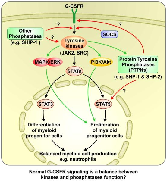 |
|---|
G-CSF is a hematopoietic cytokine that stimulates the neutrophil production and mobilization from the bone marrow into the blood circulation. Binding of G-CSF to its receptor, Granulocyte colony-stimulating factor receptor (G-CSFR) on myeloid progenitors activates signaling pathways such as JAK-STAT, MAPK, and PI3K/AKT, that promote cell proliferation and differentiation (Dwivedi & Greis, 2017). JAK-STAT pathway, is a major induction pathway in which activated (phosphorylated) STAT proteins translocate to the nucleus and activate genes such as C/EBPβ and C/EBPα that control granulocyte differentiation into neutrophils (de la Luz Sierra et al., 2010) (Figure 2). Moreover, G-CSF also promotes the release of neutrophils into the blood circulation by the CXCR2,CXCR4 to CXCL12 interaction that normally keeps them in the bone marrow (Rosales, 2018). Through both neutrophil mobilization and production from the bone marrow, G-CSF acts as a centerpiece of the innate immune system (Summers et al., 2010).
In neutropenic patients affected by chemotherapies, therefore, the external dosage of G-CSF will be helpful in compensating for the loss of neutrophils by directly inducing a higher production and release of neutrophils into the bloodstream. By resisting the effect of myelosuppressive chemotherapy, inhibiting cell replication and proliferation, the extra dose of G-CSF injected would forcefully lead less affected progenitor cells to differentiate and replenish neutrophils to the normal ANC level artificially. This lowers the burden on patients of the potential infection and prevents the incidence of febrile neutropenia as a complication of cancer therapies.
Therapeutic importance of G-CSF
We chose the specific target of febrile neutropenia, since it remains a serious complication of cancer treatment, especially in patients who are receiving myelosuppressive chemotherapy. While they're designed to destroy malignant cancer cells in the body, they can also weaken the immune system by reducing neutrophil production, placing patients at a higher risk of infections and treatment interruption. This makes supportive therapies, such as additional dose of G-CSF, highly important treatments in modern oncology, as they “subsequently allow more patients to receive adequate dose intensity chemotherapy, and lead to improved overall survival” (Flanigan et al., 2024). Furthermore, we were particularly interested in recombinant G-CSF as not only does it already have clear clinical relevance, but its production is still heavily dependent on specialized biomanufacturing systems, which we aim to enhance by lowering production costs. We aim to use biotechnology to contribute to a both practical and accessible model of therapeutic G-CSF protein production. Our project aims to expand the supply of G-CSF by mass producing the recombinant proteins using an engineered expression vector transformed into E. coli. We are particularly interested in this topic because it connects a real and clinically relevant problem with a realistic synthetic biology approach.
Systems level
Our system is based on an engineered pET expression vector transformed into an E. coli BL21 (DE3) strain to enable efficient high-yield recombinant G-CSF production.
Plasmid engineering
We designed the plasmid using an Isopropyl β-D-1-thiogalactopyranoside (IPTG) -inducible pET system that contains the T7 promoter in order to maximize the efficiency of expressing recombinant genes and corresponding G-CSF proteins. More specifically, we chose a translation vector of pET-22 (+), which enables the efficient translation and purification of G-CSF with various genes: pelB, Ribosome Binding Site (RBS), ampicillin resistance (ampR) and 6xHis-tag.
Transformation
The plasmid will be transformed into the BL21 (DE3) strain of E. coli, which produces T7 RNA polymerase and hence enables the transcription of pET-22 (+) that has a T7 promoter. This promoter is essential as it's specifically recognized by T7 RNA polymerase, allowing controlled transcription of the target gene following IPTG induction to take place. The transformation of plasmid into BL21 strain will be facilitated with the application of CaCl2 and heat shock methods. This method was chosen instead of alternate methods because it is simple and effective, and does not require more expensive specialized equipment. The application of CaCl2 causes DNA to bind to the cell more efficiently by inducing the over production of outer membrane proteins including the ones that bind DNA. After efficient binding of DNA, heat shock of briefly raising the temperature to 42°C would alter the permeability of the membrane, enabling the transport of plasmid into the cell. (Brown, 2020, p.86)
Expression
Once transformed, cells would be grown on ampicillin-containing agar to screen for those that successfully incorporated the plasmid. Transformed cells would survive due to the presence of ampicillin resistance genes on the plasmid. After the growth of colonies on the antibiotics agar, selected cell colonies will be transferred to Luria-Bertani (LB) medium, where the IPTG will be added, leading to the transcription of the target genes of the cells. After translation occurs in the cytoplasm, PelB, a signal peptide included in our engineered plasmid which is expressed along with the G-CSF protein, directs it through the Sec pathway into periplasmic space (Billen et al., 2017). The recombinant protein complex will be produced following the sequence of pelB – 6xHis-tag – TEV (tobacco etch virus protease cleavage site) – G-CSF. The presence of the bacterial peptide (pelB) prevents the recombinant protein from being degraded by the host cell and simplifies the problem of purification from the culture by directing it to a certain site of E. coli (Brown, 2020, p.286). PelB (N-terminus) leads the protein to the periplasm of the cell, which might lead to a less inclusion body formation by letting the proteins fold more properly. The media will then be centrifuged, leaving the cells in the pellet, so that they can be later extracted along with the desired expressed protein.
Extraction
The cell culture would be lysed with osmotic shock, which specifically releases the periplasmic contents minimizing cytoplasmic contamination. The periplasmic space is the area between cell membrane and outer membrane. The release of recombinant protein complex (6xHis-tag – TEV – G-CSF) in the periplasm would be followed by the centrifugation to separate the cell debris pellet and supernatant, which would contain the soluble recombinant proteins of G-CSF. After the centrifugation, the supernatant will undergo immobilized metal affinity chromatography in order to filter out the G-CSF proteins from other soluble contents with the 6xHis-tag, isolating the recombinant G-CSF protein.
Device level
The proposed chassis for this system is E. coli BL21(DE3), a commonly used and chemically competent strain suitable for transformation and high level protein expression (New England Biolabs, 2026). The BL21(DE3) strain derives from the E. coli B strain, which is particularly used for recombinant protein production due to its reduced protease activity, attributed to the absence of Lon and OmpT proteases (New England Biolabs, 2026). Lon protease mediates the degradation of mutant and misfolded proteins within the cytoplasm, while OmpT protease is located extracellularly, which breaks apart proteins released from the cell after lysis (UniProt, 2026). The lack of both these proteases ultimately allows for a more stable accumulation of recombinant proteins, which may aid in improving overall G-CSF yield. Additionally, the BL21(DE3) strain contains the DNA of the DE3 prophage in its chromosome, an engineered version of bacteriophage lambda, containing T7 RNA polymerase controlled by lacUV5 promoter. Hence, a compatible plasmid specifically containing a T7 promoter and lac operator (lacO), along with other basic necessary elements such as ribosome binding site (RBS), Origin of replication (Ori), T7 terminator, and a resistance marker is crucial.
Along with the genetic components of E. coli BL21(DE3), its cellular structure may contribute to the effective production and folding of G-CSF by providing specialized environments for recombinant protein development. After translation occurs in the cytoplasm, the protein should be transported to the periplasm because of the PelB.The pe riplasm offers an oxidizing environment which overall promotes the correct folding of G-CSF (Davies, 2001). As evident, the E. coli BL21(DE3) strain possess qualities that make it an exceptional candidate for the production of recombinant G-CSF as well as in the context of mass production for therapeutic processes.
The system achieves its intended functionality through our pET-based engineered plasmid. This device is effective through external chemical induction using isopropyl 𝛽-D-1-thiagalatopyranoside (IPTG), which introduces events such as transcription, mRNA synthesis, translation, protein transportation, folding and purification which ultimately result in the production of G-CSF.
Protein induction
In order to control the timing of gene expression, the integration of a LacO-Lacl system is implied in our plasmid. The Lacl repressor binds to the lac operator sequence, which being adjacent to the T7 promoter blocks transcription from the T7 lac promoter until IPTG induction (Lewis & Bell, 2000). Once IPTG is added externally through the growth medium, it binds to the Lacl repressor, and allosterically inhibits it, causing structural change to the repressor and ultimately forcing it to release from DNA. This allows the host RNA polymerase to transcribe the T7 polymerase gene into mRNA, which subsequently is translated by ribosomes into T7 RNA polymerase protein.
Transcription
The synthesized protein then binds to the T7 promoter, initiating transcription of the G-CSF gene. T7 RNA polymerase is selective, therefore only binds to the T7 promoter sequence due to a specificity loop, or a structural element in the enzyme that only allows it to link on to certain molecules (Cole & Eastoe, 1988). Due to this quality, T7 RNA polymerase does not mostly recognize other bacterial promoters and focuses on transcribing G-CSF only. The initiation of transcription produces G-CSF mRNA that is translated by E. coli ribosomes into recombinant G-CSF protein. The attached signal peptide directs the protein into the periplasmic space, where proper folding and disulfide bond formation occur before purification.
Translation, translocation and periplasmic development
The G-CSF mRNA produced by T7 RNA polymerase contains a ribosome binding site (RBS) that kickstarts the E. coli’s ribosomes, initiating translation of the desired protein. The initial part of the amino acid sequence created is PelB sequence. Sequentially, the sequence is recognized by Sec translocase, a protein pump which drives the unfolded chain into periplasmic space (Lu et al., 1990). Throughout the end or after translocation, PelB would be cleaved by a signal peptidase, therefore would be removed from the G-CSF protein. The periplasm is particularly important to development due to oxidation, while the cytoplasm does not offer this, the periplasm allows disulphide bonds to form which are necessary for proteins such as G-CSF to fold into their tertiary structure (Manta et al., 2019). Additionally, the periplasm contains Disulfide bond formation proteins (DsbA) which help with the folding of proteins. This is important as the production of the protein may lead to the formation of inclusion bodies, which is exactly due to the improper folding of such proteins as well as endotoxin contamination. This would lead to the requirement of a complex refolding process during purification, including harsh denaturing agents to break about the insoluble clumps of unfolded protein.
Termination
The T7 terminator sequence is responsible for the last stage of the process, which is the termination of the gene expression. When the sequence is transcribed into RNA, via T7 RNA polymerase it forms a structure that stops T7 RNA polymerase from progressing along the DNA strand and ultimately stopping the transcription of G-CSF (Part:BBa_K731721 – Parts.Igem.Org, 2019).
Centrifuge process
Once protein production ends, bacterial culture would be harvested via centrifugation. The process is a physical dewatering, or separation method, that allows components to be separated by density and the speed of the rotational forces (ref). Hence, the bacterial pellet will be formed at the end of the tube as it is denser than its surrounding liquid (Luria- Bertrani Medium), and can be isolated by simply removing the remaining liquid (supernatant). This would be repeated before immobilized metal affinity chromatography (IMAC), following osmotic shock to ensure that the sample will be separated from the soluble periplasmic proteins from insoluble material in the supernatant and cell debris which will be left in pellet form. This is because we're assuming that the G-CSF would fold properly in the periplasm in a soluble state rather than forming inclusion bodies, so the additional membrane fragments would form in the pellet. The collected supernatant can then be purified by IMAC, where the 6xHis tagged G-CSF would bind to the nickel resin.
Cell lysis
After isolation, the cells must be lysed to obtain G-CSF. As G-CSF proteins develop in the periplasmic space,osmotic shock should allow the release of periplasmic contents without disrupting the cytoplasm, reducing contamination from other proteins and, therefore, simplifying the subsequent purification process (Green & Mecsas, 2016). First cells would go under hypertonic shock, by incubating them with a high sucrose buffer (EDTA), which destabilizes the outer membrane. Cell culture would then be immediately moved to ice-cold water, which would allow water to reach the periplasm, and the outer membrane to burst and release G-CSF (Lu et al., 1990).
Purification
In order to specifically obtain recombinant G-CSF, it must be separated from other cellular components that the bacteria naturally possesses and produces. This would be done via affinity-based purification and enzymatic processing.
Immobilized Metal Affinity Chromatography (IMAC) would use the six Histidine residues (6xHis-Tag) embedded in the plasmid, in order to distinguish G-CSF from other proteins. Since the His-tags are incorporated into the protein, they allow it to be recognised and isolated specifically. Identification is possible due an imidazole side chain included in the Histag, which can bond to metal ions (Bornhorst & Falke, 2000). The IMAC chromatography column contains resin beads with immobilized nickel ions inside them. When the protein mixture passes through, only the proteins with the His-tag (G-CSF, our targeted protein), sticks to the nickel ions, and the other proteins pass through (Bornhorst & Falke, 2000). After passing through, the His-tagged G-CSF is washed away from the column by adding a high concentration of imadazole. This solution will compete with the histidine residues to bind to the nickel ions, therefore leading to the release of the protein from the resin.
Once G-CSF is separated from other bacterial proteins, the His-tag residues would be removed by enzymatic proteolysis.could influence the function of the protein itself. This would be done through TEV protease activity, which distinguishes the TEV protease cleavage site located between the His-tag and G-CSF protein. The enzyme TEV protease, cuts between Histag and G-CSF by recognizing the amino acid sequence of TEV protease cleavage site, and separates Histag from our designated protein (His-Tagged Proteins–Production and Purification | Thermo Fisher Scientific – ES, 2026).
Parts level
As previously mentioned in the Device level, our E. coli BL21(DE3) chassis requires a compatible plasmid to successfully produce the G-SCF recombinant protein. In the following section, the functions of each genetic component responsible for the G-CSF expression and purification will be described according to their specific roles in the context of our investigation.
Table 1: fGenetic components of the engineered plasmid system and their roles in the production of recombinant G-CSF.
| Part | Function |
|---|---|
| Control & Transcription Module | Lacl: The Lacl repressor protein is a homotetrameric (homodimer) transcription factor that regulates DNA expression by binding to Lac Operator sequence (LacO), stopping RNA polymerase from initiating transcription (Khan Academy, 2026). At the N-terminal headpiece, the protein contains a helix-turn-helix (HTH) motif. HTH inserts and latches itself onto the major groove of operator DNA through mostly hydrogen bonding and electrostatic attractions, ultimately allowing it to recognise the operator. Hence, the protein experiences very high affinity (10-13– 10-14M range in Kd ), which is vital because it signifies that the Lacl repressor protein forms strong connections with operator DNA, resulting in more efficiency in the context of the identification of the operator (Fidabio, 2022). Additionally, Lacl repressor protein is inhibited allosterically, through the introduction of IPTG, which alters its configuration and significantly reduces its affinity, causing it to latch off and allow RNA polymerase to transcribe the desired G-CSF gene. LacO: The Lac Operator sequence is a 27 base pair sequence which allows the binding of Lacl repressor protein, to control G-CSF transcription (Lu et al., 1990). The Lac Operator sequence is approximately palindromic, allowing both monomers that make up the Lacl repressor protein to bind on each nearly identical side (Zuo et al., 2015). Once binding occurs (in both major and minor grooves), the central CG base steps in the sequence are bent, due to the insertion of Lacl repressor proteins alpha helixes in the minor groove. Hence, when Lacl binds to two specific sites, This causes stable protein- DNA complex formation, resulting in an approximate 40-50° kink in the DNA structure (Kalodimos, 2002), contributing to the high affinity bonding, efficient attachment of the Lacl and LacO sequence and minimisation of basal expression (Wu et al., 2024). T7 promoter: The T7 promoter is a sequence of 18 DNA base pairs recognised by T7 RNA polymerase. The promoter binds to T7 RNA polymerase (which is produced by the cell, as it is a derivative of the DE3 prophage which has been explained in Device level), with high affinity and initiates rapid transcription rates, such as 200-300 nucleotides per second. T7 terminator: The T7 terminator sequence is a specific DNA sequence that stops the transcription of T7 RNA polymerase. T7 terminator, like the T7 promoter, is derived from the bacteriophage T7 phage (Calvopina-Chavez et al., 2022). It is characterised by a GC base pair inverse repeat, and an A rich region on the template strand. In the GC inverse region, the RNA transcript forms a loop, making it difficult for transcription to continue (Peters et al., 2011). Additionally the U-Tract (the adenine region), is transcribed as 8 uracils from the RNA polymerase, and when the RNA polymerase encounters the “loop” the weak AU bonds break off. This is also known as a hairpin structure (Uptain & Chamberlin, 1997). Ultimately, the hairpin structure allows the RNA polymerase to get stuck while attempting to continue transcription, making it detach, and terminating transcription. |
| Translation Module | RBS (ribosome binding site): The ribosome binding site, also known as Shine-Dalgarno sequence, is a starting point for bacterial ribosomes to initiate proper translation. The RBS is characterised by containing a variety of purines, upstream from the start codon (Jacobson & Clark, 2016). Its base pairs pair with the anti-shine-Dalargo sequence, located on the 30S subunit of Escherichia coli BL21(DE3) ribosomes, providing an access point to begin translation (Beck et al., 2016). Additionally, In order for the RBS to be efficient, mRNA must not fold onto itself creating a hairpin structure which would prevent any sequencing (there is a possibility of this happening due to the GC richness) , as well as maintaining a responsible thermodynamic stability (ΔG), which ensures that the ribosome does not detach before translation starts (Salis et al., 2009). |
| Targeting Translocation Module | PelB: The PelB signal peptide is located on the N-terminal of unfolded G-CSF and functions as a targeting sequence that guides it to the periplasm through the Sec pathway. It is structurally organised with three major regions, positively charged N-terminal, alpha helix and hydrophobic core, and C-terminal including signal peptidase cleavage site (Guo et al., 2018). The positively charged N-terminal interacts with negatively charged phospholipid headgroups in order to cross SecYEG translocon through the bacterial inner membrane (Sachelaru et al., 2017). The hydrophobic core consists of 10-15 non-polar amino acids, which create an alpha helix internally, ultimately contributing to the passing of the molecule through the non-polar tails of the lipid bilayer (Lu et al., 1990). The C-terminal contains a specific amino acid sequence, Alanine -X- Alanine, which allows Signal peptidase to “cut” the PelB sequence once it is in periplasmic space (Auclair et al., 2011). This is crucial since the G-CSF would not develop properly if PelB remained attached. |
| Purification Module | 6xHistag: The 6x Histag is a peptide containing six histidine residues, which complete their function through their interactions with transition metal ions during the IMAC process, mostly based on Lewis acid-base reactions. During IMAC, the nickel ions immobilized in the resin beads act as a “Lewis acid” accepting electron pairs, while the imidazole rings present on the histidine residues act as electron donors or “Lewis Bases”, ultimately forming a coordinate bond (metal-ligand interactions)(His-Tagged Proteins–Production and Purification | Thermo Fisher Scientific – ES, 2026). This allows for G-CSF tagged proteins to be distinguished and collected, while other bacterial proteins are washed away. |
| Post – Purification Module | TEV Cleavage site: The TEV protease cleavage site is a recognition sequence, ENLYFQ↓G/S, which allows TEV protease to recognise it, and cut in that specific location in order to separate the Histags from the protein. In order to separate the two, TEV protease forms a “binding tunnel” around the sequence (Audiopedia, 2026). In the tunnel, Histidine, Aspartate and Cysteine amino acids align between Q and G/S peptide bonds (Phan et al., 2002). Additionally, the high specificity of the cleavage site drastically decreases the probability of off-target- cleavage, making the Histag removal process efficient and precise. |
| Maintenance Module | pBR322 ori: The pBR322 origin (ori) is where DNA replication begins in the plasmid. It additionally functions to control the amount of copies that are present in a single bacteria (in this context one of our chassis, Escherichia coli BL21(DE3)) which is usually about 15-20 copies per cell (Article Series: E. coli Plasmid Origins of Replication: The Origin, 2014) . Replication is initiated through RNA II, or the pre primer about 550 base pairs from the origin. It then forms a R-loop (DNA and RNA pairing) once it reaches the origin site. The enzyme RNase H cuts the RNA to form a primer, which will be used for DNA replication (Davis et al., 1986). RNA I on the other hand is a shorter RNA molecule which is on the complementary side of RNA II. Its role is to regulate RNA I, and to prevent the formation of primers (Tomizawa, 1984). Together with the Rop gene, the origin is able to control the life cycle of the plasmid. Rop protein: The Rop protein enhances the inhibition of the RNA II by stabilizing the interaction between RNA I and RNA II, making sure that they do not separate from each other, overall increasing the control of replication through the origin of replication (Cesareni & Banner, 1985). |
| Selection Module | Ampicillin: The ampicillin resistance gene (bla) encodes for the production of β-lactamase, a serine enzyme which uses serine residue in its active site to break down the antibiotic (Class A Serine Beta-Lactamase (SBL (Class A)) – Gosset, 2026). In order to break down the ampicillin, the serine reacts with the carbonyl on the ampicillin lactam ring. When the ring opens, ampicillin becomes part of the β-lactamase enzyme, forming an intermediate acyl-enzyme. Additionally, since the ring has been broken, it loses its 3D shape, and therefore cannot function properly(Türkyılmaz & Darcan, 2026). In order for the enzyme to continue breaking apart the lactam ring, it releases the degraded ampicillin, and continues to break apart others |
Safety
Safety is a crucial focus in our investigation involving the production of a recombinant therapeutic protein, G-CSF, hoping it would eventually be used as a febrile neutropenia treatment, so both laboratory and product safety are essential to address. According to the safety data sheet provided by the possible supplier of our E. coli strain (New England Biolabs), the BL21(DE3) strain is commonly handled under BSL-1 containment using standard microbiological practices, rather than other clinical E. coli strains. Recombinant DNA production would be conducted only under institutional biosafety approval while being in accordance with established guidelines. Such would include the use of Personal Protective Equipment (PPE) such as gloves, lab coats, eye protection goggles, applying proper sterilization procedures, and following appropriate disposal of biological waste to prevent accidental exposure or even environmental release (Safety DATA Sheet, 2023).
At the process level, safety applies to each step of the system. This can be seen in the usage of ampicillin selection during transformation, induction and culture growth as it helps ensure only cells containing the plasmid are handled, likewise, our closed culture handling and aseptic techniques can reduce contamination. During processes such as protein purification, steps must be performed with chemical safety precautions because buffers and reagents can be irritating or hazardous if mishandled. Furthermore, our chassis E. coli is a gram-negative bacterium, meaning that the purified product would need strong quality control to remove endotoxins like LPS. Procedures confirming correct protein folding, purification and activity would be implemented before any form of therapeutic usage. Our investigation, once performed, must be safe both during construction and experiment testing, as well as future users to ensure that the final product is free from harmful substances and produced in a controlled environment.
Discussions
Our design uses E. coli BL21(DE3) strain with a pET expression system, a widely recognized combination for high-level recombinant protein expression. In molecular biology, bacterial systems are widely used as its practical, and we understand its genetics well, being able to follow each process during the recombinant protein expression. It's cost-effective, grows rapidly under optimal conditions and it is well suited for large-scale microbial production (Ruiz & Silhavy, 2022), which is why we selected E. coli as the host organism. BL21(DE3) strain was specifically chosen because it expresses the required T7 RNA polymerase that activates the pET vector's T7 promoter driven system. Bacterial systems are such crucial tools in biotechnology as they can use plasmids as a vector, allowing foreign genes to be introduced into the bacterial cell. The host's ability for large-scale protein production aligns with our goal of producing recombinant Granulocyte Colony-Stimulating Factor (G-CSF) protein, the large quantities would be able to be used for therapeutic purposes. However, this choice also involved its own tradeoffs, including possible folding issues and the lack of eukaryotic post-translational modifications (PTM).
BL21(DE3) was selected as it's an E. coli strain that lacks the Lon and OmpT proteases, reducing degradation of recombinant proteins during expression in the bacteria (Rosano & Ceccarelli, 2014). The DE3 designation indicates that this strain has been engineered to carry a chromosomal copy of the T7 RNA polymerase gene. Although there are other strains more suitable for disulfide bond formation or solubility, BL21(DE3) was chosen to achieve strong expression in a bacterial host. This tradeoff is relevant as our gene of interest is prone to inclusion body formation, which would require solubilization and refolding after expression (Kim et al., 2013). By combining with the protease deficient background, it makes it a practical choice for high-yield G-CSF expression using the pET vector on the T7 system.
The pET vector was selected as it allows controlled recombinant protein expression in E. coli via the T7 promoter system and a selectable marker which in our design, would be the Ampicillin resistance. Once our desired gene is transformed into the host, the bacterial machinery can express foreign genes. IPTG induction uses the lac-promoter to activate the T7 system, initiating production at a desired time point. This provides a useful level of control, allowing bacterial growth before protein expression. Although such a system may be less “advantageous for the production of those recombinant proteins with toxic or growth-burdened”, it remains widely used for “high-production of hard-to-express proteins” (Du et al., 2021) such as G-CSF. However, because the pET/BL21(DE3) system is designed for high-level expression, overproduction may also increase the risk of the formation of inclusion bodies.
As we are trying to express a human gene in a prokaryotic cell, folding issues can arise due to the lack of eukaryotic PTM systems. Although we could have used an eukaryotic system, the culture itself is more expensive, slower-growing and harder to manage at a large-scale production unlike prokaryotes like bacteria. One of the relevant limitations of the lack of PTM systems in our project is glycosylation, although this is less limiting for G-CSF as filgrastim is already used clinically as a non-glycosylated recombinant human G-CSF produced in E. coli (HIGHLIGHTS of PRESCRIBING INFORMATION, n.d.). Formation of proper disulfide bonds would present challenges in protein folding in bacterial cells, probably leading to inclusion bodies which would require refolding during purification. Therefore, E. coli remains a practical host for recombinant G-CSF production.
As this project is a hypothetical design, it hasn't been experimentally validated therefore, no conclusions can be made regarding the recombinant G-CSF's expression yield, purity or biological activity. Furthermore, expression in E. coli may present challenges such as the formation of inclusion bodies due to the improper folding and endotoxin contamination, all of which would need to be addressed before therapeutic application. Such inclusion bodies or insoluble clumps of unfolded protein require harsh denaturing agents and a complex refolding process, therefore the production yield may not express fully functional G-CSF protein without optimization. Meanwhile, since E. coli is a gram-negative bacteria, it can contaminate the product during cell lysis with endotoxins, usually lipopolysaccharides (LPS)c(Lin et al., 2004). LPS are highly toxic to humans even at very low levels, triggering strong immune responses such as fever, inflammation, and even septic shock. This can be addressed using endotoxin-removal methods such as resins, anion-exchange chromatography and other methods (ref?). One example is Pierce High Capacity Endotoxin Removal Resin from ThermoFisher, which is designed to remove LPS from purified protein samples. Furthermore, even after recombinant G-CSF is successfully expressed, the protein must demonstrate correct folding, stability, purity, and biological activity compared to such clinically approved G-CSF products before it can be applied in febrile neutropenia treatment. Any inconsistencies could affect clinical efficacy in patients, while their translational challenges such as regulatory approval and quality control will help by ensuring that the final product is safe enough for patient usage. Therefore, while our investigation hopes to offer a practical low-cost production method of the protein, fine tuning would still be required before the product could be used in febrile neutropenia management.
Next steps
Future work would include converting the conceptual design into a carried out experimental framework, including experimental cloning of the target gene, transformation into E. coli, induction of the protein's expression, and purification. The bacteria could be grown under optimal conditions (fed-batch culture) in order to increase protein yield and high cell density. Experimental validation would also be required to confirm successful protein expression and to assess the quality of the G-CSF product. By using assay methods such as sodium dodecyl sulfate-polyacrylamide gel electrophoresis (SDS-PAGE), we could confirm whether G-CSF is being expressed at the expected molecular weight at around 19 kDa, whether the protein remained intact rather than degraded, and to evaluate the protein's purity by determining whether non-target bacterial proteins have been removed. Beyond expression, future studies should compare the purity and yields with existing G-CSF products to evaluate the safety, biological activity, and its practical value in the feasibility of the system to develop a cost-effective design for the production of a recombinant G-CSF to improve the accessibility of cancer therapies. Furthermore, the project should go deeper into more fundamental problems within the proper foldings of proteins in E.coli. Currently, the protein mass-production cost is highly increased by the additional processing step of the post-translational modifications, including the glycosylation, which bacterial host cells lack when it is essential for most proteins of therapeutic use in humans. Therefore, the next step would include the discussion about the protein modifications and the proposal of more efficient modifying system in order to fundamentally lower the production cost for real therapeutic applications.
Author contributions
Abstract by SH, Video by SH, Background by RLD and SH, Systems level by SH, Device levels by MF, Parts level by MF, Safety by VW, Discussion by VW, Next-steps by VW.
Acknowledgements
We are very grateful for the support from the American School of Madrid.
Firstly, we would like to thank our mentor Dr. Michael Sheets for his invaluable guidance, and for supporting us since the start of BioBuilders. His valuable insight regarding our ideas and choices, helped us succeed in writing our Biotreks article, and ultimately helped us develop a final conceptual design.
Above all, we would like to express our sincere gratitude to our teacher leader, Ms. Meghan Perks, whose unwavering support and dedication throughout this process helped us navigate the challenges of this research. By allowing us the opportunity to further our understanding of Biology and applying it to synthetic biology, we were able to triumphantly pursue our passion for scientific exploration.
A special thanks to each author whose consistent effort and commitment contributed to the completion of this project. We appreciate the encouragement from our peers Katharina, Edith, Alexis and Fay, who provided both their valuable insights and motivation throughout this journey.
References
Billen, B., Vincke, C., Hansen, R., Devoogdt, N., Muyldermans, S., Adriaensens, P., & Guedens, W. (2017). Cytoplasmic versus periplasmic expression of site-specifically and bioorthogonally functionalized nanobodies using expressed protein ligation. Protein Expression and Purification, 133, 25–34. https://doi.org/10.1016/j.pep.2017.02.009
Borgeaud, M., Perano, S., Addeo, A., & Tsantoulis, P. (2024). Rates of febrile neutropenia and its causes in the real world. Future Oncology, 20(29), 2203-2212. https://doi.org/10.1080/14796694.2024.2349510
Bornhorst, J. A., & Falke, J. J. (2000). [16] Purification of proteins using polyhistidine affinity tags. Methods in Enzymology, 245–254. https://doi.org/10.1016/s0076-6879(00)26058-8
Bray, F., Laversanne, M., Sung, H., Ferlay, J., Siegel, R. L., Soerjomataram, I., & Jemal, A. (2024). Global cancer statistics 2022: GLOBOCAN estimates of incidence and mortality worldwide for 36 cancers in 185 countries. CA: A Cancer Journal for Clinicians, 74(3), 229–263. https://doi.org/10.3322/caac.21834
Casale, J., & Patel, P. (2024). Fluorouracil. In StatPearls [Internet]. StatPearls Publishing. Retrieved April 8, 2026, from https://www.ncbi.nlm.nih.gov/books/NBK549808/
Centers for Disease Control and Prevention. (2025, February 13). Neutropenia and risk for infection. https://www.cdc.gov/cancer-preventing-infections/patients/neutropenia.html
Cole, A. S., & Eastoe, J. E. (1988). Enzymes. Biochemistry and Oral Biology, 71–89. https://doi.org/10.1016/b978-0-7236-1751-8.50013-2
Daniel, D., & Crawford, J. (2006). Myelotoxicity from chemotherapy. Seminars in Oncology, 33(1), 74–85. https://doi.org/10.1053/j.seminoncol.2005.11.003
Davies, M. J. (2001). Periplasmic protein screening. Trends in Biotechnology, 19(8), 286. https://doi.org/10.1016/s0167-7799(01)01763-2
de la Luz Sierra, M., Sakakibara, S., Gasperini, P., Salvucci, O., Jiang, K., McCormick, P. J., Segarra, M., Stone, J., Maric, D., Zhu, J., Qian, X., Lowy, D. R., & Tosato, G. (2010). The transcription factor Gfi1 regulates G-CSF signaling and neutrophil development through the Ras activator RasGRP1. Blood, 115(19), 3970–3979. https://doi.org/10.1182/blood-2009-10-246967
Du, F., Liu, Y.-Q., Xu, Y.-S., Li, Z.-J., Wang, Y.-Z., Zhang, Z.-X., & Sun, X.-M. (2021). Regulating the T7 RNA polymerase expression in E. coli BL21 (DE3) to provide more host options for recombinant protein production. Microbial Cell Factories, 20(1).
Dwivedi, P., & Greis, K. D. (2017). Granulocyte Colony Stimulating Factor Receptor (G-CSFR) signaling in severe congenital neutropenia, chronic neutrophilic leukemia and related malignancies. Experimental Hematology, 46, 9–20. https://doi.org/10.1016/j.exphem.2016.10.008
Flanigan, J. A., Yasuda, M., Chen, C.-C., & Li, E. C. (2024). Chemotherapy-induced febrile neutropenia (FN): healthcare resource utilization (HCRU) and costs in commercially insured patients in the US. Supportive Care in Cancer, 32(6). https://doi.org/10.1007/s00520-024-08492-5
Freifeld, A. G., Bow, E. J., Sepkowitz, K. A., Boeckh, M. J., Ito, J. I., Mullen, C. A., Raad, I. I., Rolston, K. V., Young, J.-A. H., & Wingard, J. R. (2011). Clinical practice guideline for the use of antimicrobial agents in neutropenic patients with cancer: 2010 update by the Infectious Diseases Society of America. Clinical Infectious Diseases, 52(4), e56–e93. https://doi.org/10.1093/cid/cir073
Green, E. R., & Mecsas, J. (2016). Bacterial Secretion Systems: An Overview. Microbiology Spectrum, 4(1). https://doi.org/10.1128/microbiolspec.vmbf-0012-2015
Hampton, M. B., & Dickerhof, N. (2023). Inside the phagosome: A bacterial perspective. Immunological Reviews, 314(1), 197–209. https://doi.org/10.1111/imr.13182
HIGHLIGHTS OF PRESCRIBING INFORMATION. (n.d.). https://www.pi.amgen.com/-/media/Project/Amgen/Repository/pi-amgen-com/Neupogen/neupogen_pi_hcp_english.pdf
His-tagged Proteins–Production and Purification | Thermo Fisher Scientific – ES. (2026). Thermofisher.com. https://www.thermofisher.com/es/es/home/life-science/protein-biology/protein-biology-learning-center/protein-biology-resource-library/pierce-protein-methods/his-tagged-proteins-production-purification.html
International Agency for Research on Cancer. (2024, April 4). New report on global cancer burden in 2022 by world region and human development level. https://www.iarc.who.int/news-events/new-report-on-global-cancer-burden-in-2022-by-world-region-and-human-development-level/
Jagannathan-Bogdan, M., & Zon, L. I. (2013). Hematopoiesis. Development, 140(12), 2463–2467. https://doi.org/10.1242/dev.083147
Kim, C. K., Lee, C. H., Lee, S.-B., & Oh, J.-W. (2013). Simplified Large-Scale Refolding, Purification, and Characterization of Recombinant Human Granulocyte-Colony Stimulating Factor in Escherichia coli. PLoS ONE, 8(11). https://doi.org/10.1371/journal.pone.0080109
Lewis, M., & Bell, C. E. (2000). Nature Structural Biology, 7(3), 209–214. https://doi.org/10.1038/73317
Lin, M.-F., Williams, C., Murray, M. V., & Ropp, P. A. (2004). Removal of lipopolysaccharides from protein–lipopolysaccharide complexes by nonflammable solvents. Journal of Chromatography B, 816(1-2), 167–174. https://doi.org/10.1016/j.jchromb.2004.11.029
Lu, S., Reyes, V. E., Lew, R. A., Anderson, J., Mole, J., Humphreys, R. E., & Ciardelli, T. (1990). Role of recurrent hydrophobic residues in catalysis of helix formation by T cell-presented peptides in the presence of lipid vesicles. Journal of Immunology (Baltimore, Md. : 1950), 145(3), 899-904. https://pubmed.ncbi.nlm.nih.gov/2373862/
Manta, B., Boyd, D., & Berkmen, M. (2019). Disulfide Bond Formation in the Periplasm of Escherichia coli. EcoSal Plus, 8(2). https://doi.org/10.1128/ecosalplus.esp-0012-2018
Mayo Clinic Staff. (2025, January 23). Neutropenia (low neutrophil count): Causes. Mayo Clinic. https://www.mayoclinic.org/symptoms/neutropenia/basics/causes/sym-20050854
National Cancer Institute. (2026). NCI Dictionary of Cancer Terms. Cancer.gov. http://cancer.gov/publications/dictionaries/cancer-terms/def/bone-marrow
National Institute of Diabetes and Digestive and Kidney Diseases. (2020, September 15). Carboplatin. In LiverTox: Clinical and research information on drug-induced liver injury [Internet]. National Library of Medicine. https://www.ncbi.nlm.nih.gov/books/NBK548565/
New England Biolabs. (2026). Neb.com. https://www.neb.com/en/products/c2527-bl21de3-competent-e-coli
Nielson, C. M., Bylsma, L. C., Fryzek, J. P., Saad, H. A., & Crawford, J. (2021). Relative dose intensity of chemotherapy and survival in patients with advanced stage solid tumor cancer: A systematic review and meta-analysis. The Oncologist, 26(9), e1609–e1618. https://doi.org/10.1002/onco.13822
Part:BBa_K731721 – parts.igem.org. (2019). Igem.org. https://parts.igem.org/Part:BBa_K731721
Punnapuzha, S., Edemobi, P. K., & Elmoheen, A. (2023, March 30). Febrile Neutropenia. PubMed; StatPearls Publishing. https://www.ncbi.nlm.nih.gov/books/NBK541102/
Rosales, C. (2018). Neutrophil: a Cell with Many Roles in Inflammation or Several Cell Types? Frontiers in Physiology, 9(113). https://doi.org/10.3389/fphys.2018.00113
Rosano, G. L., & Ceccarelli, E. A. (2014). Recombinant protein expression in Escherichia coli: Advances and challenges. Frontiers in Microbiology, 5(172). https://doi.org/10.3389/fmicb.2014.00172
Rout, P., Reynolds, S. B., & Zito, P. M. (2024). Neutropenia. In StatPearls [Internet]. StatPearls Publishing. Retrieved April 8, 2026, from https://www.ncbi.nlm.nih.gov/books/NBK507702/
Ruiz, N., & Silhavy, T. J. (2022). How Escherichia coli Became the Flagship Bacterium of Molecular Biology. Journal of Bacteriology, 204(9). https://doi.org/10.1128/jb.00230-22
SAFETY DATA SHEET. (2023). https://www.neb.com/-/media/9d078c75306d47e798441d734840fb69.pdf?srsltid=AfmBOoqyPIhAeXSNHB4ksRseIPM6uUIt7t22dJFs15xWIOdO3V8gsGFE
Sagar Paudel, Laxman Ghimire, Jin, L., Jeansonne, D., & Samithamby Jeyaseelan. (2022). Regulation of emergency granulopoiesis during infection. Frontiers in Immunology, 13. https://doi.org/10.3389/fimmu.2022.961601
Schultz, G. S., Chin, G. A., Moldawer, L., Diegelmann, R. F., Fitridge, R., & Thompson, M. (2011). Principles of Wound Healing. In Mechanisms of Vascular Disease: A Reference Book for Vascular Specialists (pp. 423–450). University of Adelaide Press. http://www.jstor.org/stable/10.20851/j.ctt1sq5w94.27
Summers, C., Rankin, S. M., Condliffe, A. M., Singh, N., Peters, A. M., & Chilvers, E. R. (2010). Neutrophil kinetics in health and disease. Trends in Immunology, 31(8), 318–324. https://doi.org/10.1016/j.it.2010.05.006
UniProt. (2026). UnProt. https://www.uniprot.org/uniprotkb/P0A9M0/entry
van den Boogaard, W. M. C., Komninos, D. S. J., & Vermeij, W. P. (2022). Chemotherapy side-effects: Not all DNA damage is equal. Cancers, 14(3), 627. https://doi.org/10.3390/cancers14030627
Zhang, F., Xia, Y., Su, J., Quan, F., Zhou, H., Li, Q., Feng, Q., Lin, C., Wang, D., & Jiang, Z. (2024). Neutrophil diversity and function in health and disease. Signal Transduction and Targeted Therapy, 9(1). https://doi.org/10.1038/s41392-024-02049-y