
Holden Benbrook, Ben Boyd, Isadora Conour, Gayatri Gadhvi, Nathan Kralik, Audrey J. Latham, Ava M. Lebovitz, and Keaton Roberts, Oak Park and River Forest High School, Oak Park, Illinois, United States of America
Reviewed on 9 May 2026; Accepted on 22 June 2026; Published on 26 October 2026
With help from the 2026 BioTreks Production Team.
Polyvinyl chloride (PVC) is one of the most widely used plastics, accounting for 10% of global plastic production. As PVC degrades, microplastics–small plastic particles less than 5 millimeters in length–are produced. These microplastics are increasingly recognized as an environmental and public health concern due to their widespread presence and potential biological harm. Studies suggest that ingesting PVC microplastics may damage gut structure, promote carcinogenesis in humans, and induce in vitro toxicity in human pulmonary cells. Despite these risks, the lack of medically relevant methods of detecting microplastic exposure limits our ability to fully understand the extent of microplastic exposure in humans. Current detection methods largely rely on physical or chemical characterization techniques that are costly, time-consuming, and poorly suited for assessing biological impact. In this project, we refine a proposal by students at Brookline High School to genetically engineer an E. coli-based biosensor that produces a measurable visual signal in response to PVC microplastic exposure. The design stems from the hypothesis that exposure to PVC induces oxidative stress in E. coli, leading to upregulation of the OxyR regulatory pathway. By coupling this pathway with a luminescent reporter system, we aim to establish a connection between the input of PVC and the output of luminescence, allowing us to determine the feasibility and limitations of this biosensor as a practical PVC detection tool.
Keywords: Polyvinyl chloride, biosensor, oxidative stress pathway, katG, NanoLuc
Authors are listed in alphabetical order. Matthew Kirkpatrick mentored the group. Please direct all correspondence to mailto:Mkirkpatrick@oprfhs.org.



Background
The global production and degradation of plastics have led to the widespread accumulation of microplastics across ecosystems. Microplastics, defined as plastic particles less than 5 millimeters long, originate from the breakdown of large plastic debris and the direct release of small plastic particles from plastic products (National Oceanic and Atmospheric Administration, 2024). As a result, plastic pollution has widely permeated ecological systems and is consistently detected in freshwater and marine water systems, soil, air, and food chains. Microplastics derived from PVC, in particular, represent a significant concern due to the high production volume and resistance to degradation of PVC, which accounts for approximately 10% of global plastic production (Kudzin et al., 2023).
In addition to their environmental persistence, recent studies have demonstrated that microplastics are biologically active particles. Microplastics have been detected in human tissues, including within organs and skeletal systems, suggesting that microplastic exposure is widespread and may potentially lead to systemic bioaccumulation (Roslan et al., 2024; Yang et al., 2025). Minimal microplastic exposure has been associated with negative effects at the cellular level, such as oxidative stress, inflammation, and toxicity across multiple organ systems (Banerjee & Shelver, 2021; Lee et al., 2023). This indicates that microplastics pose long-term health risks, such as disease progression or tissue dysfunction (Goswami et al., 2024).
However, current microplastic detection methods are less accessible for routine use to assess biologically relevant exposure. Techniques such as Raman imaging and chemical analysis primarily focus on identifying and confirming the presence of microplastics but do not assess the biological impacts and interactions with living systems (Luo et al., 2023). Although these methods effectively confirm the presence of microplastics, they are inaccessible due to the cost, time-intensive nature, and requirement of specialized equipment. Furthermore, they fail to highlight or provide insight into the biological impact of microplastic exposure on humans at the cellular level.
To address the gap, researchers have begun exploring alternative microbial-based biodetection methods. Microbial biosensors, particularly sensors utilizing genetically engineered bacteria, provide a promising approach for measuring biological outputs. These systems utilize natural cellular pathways that respond to environmental stressors, enabling the detection of contaminants through functional biological responses rather than solely physical presence (Choi et al., 2025; American Chemical Society, 2025). By integrating environmental pressures with cellular response mechanisms, biosensors have the potential to greatly impact how microplastic exposure is monitored. Instead of relying solely on detecting plastic particles, this biosensor can provide insight into the biological effects of exposure.
As microplastic pollution continues to increase, there is a growing need for cost-effective and biologically meaningful detection tools. A biosensor could be used in a clinical or diagnostic setting to detect biomarkers of microplastic-induced cellular stress once proper certification and validation are achieved. This would enable real-time environmental monitoring and improve the assessment of human exposure. Additionally, these systems may contribute to regulatory efforts by providing more accurate data on exposure risks, ultimately informing policy decisions and mitigation strategies. Integrating biosensor technology into clinical research could improve the understanding of how environmental pollutants impact human health outcomes and help guide the development of targeted prevention and treatment strategies.
Systems level
To create our sensor, we would first insert our plasmid, containing the katG promoter, NanoLuc reporter, pUC19 backbone, and ampicillin resistance marker, into our E. coli using heat-shock methods.
PVC is hypothesized to induce oxidative stress in E. coli by adhering to and disrupting the cell membrane, since similar results are viewed upon exposure to polystyrene microplastics. This oxidative stress upregulates the OxyR regulatory pathway. The activation of OxyR will then activate the katG promoter which will initiate NanoLuc expression.
To test the sensor, we will then put the E. coli into a cuvette containing furimazine, the substrate, and PVC. In theory, the presence of PVC in the cuvette should induce a pathway that leads to the transcription of the luciferase protein NanoLuc. This protein will then react with the furimazine to create furimamide and luminescence.
To determine whether or not the experiment yielded promising results, we will analyze the sample using a luminometer. If the experiment is successful, we will have established that PVC induces oxidative stress in E. coli. Verifying this hypothesis could allow us to augment a new sensor for measuring PVC in water, soil, humans, and even air.
Since the output of the experiment is luminescence, at this stage, the sensor will not be sufficient to integrate into clinical research to test PVC exposure in humans. However, the current sensor has potential for many applicable areas. The possibilities for the technology could have several future applications. By adapting the sensor to have a different output suitable for the sample tested, the sensor could be very useful.
| Figure 1: Overview of device: PVC interacts with E. coli that has been transformed with our plasmid and is expected to activate oxidative stress signaling, therefore producing the NanoLuc protein. NanoLuc then catalyzes the reaction of Furimazine to Furimamide to produce measurable light. | 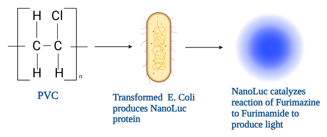 |
|---|
Device level
This biosensor device consists of an E. coli K12 chassis containing a plasmid engineered with an oxidative stress-sensing system coupled to a luminescent reporter module. We chose E. coli K12 as our chassis because our chosen promoter, katG, can be activated by the increase in reactive oxygen species in E. coli caused by PVC mentioned above (Jin et al., 2024). Additionally, E. coli’s genetic code is well-documented, and the K12 strain poses minimal danger, thus making it the most feasible and safe chassis for our experiment.
We designed a plasmid with a pathway that responds to an upregulation of hydrogen peroxide caused naturally by PVC in E. coli. The upregulated hydrogen peroxide oxidizes the OxyR protein already present in the bacteria. Once oxidized, the OxyR initiates transcription of the engineered plasmid, leading to the transcription of the Luciferase protein, NanoLuc. NanoLuc reacts with furimazine to create furimamide and luminescence, which we can measure and use to determine the presence of PVC in a sample.
Overall, this device connects the input of PVC to a luminescent output, allowing for the detection of microplastics by utilizing this system.
Parts level
To create the biosensor, it was necessary to identify a promoter sequence that would be activated in the presence of PVC, and a reporter system that could validate the activation of the promoter.
For the promoter, katG was identified as a promising candidate due to its association with oxidative stress responses (Zhang et al., 2022). A study from Zhang et al. (2022) revealed that when the bacteria Klebsiella sp. EMBL-1 was fed PVC, there was increased production of a protein associated with the katG gene. This supports our hypothesis because it reveals that the katG gene is upregulated in the presence of PVC.
The katG promoter can be activated through natural oxidative stress pathways in E. coli. Italiani et al. (2011) found that the transcription factor OxyR responds to hydrogen peroxide, a reactive oxygen species (ROS), and activates the transcription of the katG gene. Furthermore, Jin et al. (2024) found that PVC caused the production of ROS, including hydrogen peroxide, in human lung cells. Therefore, we hypothesize that PVC exposure may increase oxidative stress in E. coli cells, similarly to how it does in human lung cells. The hydrogen peroxide levels would increase and bind to the OxyR already present in E. coli, leading to the activation of the katG promoter.
We chose to incorporate a NanoLuc luciferase reporter downstream of the katG promoter, which would be transcribed in the presence of PVC to allow for visualization of the plastic. Mechanistically, the NanoLuc reporter produces the NanoLuc protein, which catalyzes the breakdown of the substrate, furimazine, into furimamide, carbon dioxide, and light. According to England et al. (2016), the NanoLuc luciferase reporter produces a luminescent output greater than 150 times brighter than other traditional luciferase reporters like Firefly and Renilla luciferases. This increased luminescence would therefore be easier to visualize and quantify. The mentioned natural biological pathway in response to PVC would lead to the activation of the katG promoter, which would, in turn, activate the transcription of the NanoLuc gene and produce luminescence.
While it has been established that E. coli contains the oxidative stress pathway utilizing OxyR and katG, PVC-specific activation must be validated experimentally. We propose a plasmid with the katG promoter and NanoLuc reporter inserted into a pUC19 backbone, as shown in Figure 2.
| Figure 2: pUC19-based Biosensor Plasmid: The AmpR gene acts as a selective marker to ensure only transformed bacteria are accounted for during quantification. The katG promoter activates the transcription of the NanoLuc reporter gene in the presence of PVC. When the NanoLuc reporter is activated, it produces the NanoLuc protein, which produces luminescence. | 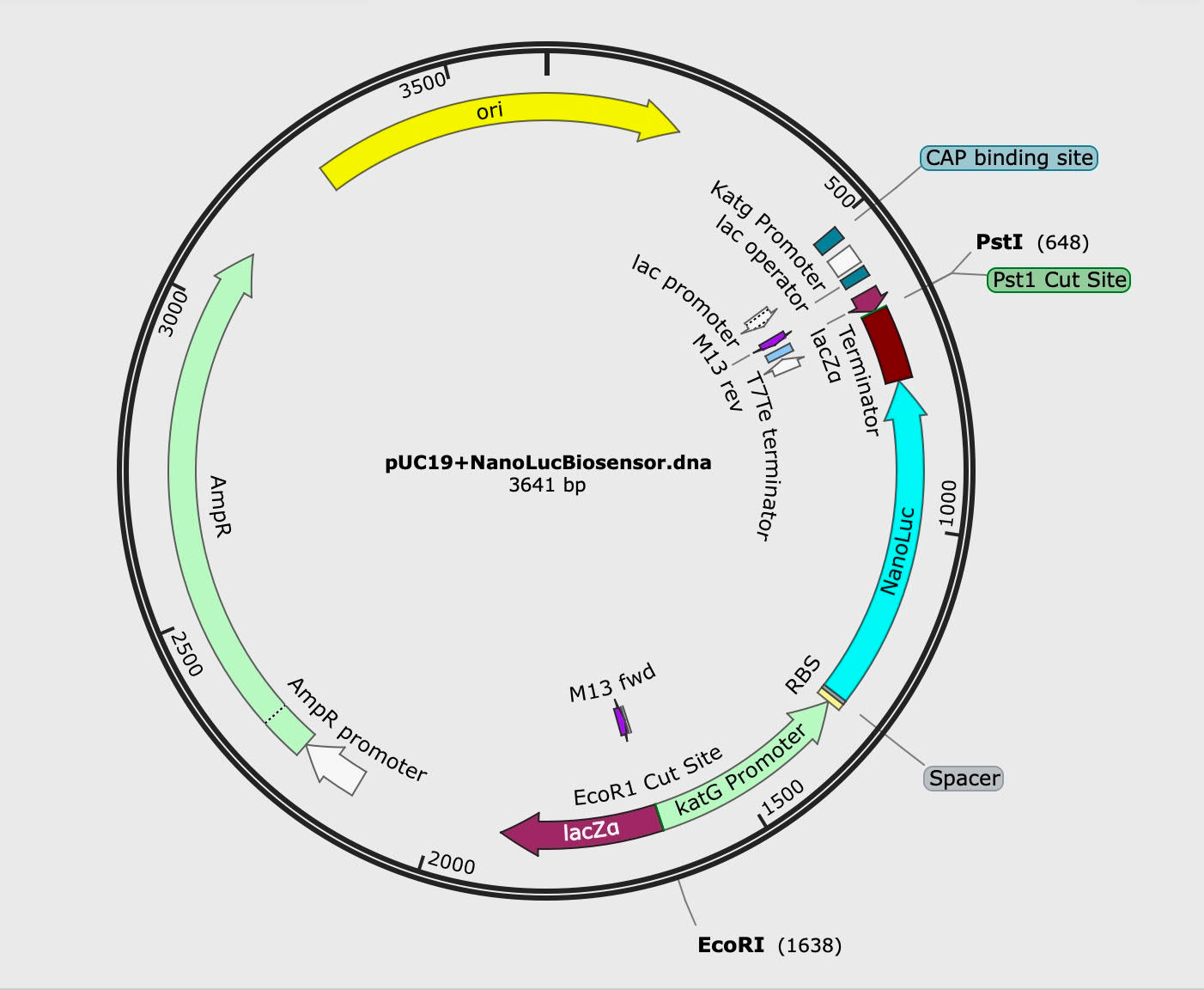 |
|---|
We utilized a pUC19 backbone to encode for ampicillin resistance, which would act as a selective marker. We inserted the katG promoter and luciferase reporter within the lac operon to allow blue-white screening during the transformation and cultivation procedures. Blue colonies would indicate our promoter and reporter were not successfully incorporated and that the lac operon was activated instead, while white colonies would suggest successful incorporation of our plasmid. We included a ribosomal binding site downstream of the katG promoter to support transcription as well, and a terminator downstream of the NanoLuc reporter to end transcription. Overall, this plasmid would allow for the isolation of transformed bacteria and the visualization of luminescence resulting from the activation of the engineered reporter system.
Safety
If we were to conduct an experiment, we would first create our biosensor. To do this, our first step is to use heat shock transformation methods to insert our plasmid into our E. coli. We will then grow this E. coli in lysogeny broth (LB) with Ampicillin to select for transformation.
To test if our device works, we will have one control group and one experimental group. The first group will contain 250 mL of distilled water, and the other will contain the PVC plastic concentration in 500 mL of distilled water. The first group is our negative control, with an expected output of no luminescence. The second group is expected to have an output of luminescence and is our experimental group. We will use a luminometer to measure the luminescence of the groups. Analyzing the results of our experiment could lead us to the confirmation, or partial confirmation, of the hypothesis that PVC does induce oxidative stress in E. coli.
When testing our proposed E. coli system, it is imperative to take proper precautions to ensure the safety of ourselves, others, and the local environment. To guarantee this safety, every step will be outlined by a detailed procedure and will be under the supervision of experienced individuals. We would also work in a sterile and well-managed Biosafety Level 1 (BSL-1) lab to ensure that any incident that may occur would not impact the local environment.
While studying E. coli, proper biosafety attire (lab coat, safety goggles, nitrile gloves, etc.) would be worn, and precision would be used while following the carefully crafted procedure to ensure safety. In order to apply our engineered E. coli in a human or in the environment, we would need to go through additional biosafety evaluations, testing, and regulatory approval before enactment. To keep the surrounding environment safe, proper disposal of the E. coli would require a bleach treatment and storage in proper biohazard bags for autoclaving.
Discussions
If laboratory testing reveals that PVC induces the katG promoter, then this design could provide a novel, biological method for plastic detection. This method of plastic detection can be applied both environmentally and medically. As evidenced by Huang et al., this sensor could allow environmental detection of PVC in waterways, determining the extent of plastic contamination present and the safety of water from specific sources (Huang et al., 2022). This design could also be implemented for the detection of microplastics in the body, elucidating the prevalence and extent of the harm of microplastics on human health. By designing and implementing this biosensor in a low-cost and widely accessible way, we would be able to more effectively raise concerns about microplastic exposure in society overall by making tests available to the public and revealing the extent to which people have been exposed.
The primary limitation of the current design of the biosensor is the lack of medically relevant detection methods for the output. The biosensor luminescence can be observed visually but requires a luminometer for quantitative measurement. It would be challenging and unfeasible to observe and quantify luminescence in humans, revealing the importance of a more medically relevant reporter that produces an easily detectable output. One possibility for this reporter would be violacein, which produces a purple dye when translated. It is possible to detect violacein in fecal matter, allowing for the detection and quantification of the dye in relation to microplastic levels in the body (Pauer et al., 2018). The human subject would potentially need to ingest the engineered E. coli in order for the pathway to detect the presence of PVC and produce a violacein output. While this method would likely be costly and inaccessible, future studies may be able to optimize this design for accessibility in clinical settings. Such clinical tests could reveal the prevalence of microplastics in humans and highlight the importance of reducing plastic use and production.
One challenge presented in this design is the possible lack of specificity of the biosensor. Because the katG promoter responds to oxidative stress, other environmental stressors could potentially cause the biosensor to respond and trigger a false positive result. To test this, the transformed E. coli can be exposed to other substances known to cause oxidative stress, and their luminescence can be measured. We aim to refine the genetic circuit to distinguish between PVC-induced oxidative stress and general environmental stress. Integrating a second biological system that reduces the impact of other stressors besides PVC on oxidative stress is a possible method to control for non-PVC-induced stress, improving the accuracy of our biosensor.
Next steps
After designing the plasmid using SnapGene, the next step is to validate whether PVC exposure induces oxidative stress in E. coli by testing the Brookline hypothesis that PVC increases OxyR activation. Measuring luminescence across varying PVC concentrations will allow us to establish a dose-response relationship, confirming whether OxyR activation drives katG-dependent reporter expression.
Following validation, the engineered gene pathway should be optimized to improve sensitivity and reduce background noise, potentially through promoter strength adjustment or incorporating a secondary reporter to control for overall stress responses in E. coli. The environmental robustness should also be evaluated by testing the biosensor under a range of environmental conditions: varying temperatures, pH, salinity, exposure period, etc. This will determine the biosensor’s durability outside controlled laboratory settings.
To increase the applicability of the biosensor, other plastics should be tested in comparison to PVC to evaluate the biosensor’s specificity and durability and to determine its ability to detect general microplastic-induced stress. Future engineering may be needed to improve overall reactivity across many different plastic types, depending on the result of these comparison tests.
Finally, to enable real-world application, the luminescent output should be replaced or supplemented with an easily detectable signal that can be utilized in field or clinical contexts. Long-term goals would include integrating the biosensor into human detection platforms to enable real-time monitoring of microplastic exposure in environmental or biological samples and specimens. Additionally, future research should explore alternative output systems beyond luminescence. One promising approach is the use of a violacein-based reporter, which produces a visible purple pigment that can be more easily detected without specialized equipment. This type of output could enable simpler, cost-effective detection methods in environmental or clinical settings, supporting the development of accessible biosensors.
Author contributions
Author contributions: AJL – discussions, next steps, background research, abstract; AML – “Device level” section, background research, diagrams for video and “Device level”; BB – background research, video; GG – group leader, plasmid research, plasmid design on Benchling, coordination with remote mentor, background research, narrowing research focus, “Background” section, “Next steps” section, manuscript editing, abstract editing; HB – background research, safety, references; IC – acknowledgements, background research, culturing E. coli procedure, discussions, system levels; KR – group leader, organized meetings, background research, narrowing of research focus, plasmid design, “Parts levels” section, abstract writing, mentor correspondence; NK – background research, next steps, discussions.
Acknowledgements
We would like to thank our supervising teacher, Mr. Kirkpatrick, and our mentor, Dr. Eric Young, for their invaluable support and guidance throughout this project. We would also like to express our sincerest gratitude to BioBuilders for allowing us the privilege of this research opportunity.
Additionally, we would like to acknowledge a team of students from Brookline High School for their innovative project proposal, Visual quantification of PVC levels via luciferase and the catalase-peroxidase promoter: A unique solution to an under-discussed issue, which inspired this project.
References
American Chemical Society. (2025). Bacteria that “shine a light” on microplastic pollution. https://www.acs.org/pressroom/presspacs/2025/september/bacteria-that-shine-a-light-on-microplastic-pollution.html
Banerjee, A., & Shelver, W. L. (2021). Micro- and nanoplastic induced cellular toxicity in mammals: A review. Science of the Total Environment, 755, 142518. https://doi.org/10.1016/j.scitotenv.2020.142518
Choi, Y., Ma, Y., Wei, W., Wing, Y., Tsz, J., & Chua, S. L. (2025). Detection of microplastics pollution using a green fluorescent protein-based microbial biosensor coupled with Raman spectroscopy. ACS Sensors. https://doi.org/10.1021/acssensors.5c01120
England, C. G., Ehlerding, E. B., & Cai, W. (2016). NanoLuc: A small luciferase is brightening up the field of bioluminescence. Bioconjugate Chemistry, 27(5), 1175–1187. https://doi.org/10.1021/acs.bioconjchem.6b00112
Goswami, S., Adhikary, S., Bhattacharya, S., Agarwal, R., Ganguly, A., Nanda, S., & Rajak, P. (2024). The alarming link between environmental microplastics and health hazards with special emphasis on cancer. Life Sciences, 355, Article 122937. https://doi.org/10.1016/j.lfs.2024.122937
Huang, Z., Hu, B., & Wang, H. (2022). Analytical methods for microplastics in the environment: a review. Environmental Chemistry Letters, 21(1), 383–401, https://doi.org/10.1007/s10311-022-01525-7
Italiani, V. C. S., da Silva Neto, J. F., Braz, V. S., & Marques, M. V. (2011). Regulation of catalase-peroxidase katG is OxyR dependent and fur independent in Caulobacter crescentus. Journal of Bacteriology, 193(7), 1734–1744. https://doi.org/10.1128/jb.01339-10
Jin, W., Zhang, W., Tang, H., Wang, P., Yan, Z., Liu, S., Qiu, J., Chen, H., Wang, L., Wang, R., Sun, Y., Liu, P., Tang, H., & Zhu, Y. (2024). Microplastics exposure causes the senescence of human lung epithelial cells and mouse lungs by inducing ROS signaling. Environment International, Article 108489. https://doi.org/10.1016/j.envint.2024.108489
Kudzin, M. H., Piwowarska, D., Festinger, N., & Chruściel, J. J. (2023). Risks associated with the presence of polyvinyl chloride in the environment and methods for its disposal and utilization. Materials, 17(1), Article 173. https://doi.org/10.3390/ma17010173
Lee, Y., Cho, J., Sohn, J., & Kim, C. (2023). Health effects of microplastic exposures: Current issues and perspectives in South Korea. Yonsei Medical Journal, 64(5), 301–308. National Library of Medicine. https://doi.org/10.3349/ymj.2023.0048
Luo, Y., Awoyemi, O. S., Naidu, R., & Fang, C. (2023). Detection of microplastics and nanoplastics released from a kitchen blender using Raman imaging. Journal of Hazardous Materials, 453, Article 131403. https://doi.org/10.1016/j.jhazmat.2023.131403
National Oceanic and Atmospheric Administration. (2024, June 16). What are microplastics? Noaa.gov; National Ocean Service. https://oceanservice.noaa.gov/facts/microplastics.html
Nur Sakinah Roslan, Yeong Yeh Lee, Yusof Shuaib Ibrahim, Sabiqah Tuan Anuar, Ku, Lisa Ann Lai, & Brentnall, T. (2024). Detection of microplastics in human tissues and organs: A scoping review. Journal of Global Health, 14. https://doi.org/10.7189/jogh.14.04179
Pauer, H., Hardoim, C. C. P., Teixeira, F. L., Miranda, K. R., Barbirato, D. D. S., de Carvalho, D. P., Antunes, L. C. M., Leitão, Á. A. D. C., Lobo, L. A., & Domingues, R. M. C. P. (2018). Impact of violacein from Chromobacterium violaceum on the mammalian gut microbiome. PLOS ONE, 13(9), https://doi.org/10.1371/journal.pone.0203748
Yang, Q., Peng, Y., Wu, X., Cao, X., Zhang, P., Liang, Z., Zhang, J., Zhang, Y., Gao, P., Fu, Y., Liu, P., Cao, Z., & Ding, T. (2025). Microplastics in human skeletal tissues: Presence, distribution and health implications. Environment International, 196, Article 109316. https://doi.org/10.1016/j.envint.2025.109316
Zhang, Z., Peng, H., Yang, D., Zhang, G., Zhang, J., & Ju, F. (2022). Polyvinyl chloride degradation by a bacterium isolated from the gut of insect larvae. Nature Communications, 13(1). https://doi.org/10.1038/s41467-022-32903-y