
Irene Pang and Iris Pang, Newton South High School, Newton, Massachusetts, United States
Reviewed on 9 May 2026; Accepted on 22 June 2026; Published on 26 October 2026
With help from the 2026 BioTreks Production Team.
Lung cancer remains one of the leading causes of cancer-related mortality worldwide, with late-stage diagnosis and the limited regenerative capacity of lung tissue posing significant challenges to effective treatment options. As a result, lung tissue engineering has emerged as a promising path for developing regenerative therapies, creating lung models, and repairing tissue damage. Among current approaches, electrospinning and three-dimensional (3D) bioprinting have gained attention for their ability to fabricate biomimetic scaffolds, which are engineered 3D structures designed to replicate biological properties, that support cellular growth and tissue organization. Electrospinning produces nanofibrous structures in the extracellular matrix that promote structural support for lung cells and model lung tissue structures, providing valuable information about cell-cell interactions in cancer. However, limitations in controlling tissue structure and in preventing cell damage restrict its ability to fully replicate the lungs. In contrast, 3D bioprinting, where researchers use 3D bioprinters equipped with bioink containing lung cells, can replicate spatially organized constructs like vascularized tissue models and airway structures for modeling, yet challenges remain. Limited resolution, bioink constraints, and the inability to reproduce fully functional lung systems are significant barriers. Therefore, it is important to evaluate the strengths and limitations of electrospinning and 3D bioprinting in the context of lung tissue engineering. This requires considering the technical and biological risks associated with these methods in clinical applications, and how these issues can be addressed through further scientific advancements.
Keywords: Lung cancer, electrospinning, bioprinting, 3D modeling
Authors are listed in alphabetical order. Bo Pang mentored the group. Please direct all correspondence to mailto:madradical@gmail.com.
Body
Lung cancer origin and development
Lung cancer is the second most commonly occurring cancer and the primary cause of cancer-related deaths worldwide, with 2.2 million new cases and 1.8 million deaths as a result every year (World Health Organization 2026). Certain factors increase the incidence of carcinogenesis, the biological process by which healthy cells are transformed into cancer cells, depending on the country a patient is located. For example, in more developed nations, cultural smoking patterns have resulted in rising rates of lung cancer in women, while developing economies struggle with environmental exposure to air pollution. Studying lung cancer epidemiology will identify which populations are most affected by lung cancer, and where to prioritize novel treatment methods (Barta et al., 2019). Unfortunately, almost 60% of patients are diagnosed at advanced stages where treatment is difficult and lung transplant is unfeasible (Polonio-Alcalá et al., 2022). Furthermore, because lungs struggle to regenerate tissue, treating severe lung injury is complicated. Thus, ongoing studies emphasize lung tissue engineering not only as a path toward future bioengineered lung transplant through ex vivo-engineered lung tissues, but also for repairing localized damage as a result of lung cancer and developing reliable in vitro lung cancer models (Torkashvand et al., 2024, Polonio-Alcalá et al., 2022). Along with lung cancer’s complications themselves, lung cancer metastasis creates further harms, which emphasize the importance of comprehensive treatment. Lung cancer metastasis is the process in which the primary tumor in the lung spreads to other areas in the body, with the most common sites being brain, bone, lymph nodes, and liver. Lung cancer metastasis is controlled by a variety of factors, including interactions in the lung cancer microenvironment, epithelial mesenchymal transition transformation, and metastasis through the blood or lymph system (Xie et al., 2021). Lung cancer is one of the most frequent tumors to metastasize to the brain, with brain metastasis being the major cause of patient morbidity and mortality; therefore, addressing lung cancer at its root is crucial to preventing many more additional deaths (Souza et al., 2023).
Lung cancer has generally been classified into two principal groups: small versus non-small cell type, where further immunohistochemical markers (specific proteins or antigens pathologists find and identify in tissue samples to understand more about a disease) have helped specify the differences between invasive adenocarcinomas, which are cancers that originate in the glandular cells, and pre-invasive lesions (Siddique et al., 2024). Since then, scientists have identified over 50 histomorphological subtypes, but the most well-known are non-small cell lung cancer (NSCLC) and small cell lung cancer (SCLC), with NSCLC being more prevalent, making up 85% of total lung cancer cases. However, the remaining SCLC cases have a poorer prognosis, with high relapse rates and low overall survival. Of NSCLC, the three subtypes include adenocarcinoma, 38.5% of all lung cancer, squamous cell carcinoma—20%—and large cell carcinoma, at less than 3% (Barta et al., 2019; Siddique et al., 2024).
Electrospinning
Currently, biomarker detection, which is the process of testing blood, bodily fluids, or tissues to detect circulating tumor cells, proteins, and biological molecules indicating the presence of cancer, is standard for lung cancer treatment. These include autoantibodies, microRNA, methylated DNA, and proteins. However, despite being specific, they are not sensitive, meaning testing has a high rate of false negatives. Furthermore, biopsy tissue samples are often not suitable for biomarker testing—advancing blood marker testing or exhaled breath biomarker testing could potentially obviate the need for tissue tests (Zarinshenas et al., 2023). Another lung cancer treatment approach lies in lung tissue regeneration. Though this approach is limited, one method involves lung regeneration stem cell therapies, which aim to repair damaged respiratory tissue by using stem cells to reduce inflammation and stop tissue scarring. These therapies are currently restricted to clinical trials, but the complex architecture of lung tissue and issues integrating treatments into existing tissue pose difficulties rebuilding lung tissue (STEMS Health Regenerative Medicine, 2025). Electrospinning offers solutions to many barriers of current treatments. Electrospinning, which uses electrical forces to draw charged threads from polymer solutions or melts, produces ultrafine filaments called electrospun fibers with diameters ranging from nanometers to micrometers (Figure 1). These fibers can create materials with superior characteristics since they can closely resemble human tissues (Fluidnatek, 2023). Electrospinning is unique in that their scaffolds can be fabricated to a large range of sizes, making it ideal for mimicking lung tissue’s fiber and mechanical properties. To encourage successful tissue transplantation, researchers can also combine growth factors into these scaffolds, which increase the rate of tissue formation. Finally, electrospinning is relatively simple, low-cost, and very adaptable compared to other tissue engineering methods (Shakir et al., 2022).
| Figure 1. Electrospinning mechanism. Electrospinning uses a high-voltage power supply to create electrical forces, drawing charged polymer threads from a syringe. The syringe pump is a motorized device that secures a syringe and controls the plunger to draw polymer threads at a specific, constant rate. The nanofibers are typically collected on a plate, but can also be collected on rotating drums or disks to enable specific fiber alignments. | 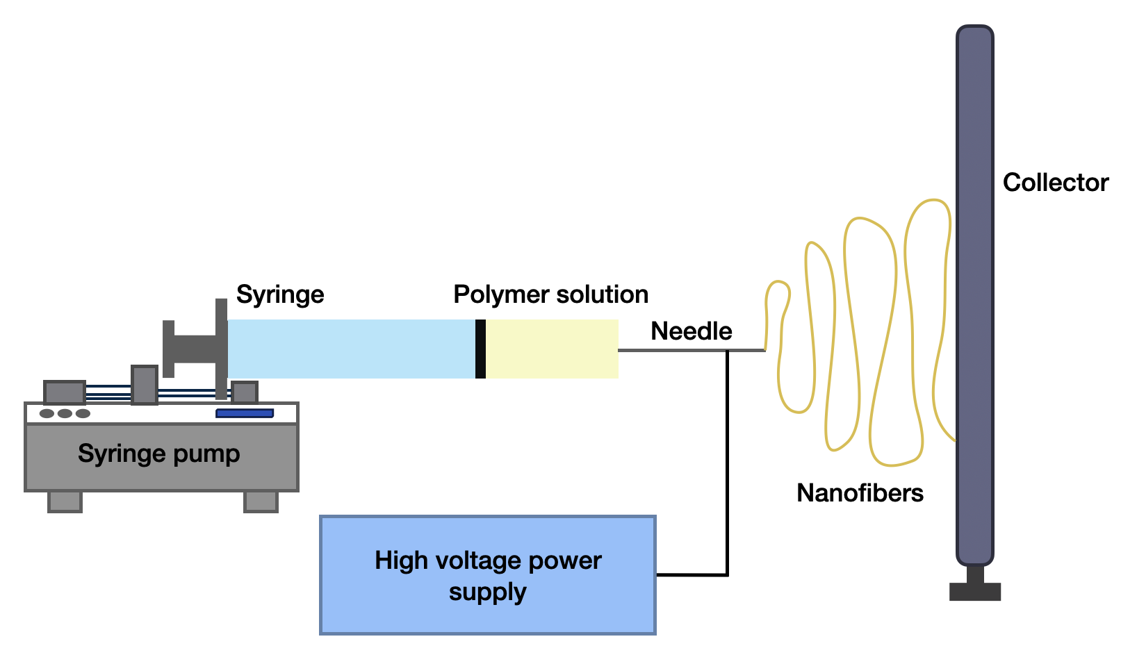 |
|---|
Electrospinning has multiple applications in furthering lung cancer treatment. By developing an accurate in vitro lung cancer model, researchers can more effectively identify new biomarkers and develop new therapies for lung cancer. That is because electrospinning can cost-effectively produce scaffolds that resemble the extracellular matrix (ECM) of the lung (Polonio-Alcalá et al., 2022). Nanofibers stand out among bioactive molecules because of their compositional diversity, structural flexibility, and exceptional functionality -(Flores-Rojas et al., 2023). While researchers have obtained a range of valuable information from conventional in vitro 2D studies, which are laboratory experiments where living cells are grown in a flat, single layer on a rigid surface (typically a glass or plastic Petri dish), 3D models are becoming increasingly necessary to capture the cell-cell communications and cell-matrix interactions in cancer. Electrospun nanofiber scaffolds offer an alternative artificial 3D matrix because they can mimic the composition of ECM and have high biochemical stimuli versatility (Chen et al., 2019).
In the realm of cancer biomarker detection, electrospun nanofibers are useful for biosensor development due to their high surface-to-volume ratio and low molecular-level defects (Chen et al., 2019). Traditional diagnostic methods typically lack the sensitivity to detect low-abundance biomarkers, but electrospun nanofibers can mimic the ECM’s architecture, enabling researchers to functionalize them with biomolecular probes (such as antibodies or aptamers) to improve sensitivity to specific markers (Fluidnatek, 2023). For example, hollow electrospun fibers have a larger surface area and porosity, and are frequently used to prepare gas sensors when combined with gas-sensitive materials. The hollow structure of these fibers not only aids in gas sensing but also permits the loading of other sensing elements to improve performance. In lung cancer, acetone in exhaled breath can serve as a biomarker, and these fibers can significantly improve detection. In comparison to film-based systems for biosensing, nanofiber-based systems improve performance nearly ten times, demonstrating more effective analytical capabilities. Indeed, electrospinning positions nanofibers to have broader applications in cancer detection by addressing equipment and procedure costs while also providing high reliability (Zhou et al., 2024).
For example, folate receptor alpha is expressed in 74% of lung adenocarcinomas and serves as a useful diagnostic marker for NSCLC (O’Shannessey et al., 2012). In 2018, Wang et al. prepared one-dimensional CuO-NiO/C nanofibers using electrospinning, along with other heat treatment techniques. These nanofibers, conjugated with folic acid, successfully detected folate receptor-positive cancer cells, exhibiting suitable clinical applicability and high sensitivity for detecting cancer cells in whole blood (Zhou et al., 2024, Wang et al., 2018).
Furthermore, electrospinning facilitates advances in lung tissue regeneration. In 2011, Lee et al. discovered that hybrid scaffolds could create an environment instrumental in lung tissue growth and regeneration. However, the complex 3D structure and function of the natural ECM, providing a microenvironment of the cell mechanism, have been historically difficult to replicate with these scaffolds. Electrospinning’s ability to closely fabricate cell activity and tissue integrity played a role in advancing hybrid scaffolding due to nanofibers’ versatile and efficient manufacturing technique, high porosity, and pore interconnectivity (Torkashvand et al., 2024; Abel et al., 2020). Most importantly, ideal tissue-engineered scaffolds should behave similarly to targeted tissue ECM to facilitate growth, and electrospinning can recreate nanofibers almost identical to native nanofibrillar because of their similar aspect ratios (over 1:20) and size-scale (Xing et al., 2023).
3D bioprinting
Tissue engineering hinges heavily on in vitro modeling of the airways and respiratory system, crucial for research in lung cancer. While the construction of biomimetic native airways with tissue-specific function is difficult, bioprinting is a promising technology for the development of lung models. As shown in Figure 2, 3D printing uses bioink to accurately deposit cells and other molecules into printed constructs (Zhang et al., 2024). In addition, 3D bioprinting has been more successful in replicating the architecture and physicochemical properties of the tumor microenvironment of lung cancer than traditional 2D cell culture systems (Xu et al., 2025). Bioinks are generally natural, synthetic, or compound, each consisting of different materials (Zhang et al., 2024). Natural bioinks are made up of various natural sources that are not native to the ECM, such as alginate, cellulose, collagen, gelatin. The properties of these materials allow for cellular interaction and motility (the ability to move autonomously using metabolic energy), have little host response, and are also biodegradable and bioabsorbable. Natural biomaterial-based bioinks can recreate the viscoelasticity of the lungs, especially the alveolar area. However, these bioinks “have lower printability compared to synthetic polymers and may not transition from ink to gels efficiently” (Dabaghi et al., 2023). On the other hand, synthetic biomaterials have high flexibility, beneficial for tuning and chemical modification. Materials include Polycaprolactone (PCL), Polyurethane (PU), poly(sebacoyl Diglyceride) (PSeD), and polylactic acid (PLA). When constructing tracheal tissue, PCL can be printed into fibrous networks to mimic the hollow tissue, where microscale pores support nutrient diffusion. However, though synthetic bioinks are easy to print and customize, they do not fully imitate native tissues due to their lack of bioactivity. Limited cellular interactability creates challenges replicating the functions of native tissues, reducing its stability and functionality. Lastly, compound bioinks blend natural and synthetic polymers to create stable structures with the benefits of both previously mentioned bioinks, supporting cellular adhesion and 3D bioprinting’s mechanical qualities. As 3D bioprinting technology improves, more strategies will continue to enhance the potential to model functional tissues and organs (Zhang et al., 2024).
| Figure 2. Extrusion vs. DLP bioprinting mechanisms. Both extrusion and DLP 3D bioprinting utilize bioink consisting of living cells, biocompatible materials (hydrogels), and biomolecules. In extrusion bioprinting, a computer-controlled extruder pumps out bioink through a nozzle to build layers upon layers of filament. The computer controls both the rate of material deposition and the nozzle position to achieve the print design. DLP bioprinting also uses a layer-by-layer process, but a light source projects a still image of each layer onto a light-sensitive liquid, creating a chemical reaction that causes liquid to cure where the light is illuminated. | 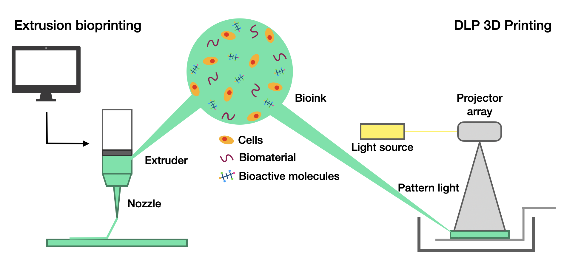 |
|---|
There are several techniques used in 3D bioprinting, each offering their own unique advantages to generating living tissue constructs. Extrusion-based bioprinting delivers bioink layer by layer through a needle-like nozzle into the lungs. This method can be applied to a broad range of cells and growth factors, but it can potentially damage cells due to the stress of the extrusion process. Consequently, the design of bioinks, including their “rheological properties, viscoelasticity, surface tension, gelation mechanisms” needs to be considered, as well as the pressure, speed, and temperature of the nozzle used (Zhang et al., 2024).
In a 3D model printed by Wang et al., it was shown that the overexpression of vimentin, α-SMA, and the loss of E-cadherin -, promotes Epithelial-to-Mesenchymal Transition (EMT), where cancer cells lose rigidity and move to invade surrounding tissues, in lung cancer. This – reveals that 3D models can effectively simulate the interactions between tumor cells and the matrix. Specifically, it was noted that a SA-GL hydrogel-based bioprinting model of NSCLC patient-derived xenograft cells can reduce severe damage to cancer cells caused by temperature and pressure changes. Aragaw Gebeyehu et al. built on these findings and demonstrated that bioink using a non-xenogeneic VitroGel system could print modified ink H4-RGD at extrusion pressures of 25–40 kPa, displaying 90% cell viability and quick recovery properties, preventing the need for UV curing or temperature regulation (Li et al., 2023).
Digital light procession (DLP) bioprinting, contrarily, cures a liquid using a digitally controlled ultraviolet light source, enabling more precisely printed structural features compared to other methods. The resolution in light-based bioprinting is high, even in sub-micrometer scales depending on the characteristics of the photopolymerizable materials (photoresins). However, while high intensities may speed up polymerization, they generate heat harmful to cell viability. Luckily, these effects can be balanced by optimizing photo-initiator type, light exposure time and intensity, and bioink components (Zhang et al., 2024).
One such example of a 3D model was used to replicate the interfacial structures and matrix stiffness of NSCLC lung cancer tissue and adjacent normal tissue. Xu et al. wondered whether a reduction in CCN1 expression within soft matrices could reduce the migratory and proliferative capabilities of tumor cells. As a result of 3D bioprinting, researchers were able to observe how the matrix stiffness of normal tissue compared to cancerous tissue impacted tumor proliferation, drug sensitivity, and the role of the CCN1 gene. Using DLP-based 3D bioprinting, a bioink was formulated to mimic the ECM components and stiffness of the two tissue types. It was demonstrated that a reduction in CCN1 expression within soft matrices can reduce tumorigenesis, validating the potential of chemotherapeutic and immunotherapeutic treatment options (Xu et al., 2025).
Next steps
In the future, the combination of 3D printing technology and electrospinning could be a path forward toward more accurate and complex 3D cancer models, facilitating the translation of therapeutic drugs into clinical trials (Chen et al., 2019). However, several limitations exist that impact the viability of these techniques.
Though electrospun 3D models have great potential, they are limited by their inability to mimic certain cell activities, including heterotypic cell-cell signaling, cell motility, tumor cell metabolism, and biomechanical stimulation, which is critical in tumor progression and metastasis. Because cancer can spread throughout the body, breaking away from the primary tumor to form tumors in other parts of the body, a static model might not fully represent how lung diseases might progress. Furthermore, they are unable to be scaled up and have high levels of variability between batches, limiting their wide employment for high-throughput drug screening. Because they cannot replicate a native tumor microenvironment either, especially their complexity and diversity, further research is necessary (Chen et al., 2019). Nevertheless, there has been some progress: researchers have discussed using native ECM in the solution or fusion of electrospinning, incorporating bioactive molecules, and biofunctionalization postprocessing. These approaches offer more similarity to ECM components and better support cell adhesion and proliferation (Politi et al., 2020).
Furthermore, electrospun biosensors are limited by their sensitivity, size distribution unevenness, morphology control, experimental conditions, cost, and biocompatibility. In the future, scientists can research new nanomaterials to enhance biosensor sensitivity, multi-marker detection, improved communication speed, and further integration into biomedical engineering. For example, wearable devices could monitor real-time symptoms. Finally, these sensing platforms are expected to be commercialized for detection of diseases, hopefully alleviating the cost burden of electrospun biosensing (Zhou et al., 2024).
Electrospinning’s role in scaffolding and tissue generation is also limited by their limited thickness, small pore size, and poor mechanical properties, meaning they do not effectively recapitulate cellular organization and cell interactions in vivo despite their success in recreating morphological and nanotopographical features. Moreover, their lack of vascularization limits clinical translation, leading to weak tissue repair and regeneration, along with their inability to repair tissues with higher dimensions (millimeters to centimeters) (Abel et al., 2020).
However, advancements have been made using “diverse methodologies involving one or more materials and phases” to increase flexibility, biocompatibility, and programmability. These solutions involve broadly exploring biocompatible and biodegradable polymeric materials that can better replicate living tissue function (Flores-Rojas et al., 2023). For example, Abel et al. 2020 outlines several techniques to enhance industrial scaling and novel applications: porogen leaching can create larger pores in fibers using salt particles, polymers, or ice crystals; and gas foaming utilizes a blowing/foaming agent to cost-effectively create 3D porous matrices with a controllable architecture. Other techniques can also increase the variability of micro/nanofibrous materials, including electrospraying (Abel et al., 2020). Indeed, the combination of natural and synthetic polymers can enhance the biocompatibility and structural properties of scaffolds, thereby promoting tissue regeneration and enhancing cell adhesion and growth (Politi et al., 2020). These approaches can aid in the clinical translation of electrospinning technology.
In terms of 3D bioprinting, some theoretical bottlenecks include how alveoli structures are extremely complex and thin-walled, requiring precise implementation of alveolar and capillary endothelial cells (Zhang et al., 2024). Due to these dimensions, there are currently no 3D bioprinting technologies that can generate functional alveoli-like 3D structures with cells, but strategies are being pursued on other models larger than the human lung (Dabaghi et al., 2023). Another difficulty is how challenging it is to accurately reproduce the branching morphology of airways, which are lined with epithelial cells. Lastly, bioprinted tissues often need to be compatible with the immune system to avoid rejection, which is the biggest barrier currently impacting bioprinting’s workability. When Kaye et al. attempted to implant tracheal replacement grafts into several rabbits (chosen for relatively similar dimensions compared to the human lungs), most developed respiratory issues, demonstrating that immunocompatibility still poses a barrier to in vivo implantation (Kaye et al., 2019). Additional technical bottlenecks include how current bioprinting techniques have inadequate resolution and struggle to print multiple cell types at once. Moreover, bioprinted products need to mimic lungs’ mechanical functions like elasticity and compliance, which has not yet been achieved. Finally, scaling up the bioprinting process needs to account for reproducibility and quality control, which poses a significant obstacle to clinical translation (Zhang et al., 2024).
Indeed, 3D bioprinting still has a long way to go in achieving clinical translation. The source of cells is a deep concern relating to immune rejection. While patient-derived cells present the best-case scenario in terms of immune response, the feasibility and time taken to expand the cells in the laboratory at scale is an extensive process. An alternative is induced pluripotent stem cells (iPSCs), which are isolated from skin cells and isolated into stem cells to differentiate into different types of cell. However, this method still does not solve how long the process would take in a clinical setting. If scientists manage to achieve a biological breakthrough by establishing a synthetic or genetically modified iPSC that is compatible with most humans, then bioprinted tissues may very much be commercially achievable, solving duration and immune response challenges (Vijayavenkataraman, 2023).
Last, but not least, ethical concerns must be taken into account and suitably addressed. Bioprinted products are still not subject to specific regulatory guidelines for their use. The US FDA released a draft policy in 2016, but additional laws must be put into place. It is difficult to classify what bioprinting exactly is, as its fabrication involves multiple, complex moving parts. The product could include a 3D-printing scaffold made out of synthetic biopolymer functionalized with certain drugs, complete with human-derived cells. Given these possibilities, it becomes hard to approve such a product. Moreover, a perfect recipe for bioinks has not yet been developed. While there have been many attempts, a solution is still far away (Vijayavenkataraman, 2023). In addition, many clinical trials would be necessary to eliminate the associated risks of 3D bioprinting. However, each bioproduct would be fabricated for a specific patient, meaning that standard double-blind randomization studies cannot be applied to testing, and results could not necessarily be extrapolated into future treatments. Safety cannot be tested on other individuals, so each volunteer would become the first patient (Kirillova et al., 2020).
To overcome many of these challenges, artificial intelligence (AI) and big data are crucial to the field. As doctors, biologists, and engineers work with material scientists to develop new “multi-material composite bioinks, mix functional materials and cells in specific proportions to create bionic structures and enhance the physiological functions of tissues”, AI can analyze large swaths of data to quickly design rational designs for airways and lungs, allowing for the quick and accurate manufacturing of bioprinted tissues. Altogether, further advancing 3D bioprinting requires the efforts of multiple fields and interdisciplinary collaboration to break into a new stage of clinical application. The faster technical barriers are overcome, the easier it will be to classify 3D bioprinting and create a set of regulatory guidelines (Zhang et al., 2024). However, it is worth noting that AI has multiple drawbacks. In addition to the fact that the existing lung datasets are mostly derived from tissue or pathological sections with a lack of stem cell–differentiation/proliferation process datasets, “limited experimental results with narrow sources and small scales” result in high variability, “reducing the generalization capacity of AI models.” This significantly lowers the reliability of big data in a clinical setting, which requires high precision. While solutions are not perfect, establishing models capable of adapting to various working scenarios will reduce the cost of AI and increase accuracy (Zhang et al., 2025).
Author contributions
Iris P. researched 3D bioprinting techniques, including light-based bioprinting and extrusion-based bioprinting, as well as their advantages and drawbacks. She also listed a few examples of 3D printing’s findings, risks, and next steps to improving viability. Irene P explored electrospun fibers’ roles in creating 3D models of the lung, facilitating biomarker detection, and promoting lung tissue regeneration. She also researched electrospinning’s advantages and drawbacks, along with its potential for future improvement. Both authors contributed to researching the origins and population impacts of lung cancer, including its epidemiology.
Acknowledgements
The authors would like to acknowledge Newton South High School, which deepened their understanding of the scientific field, as well as the Boston Leadership Institute Summer Program, for bringing this journal to their attention.
References
Abel, S. B., Ballarin, F. M., Abraham, G. A. (2020). Combination of electrospinning with other techniques for the fabrication of 3D polymeric and composite nanofibrous scaffolds with improved cellular interactions. Nanotechnology, 31(17), 172002. https://doi.org/10.1088/1361-6528/ab6ab4
Barta, J. A., Powell, C. A., & Wisnivesky, J. P. (2019). Global Epidemiology of Lung Cancer. Annals of Global Health, 85(1), 8. https://doi.org/10.5334/aogh.2419
Chen, D. S., Kumar Boda, D. S., K Batra, P. S., Li, P. X., & Xie, P. J. (2017). Emerging Roles of Electrospun Nanofibers in Cancer Research. Advanced Healthcare Materials, 7(6), e1701024. https://doi.org/10.1002/adhm.201701024
Dabaghi, M., Carpio, M. B., Moran-Mirabal, J. M., Hirota, J. A. (2023). 3D (bio)printing of lungs: past, present, and future. European Respiratory Journal, 61(1), 2200417. https://doi.org/10.1183/13993003.00417-2022
Flores-Rojas, G. G., Gómez-Lazaro, B., López-Saucedo, F., Vera-Graziano, R., Bucio, E., & Mendizábal, E. (2023). Electrospun Scaffolds for Tissue Engineering: A Review. Macromol, 3(3), 524-553. https://doi.org/10.3390/macromol3030031
Fluidnatek. (2023). Cancer Detection and Diagnosis Using Electrospun Fibers. https://fluidnatek.com/cancer-detection-electrospun-fibers/
Kirillova, A., Bushev, S., Abubakirov, A., & Sukikh, G. (2020). Bioethical and Legal Issues in 3D Bioprinting. International Journal of Bioprinting, 6(3), 272. https://doi.org/10.18063/ijb.v6i3.272
Li, Y., Liu, J., Xu, S., & Wang, J. (2023). 3D Bioprinting: An Important Tool for Tumor Microenvironment Research. International Journal of Nanomedicine, 18, 8039. https://doi.org/10.2147/IJN.S435845
O’Shannessy, D. J., Yu, G., Smale, R., Fu, Y. S., Singhal, S., Thiel, R. P., Somers, E. B., & Vachani, A. (2012). Folate Receptor Alpha Expression in Lung Cancer: Diagnostic and Prognostic Significance. Oncotarget, 3(4), 414. https://doi.org/10.18632/oncotarget.519
Politi, S., Carotenuto, F., Rinaldi, A., Nardo, P. D., Manzari, V., Albertini, M. C., Araneo, R., Ramakrishna, S., & Teodori, L. (2020). Smart ECM-Based Electrospun Biomaterials for Skeletal Muscle Regeneration. Nanomaterials, 10(9), 1781. https://doi.org/10.3390/nano10091781
Polonio-Alcalá, E., Casanova-Batlle, E., Puig, T., & Ciurana, J. (2022). The solvent chosen for the manufacturing of electrospun polycaprolactone scaffolds influences cell behavior of lung cancer cells. Scientific Reports, 12(1), 19440. https://doi.org/10.1038/s41598-022-23655-2
Shakir, S., Hackett, T. L., & Mostaço-Guidolin, L. B. (2022). Bioengineering lungs: An overview of current methods, requirements, and challenges for constructing scaffolds. Frontiers in Bioengineering and Biotechnology, 10, 1011800. https://doi.org/10.3389/fbioe.2022.1011800
Siddique, F., Shehata, M., Ghazal, M., Contractor, S., & El-Baz, A. (2024). Lung Cancer Subtyping: A Short Review. Cancers, 16(15), 2643. https://doi.org/10.3390/cancers16152643
Souza, V. G. P., Santesso, M. R., Seneda, A. L., Minutentag, I. W., Felix, T. F., Hamamoto Filho, P. T., Pewarchuk, M. E., Brockley, L. J., Marchi, F. A., Lam, W. L., Drigo, S. A., & Reis, P. P. (2023). Advances in the Molecular Landscape of Lung Cancer Brain Metastasis. Cancers, 15(3), 722. https://doi.org/10.3390/cancers15030722
STEMS Health Regenerative Medicine. (2025, August 7). Can human lungs regenerate? What the science says. https://stemshealthregenerativemedicine.com/blog/can-human-lungs-regenerate-what-the-science-says/
Torkashvand, M., Rezakhani, L., Habibi, Z., Mikaeili, A., & Rahmati, S. (2024). Innovative approaches in lung tissue engineering: The role of exosome-loaded bioscaffolds in regenerative medicine. Frontiers in Bioengineering and Biotechnology, 12, 1502155. https://doi.org/10.3389/fbioe.2024.1502155
Vijayavenkataraman, S. (2023). 3D Bioprinting: Challenges in Commercialization and Clinical Translation. Journal of 3D Printing in Medicine, 7(2). https://doi.org/10.2217/3dp-2022-0026
Wang, Z., Hai, J., Li, T., Ding, E., He, J., & Wang, B. (2018). Pressure and Fluorescence Dual Signal Readout CuO-NiO/C Heterojunction Nanofibers-Based Nanoplatform for Imaging and Detection of Target Cancer Cells in Blood. ACS Sustainable Chemistry & Engineering, 6(8), 9921-9929. https://doi.org/10.1021/acssuschemeng.8b01166
World Health Organization. (2026). Lung cancer. https://www.who.int/news-room/fact-sheets/detail/lung-cancer
Xie, S., Wu, Z., Qi, Y., Wu, B., & Zhu, X. (2021). The metastasizing mechanisms of lung cancer: Recent advances and therapeutic challenges. Biomedicine & Pharmacotherapy, 138, 111450. https://doi.org/10.1016/j.biopha.2021.111450
Xing, J., Zhang, M., Liu, X., Wang, C., Xu, N., & Xing, D. (2023). Multi-material electrospinning: From methods to biomedical applications. Materials Today Bio, 21, 100710. https://doi.org/10.1016/j.mtbio.2023.100710
Xu, S., Sun, X., Gu, Y., Liu, T., Liu, S., Weng, Y., Zhang, W., Wang, L., Zhou, M., Lu, G., Tang, M., Wang, H., & Li, J. (2025). ECM characterization and 3D bioprinted models of NSCLC for investigating stiffness-dependent tumor behavior and drug response. Materials Today Bio, 32, 101823. https://doi.org/10.1016/j.mtbio.2025.101823
Zarinshenas, R., Amini, A., Mambetsariev, I., Abuali, T., Fricke, J., Ladbury, C., & Salgia, R. (2023). Assessment of Barriers and Challenges to Screening, Diagnosis, and Biomarker Testing in Early-Stage Lung Cancer. Cancers, 15(5), 1595. https://doi.org/10.3390/cancers15051595
Zhang, Y., Liu, Y., Shu, C., Shen, Y., Li, M., Ma, N., & Zhao, J. (2024). 3D bioprinting of the airways and lungs for applications in tissue engineering and in vitro models. Journal of Tissue Engineering. https://doi.org/10.1177/20417314241309183
Zhang, Z., Zhou, X., Fang, Y., Xiong, Z., & Zhang, T. (2025). AI-driven 3D bioprinting for regenerative medicine: From bench to bedside. Bioactive Materials, 45, 201-230. https://doi.org/10.1016/j.bioactmat.2024.11.021
Zhou, J., Zheng, J., Wang, C., Zhang, G., Yang, H., Xiong, F., Fan, M., Wang, Z., Li, Y., & Yang, C. (2024). Electrospun biosensors for biomarker detection. Colloid and Interface Science Communications, 59, 100767. https://doi.org/10.1016/j.colcom.2024.100767