
Abigail Ejoga, Temitayo Fakunle, Zion Jatta, Chloe Martin, Daniella Rico, Abigail Sanchez Chavez, and Bria Washington, South Cobb High School, Austell, Georgia, United States
Reviewed on 9 May 2026; Accepted on 22 June 2026; Published on 26 October 2026
With help from the 2026 BioTreks Production Team.
Human epidermal growth-factor receptor 2 (HER-2) is a membrane protein found overexpressed in 25% of breast cancer cases, often in the more malignant and aggressive tumors. This project aims to design a biosensor that detects HER-2 overexpression using a DNA aptamer with high specificity for the HER-2 protein in a patient sample. With early detection, the cancer can be identified in earlier stages, which reduces the chance of metastasis. Additionally, a cell-free system was chosen for its high protein yield and efficiency in the lab. Our protocol utilizes PURExpress, a simplified cell-free model that minimizes background, allowing for clear reporter signals and greater control than other extraction methods. Once the protein binds to the HER-2 aptamer, it enables the activation of the toehold switch, producing a fluorescent reporter signal that indicates HER-2 overexpression. With further experimental verification, the final design for the biosensor and research suggests that this approach may offer a low-cost alternative for detecting HER-2 overexpression in the future and improving the accessibility of cancer detection methods.
Keywords: Breast cancer, HER-2, cancer, cancer detection, cell-free system, biosensor
Authors are listed in alphabetical order. Laura Guerrero mentored the group. Please direct all correspondence to mailto:Drjonesstem@gmail.com.
Background
Currently, breast cancer (BC) is the most common carcinoma worldwide, in which 1 in 8 women are diagnosed with BC at least once throughout their lifetime (National Cancer Institute, n.d.). It is a genetic disease caused by mutations that induce prolific cell growth in breast tissues, thereby forming benign and malignant BC tumors. Generally, these tumors originate in the ducts or lobules of the breasts and later metastasize throughout the body (National Cancer Institute, 2011). Consequently, the metastasization of BC increases the risk of mortality and grim prognoses, which is further exacerbated with an increasing age of diagnosis (Taskindoust et al., 2021). To reduce the moderately high mortality of metastasized BC, scientific advancements have been made in biomarker-based diagnoses and therapeutics. As a result, over the past three decades, BC associated deaths have been reduced by 40% (Taskindoust et al., 2021).
HER2-positive breast cancer
There are several types of BC, with the major classifications being location based. For example, the two most common types of BC are invasive ductal carcinoma, which originates in the milk ducts, and invasive lobular carcinoma, which originate in the lobules. Moreover, BC is also subtyped molecularly, specifically based on associated biomarkers (e.g., luminal A, triple-negative, HER2-positive). This design study takes a notable look at HER2-positive BC, a historically aggressive BC histology (Chen et al., 2010).
Human epidermal growth-factor receptor 2 (HER-2) is a well-characterized biomarker, often used as a therapeutic target across various tumor types. Under normal circumstances, regulated HER-2 plays a role in the growth and repair of epidermal tissues, such as breast tissues (Cleveland Clinic, 2023). In HER-2 positive breast tumors and several other cancer types, the HER-2 protein is irregularly overexpressed, serving as an indicator for the presence of a tumor. Of the types of BC, overexpressed HER-2 only occurs in 25% of breast carcinomas, with HER-2 being mostly overexpressed in inflammatory BC (Menard et al., 2001). Detection of increased concentration of HER-2 is often linked to indicators of a poor prognosis, such as tumor grade III, and the presence of necrosis and lymphocyte infiltration at the tumor site. However, while HER-2 overexpression is generally associated with these indicators, they are not signs for tumor size, nor tumor vascularity (Menard et al., 2001).
HER-2 detection
While research and clinical studies have significantly improved the efficiency of BC diagnoses, this paper aims to add to the literature by providing a possible method of detecting HER2 overexpression. In the field of detecting the status of HER2 for BC screenings, the most common method of detection is a combination of immunohistochemistry (IHC) and fluorescence in situ hybridization (Dendukuri, 2007). IHC is a common stain-based, moderate-cost (i.e., $300–$3000) evaluation of cancer biomarkers spatially, within and around the tumor. As such, IHC allows pathologists to effectively assess information regarding the behavior of tumors and biomarkers (Scudeler et al., 2026). IHC, however, is generally time-consuming, often taking days for an evaluation. Moreover, IHC is considered to be invasive, as it requires a tissue sample for a biopsy. Therefore, this paper proposes an aptamer-based biosensor to detect abnormal quantities of HER-2 in BC.
Systems level
The purpose of the biosensor system is to detect the overexpression of HER-2 in cases of malignant BC (Cleveland Clinic, 2023; Menard et al., 2001). The system is cell-free, meaning it allows for rapid and controlled detection without requiring the use of living cells, which increases the safety and effectiveness of the biosensor, allowing it to be used for diagnostic purposes (Shimizu & Ueda, 2010). This process will begin with the input of a purified HER2 protein into the cell-free reaction mixture, which also includes a plasmid that was engineered with biosensor components (Yiyuan, 2019). The HeA2_3 aptamer provides high specificity to HER2, allowing the system to selectively recognize the target protein (Gijs et al., 2016). This binding causes a conformational change that controls whether the system gives a detectable signal (Yiyuan, 2019). When HER2 is not present, the system does not produce a signal and stays dormant. When the protein is present, the system produces an output signal through green fluorescent protein (GFP), which is fluorescence (Sharma, 2013). The fluorescence intensity correlates with the overexpression of HER-2, which can be quantitatively measured using fluorescence detection tools, such as a fluorometer, to determine its presence. Every part of the system depends on the others: the aptamer provides specificity, the toehold switch regulates translation, and the reporter gene gives a measurable signal through fluorescence.
Device level
The HER2-positive BC detection system detects HER-2 overexpression through a complex synthetic genetic circuit (see Figure 1). In the presence of HER2, the protein binds to the HER2-specific aptamer. The target binding then activates the aptamer-based toehold switch mechanism, which exposes the ribosomal binding site (RBS) (Vezeau et al., 2023). Consequently, the exposure of the RBS triggers the production of green fluorescent protein (GFP), creating a green fluorescent signal that indicates HER2 detection (Vezeau et al., 2023). The device centers on a modular genetic circuit consisting of a sensory and a reporter component.
| Figure 1: This image showcases the plasmid construct. The system consists of the following structures: T7 Promoter, RBS, HeA2_3 aptamer, Toehold Switch, green fluorescent protein (GFP). | 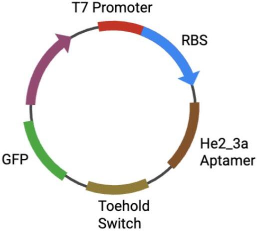 |
|---|
Sensory component
The sensory component of the system is an aptamer-based mechanism that relies on the HER2-specific aptamer: HeA2_3. When HER2 is present, the protein will bind to the aptamer sequence, which in turn activates the toehold switch within the circuit (Vezeau et al., 2023). In its default (OFF) state, the toehold switch forms a hairpin structure that hides the RBS (Wyss Institute, 2018). Upon the binding of the target RNA, initiated by the HER2-aptamer interaction, the hairpin structure unfolds and exposes the RBS, which allows translation to occur (Vezeau et al., 2023). Conversely, in the absence of the HER2 protein, the toehold switch remains in its closed hairpin structure, which will block the RBS, preventing further expression of the genetic circuit.
Reporter component
The reporter component for the system is the GFP gene, which creates a simple green fluorescent output. Following the sensory component, once the RBS is exposed, GFP translation will occur, producing a green fluorescent signal indicating HER-2 detection.
Parts level
Our planned plasmid will be ordered in fragments and constructed through Gibson Assembly. Each fragment has been strategically chosen to ensure accuracy, precision, and efficiency.
T7 promoter
Promoters regulate gene expression and begin RNA synthesis, adding efficiency to the design. The 18 bp T7 promoter was chosen for its high specificity, speed, and common use in E. coli, as that is our chosen chassis (MilliporeSigma, 2026). In typical systems, the structure drives initial transcription by interacting with the T7 polymerase of the prokaryotic host, and ensures high protein yield.
HeA2_3 aptamer
The HeA2_3 aptamer functions as the backbone of our system. An aptamer is a short strand of nucleic acids that binds with high specificity (see Figure 2). The HeA2_3 aptamer is designed to bind to HER-2 receptors in particular, allowing for an extra level of security and accuracy for our test. A 2016 study led by Marlies Gijs compared several previously discovered HER2 targeting, single-structure aptamers (HeA2_1, HeA2_6,…) and found that HeA2_3 offers the highest structural stability, acting as a key feature in maintaining system stability and function (Gijs et al., 2016). The specificity of the HeA2_3 aptamer was evaluated via flow cytometry, where in the presence of HER-2, the fluorescence of the aptamer is compared to that of a negative control parameter. By analyzing the spectral transmittance from a spectrophotometer, Gijs and her team quantitatively determined high GFP expression in SKOV3 and SKBR3 cells, and HeA2_3 indicated exclusive binding to overexpressed HER-2 cells, equating to a binding affinity coefficient (Kd) of 6.2 nM (Gijs et al., 2016).
| Figure 2: This figure presents a visual representation of the HeA2_3 Aptamer and demonstrates how the nucleic acid subunits bind with target proteins. This is the main regulatory structure in the bioengineered plasmid, as it promotes detection specificity by triggering a signal cascade only in the presence of HER2. | 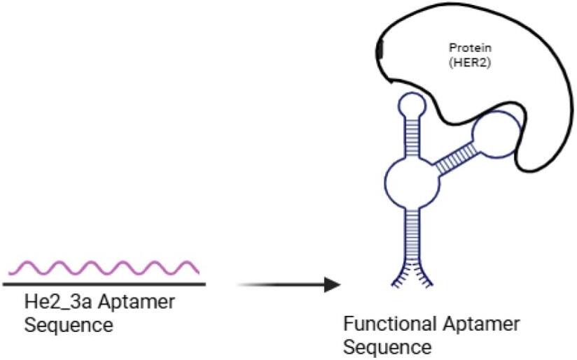 |
|---|
Toehold switch
A toehold switch is a synthetic RNA structure that forms a hairpin trigger to block the gene expression unless a complementary strand of RNA binds to the 5’ end. In this system, the toehold switch activates after the aptamer binds to a HER2 receptor. The RBS is located within the toehold switch, inside the loop formed by the hairpin trigger. Once the toehold switch is activated, the RBS is exposed (Green et al., 2014).
Ribosomal binding site
Located within the toehold switch, the RBS is only exposed after the switch activates. The RBS ensures that the ribosome is perfectly positioned before beginning protein production. This promotes both accuracy and efficiency through the process (Green et al., 2014).
Green fluorescent protein
Green Fluorescent Protein (GFP) is a reporter protein originally from the bioluminescent jellyfish Aequorea victoria, it produces a glowing green pigment under sunlight or UV rays when it encounters a target marker, which, in this project, is the HER-2 protein (Voss et al., 2013). The vibrancy of the green color serves as an indicator for the level of HER-2, which can be measured both by the naked eye and more accurately by a spectrometer.
Safety
Working with living cells is an important factor to account for in our project. Due to the limitations of BioBuilder guidelines, which only allow prokaryotic cells to be used, our team focused on a cell-free model instead. Throughout the development of our cell-free HER-2 biosensor model, safety was an essential aspect to consider. Although our team was unable to carry out wet-lab this year, the planned genetic modifications will be conducted utilizing Biosafety Level 1 (BSL-1) E. coli (DH5a). Proper PPE, waste disposal, and environmental safety will be done effectively to prevent contamination and incorrect waste disposal. In the case that the modified E. coli is accidentally released into nature, they will likely die off due to being outcompeted. This is because engineered traits such as the toehold switch can only be activated in the presence of HER-2, which indicates that our biosensor would not be activated; therefore, the modifications would have made the bacteria weaker compared to the wild strains of E. coli. Since the components of our project align with the NIH guidelines (National Institutes of Health, 2024), our cell-free biosensor system is highly unlikely to be misused or have adverse effects on the environment or people.
Discussions
The purpose of this design article was to propose the idea of a cell-free biosensor system that is designed to detect overexpression of the HER-2 protein. HER-2 plays a key role in cell division and growth. When its cells are overexpressed, it causes aggressive tumor development, making early detection imperative to reducing mortality. In the system, the aptamer specificity forces the system to respond to only the HER-2 protein, ignoring miscellaneous proteins.To control for possible false positives and negatives, the biosensor utilizes a verification system through PURExpress. To verify that the bound protein is HER2, reverse transcriptase is used to turn the mRNA to cDNA. This DNA is then compared to HER2 genetic markers for confirmation (see Figure 3). Compared to other tests, such as immunohistochemistry (IHC), it is not nearly as invasive. However, a combination of IHC and the proposed system would allow for an even higher percentage of efficiency. A potential issue within the system is the patient-to-patient variation in HER2 concentration; these variations often stem from factors like hormones, menstrual cycle, and age. The influence of this variability may have an impact on the rate of false positives and negatives, making it vital to consider HER2 differences. The level of GFP fluorescence adjusts based on the amount of HER-2. While this is useful for initial discovery, patients still need to consult a medical professional for further examination and treatment.
| Figure 3: This figure displays the HER2 detection process utilizing the constructed plasmid. An in vitro cell-free expression system followed by PURExpress allows for protein detection via fluorescence level. |  |
|---|
Next steps
Expanding on this project would require conducting essential tests to ensure that the plasmid functions reliably and efficiently in an E. coli bacterium. To begin, the designed plasmid components would need to be stitched together using Gibson Assembly. After ensuring that the E. coli absorbs the plasmid through temperature manipulation to induce a stressful environment, the E. coli would be introduced to a sample of HER2 protein, and allowed to incubate to produce GFP. GFP production will be quantitatively measured with a spectrophotometer to clearly determine the output and effectiveness of our system reacting to HER2. Upon fluorescence, reverse transcriptase would be performed using a PURExpress kit to ensure that the GFP is reacting to the HER2 protein and is not a malfunction or reacting to an external variable. Exploring this project further would include making the transition from a proof of concept model to mammalian cells as well as any adjustments necessary to ensure the system's accuracy and precision.
Author contributions
ASC contributed to the device level section and graphics. ZJ contributed to the background section. CM contributed to the parts level, next steps, and video. BW contributed to the parts level, graphics, and video. DR contributed to the systems level and abstract. AE contributed to the safety section and abstract. TF contributed to the discussion section and video.
Acknowledgements
Our team would like to thank Dr. Tiffany Jones, our teacher sponsor, as well as Laura Guerrero, our mentor, for giving us valuable advice, supporting us, and guiding us throughout the process of this project. We would also like to thank New England Biolabs, K2 Scientific, and CobbTank for funding lab supplies to give the team a hands-on experience this BioBuilderClub season.
References
Chen, H., Pimienta, G., Gu, Y., Sun, X., Hu, J., Kim, M. S., Chaerkady, R., Gucek, M., Cole, R. N., Sukumar, S., & Pandey, A. (2010). Proteomic characterization of Her2/neu-overexpressing breast cancer cells. Proteomics, 10(21), 3800–3810. doi.org/10.1002/pmic.201000297
Cleveland Clinic. (2023). HER2-Positive breast cancer. https://my.clevelandclinic.org/health/diseases/25213-her2-positive-breast-cancer
Dendukuri, N., Khetani, K., McIsaac, M., & Brophy, J. (2007). Testing for HER2-positive breast cancer: a systematic review and cost-effectiveness analysis. CMAJ: Canadian Medical Association journal, 176(10), 1429–1434. https://doi.org/10.1503/cmaj.061011
Gijs, M., Penner, G., Blackler, G. B., Impens, N. R., Baatout, S., Luxen, A., & Aerts, A. M. (2016). Improved aptamers for the diagnosis and potential treatment of HER2-positive cancer. Pharmaceuticals, 9(2), 29. doi.org/10.3390/ph9020029
Green, A. A., Silver, P. A., Collins, J. J., & Yin, P. (2014). Toehold switches: De-Novo-Designed regulators of gene expression. CellPress, 159(4), 925–939. doi.org/10.1016/j.cell.2014.10.002
Menard, S., Casalini, P., Campiglio, M., Pupa, S., Agresti, R., & Tagliabue, E. (2001). HER2 overexpression in various tumor types, focusing on its relationship to the development of invasive breast cancer. Annals of Oncology, 12(1), 15–19.
doi.org/10.1093/annonc/12.suppl_1.s15
MilliporeSigma. (2026). T7 Promoter System. Sigmaaldrich. https://www.sigmaaldrich.com/US/en/technical-documents/technical-article/genomics/cloning-and-expression/t7-promoter-system
National Cancer Institute. (n.d.). Cancer Stat Facts: Female Breast Cancer [Fact Sheet]. National Institute of Health, U.S. Department of Health and Human Services. https://seer.cancer.gov/statfacts/html/breast.html
National Institutes of Health. (2024). NIH guidelines. NIH Office of Intramural Research. https://oir.nih.gov/sourcebook/ethical-conduct/research-ethics/nih-guidelines
Scudeler, C. M., Silva, K. S. da, Silva, R. V. N., Micheletti, A. M. R., Ribeiro, K. B., Machado, J. R., Lira, R. C. P., & Pathania, A. (2026). NADPH‐related enzymes and cancer: facts and insights into the application of immunohistochemistry. Biochemistry Research International, 2026, 1–15. doi.org/10.1155/bri/1853546
Shimizu, Y., & Ueda, T. (2010). PURE Technology. In: Endo, Y., Takai, K., Ueda, T. (eds) Cell-free protein production. Methods in Molecular Biology, vol 607. doi.org/10.1007/978-1-60327-331-2_2
Taskindoust, M., Thomas, S. M., Sammons, S. L., Fayanju, O. M., DiLalla, G., Hwang, E. S., & Plichta, J. K. (2021). Survival outcomes among patients with metastatic breast cancer: Review of 47,000 patients. Annals of Surgical Oncology, 28, 7441–7449.
doi.org/10.1245/s10434-021-10227-3
Vezeau, G. E., Gadila, L. R., & Salis, H. M. (2023). Automated design of protein-binding riboswitches for sensing human biomarkers in a cell-free expression system. Nature Communications, 14(1), 2416. doi.org/10.1038/s41467-023-38098-0
Voss, U., Larrieu, A., & Wells, D. M. (2013). From jellyfish to biosensors: the use of fluorescent proteins in plants. The International journal of developmental biology, 57(6-8), 525–533. https://doi.org/10.1387/ijdb.130208dw
Wyss Institute. (2018). Toehold Switches for Synthetic Biology. https://wyss.harvard.edu/technology/toehold-switches-for-synthetic-biology/