
Chloe Dias, Siena Elliffe, Sophia Hoffmann, Aneesa Maity, Tvesha Patel, Ruby Quintiliani, Elena Schuster, and Leah Vaz, Montrose School, Medfield, Massachusetts, United States
Reviewed on 9 May 2026; Accepted on 22 June 2026; Published on 26 October 2026
With help from the 2026 BioTreks Production Team.
Second-degree burns damage the epidermis and part of the dermis, resulting in scar formation during wound healing. During the proliferative phase, activation of TGF-β1 causes fibroblasts to differentiate into myofibroblasts, which rapidly deposit type III collagen, often poorly aligned physically, and contribute to thick scar tissue. In early remodeling, type III collagen is gradually replaced by type I collagen, and fiber organization remains modifiable. Unlike post-scar treatments that cannot influence collagen alignment, our approach targets scar formation while it is actively occurring. We will use Escherichia coli, a non-pathogenic bacterial chassis, to produce RsaA S-layer proteins, which self-assemble into rigid hexagonal lattices and form a temporary, two-dimensional mesh. The proteins are then harvested and applied in an external patch. This mesh delivers nanoscale mechanical cues that provide contact guidance. By mimicking the structural features of the extracellular matrix, the lattice directs cell alignment, ensuring that as type III collagen is replaced by type I, the resulting fibers are organized. Successful protein production will be verified using a green fluorescent protein reporter, and lattice formation will be confirmed through imaging techniques such as electron microscopy. Therefore, this project presents a safe, preventative strategy to reduce scar formation following second-degree burns.
Keywords: Burn scar prevention, rsaA, S-layer protein, Escherichia coli, wound healing
Authors are listed in alphabetical order. Paola Favaretto and Sarah Hanna mentored the group. Please direct all correspondence to mailto:shanna@montroseschool.org.

Background
Second-degree burn wounds damage both the epidermis and dermis of the skin and often result in long-term hypertrophic scar formation (Hypertrophic Scar: What Is It, Causes, Treatment, 2021). Hypertrophic scars are thick, raised scars that develop when the wound healing process produces an excess of collagen and deposits it in a disorganized way. These scars can tighten the skin, reduce elasticity, limit mobility, and create lasting cosmetic concerns, which highlights the need for strategies that focus not only on scar treatment, but also on scar prevention (Jeschke et al., 2020). Current treatments primarily address scars after they have already formed, rather than preventing them during wound healing. Common non-surgical treatments include steroid injections, cryotherapy, pressure therapy, and skin grafts, which can help reduce scar thickness or improve appearance in some cases, but often do not fully prevent scar formation or restore normal tissue organization (Scar Revision, n.d.). However, these methods do not directly address the underlying cause of hypertrophic scarring, which occurs during the proliferative and remodeling phases of wound healing. Because collagen fiber organization remains modifiable during early remodeling, this stage presents an opportunity for preventive intervention (Fleischmajer et al., n.d.). Our project proposes an engineered plasmid containing the rsaA gene to produce rsaA S-layer proteins, which self-assemble into hexagonal lattice structures (Ford et al., n.d.). These lattices are intended to mimic aspects of the extracellular matrix and provide nanoscale mechanical cues that guide fibroblast alignment and collagen deposition (Hinz et al., 2007). Signal from a green fluorescent protein (GFP) reporter system will be used to confirm that the engineered plasmid is successfully producing the RsaA S-layer proteins, ensuring that the system is functioning as intended. By targeting scar formation during active wound healing rather than after the scar has formed, this project aims to develop a more preventive approach to reducing hypertrophic scarring.
| Figure 1. Proposed RsaA-based strategy for reducing burn scar formation (Phases 1-4) Panels 1-4 illustrate the design and production of the RsaA S-layer protein system. A plasmid containing the RsaA gene and a GFP reporter is introduced into a non-pathogenic strain of E. coli, which is then used to express the protein under controlled lab conditions. The harvested RsaA proteins self-assemble into a two-dimensional hexagonal lattice that is incorporated into an external patch and applied to a healing burn wound. | 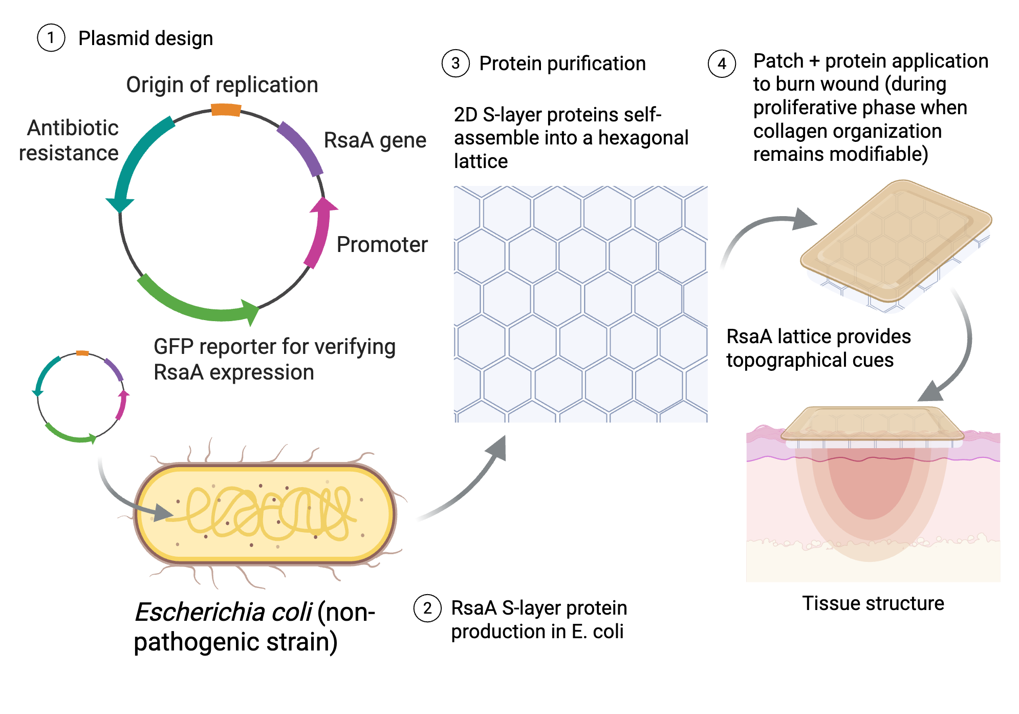 |
|---|
| Figure 2. Proposed RsaA-based strategy for reducing burn scar formation (Phase 5) Panel 5 illustrates the intended effect of the patch during wound remodeling, where the RsaA lattice provides the topographical cues that encourage fibroblast alignment and promote more organized collagen deposition, leading to reduced scar formation. In the absence of the RsaA mesh, type III collagen is more likely to remain disorganized, contributing to hypertrophic scarring. | 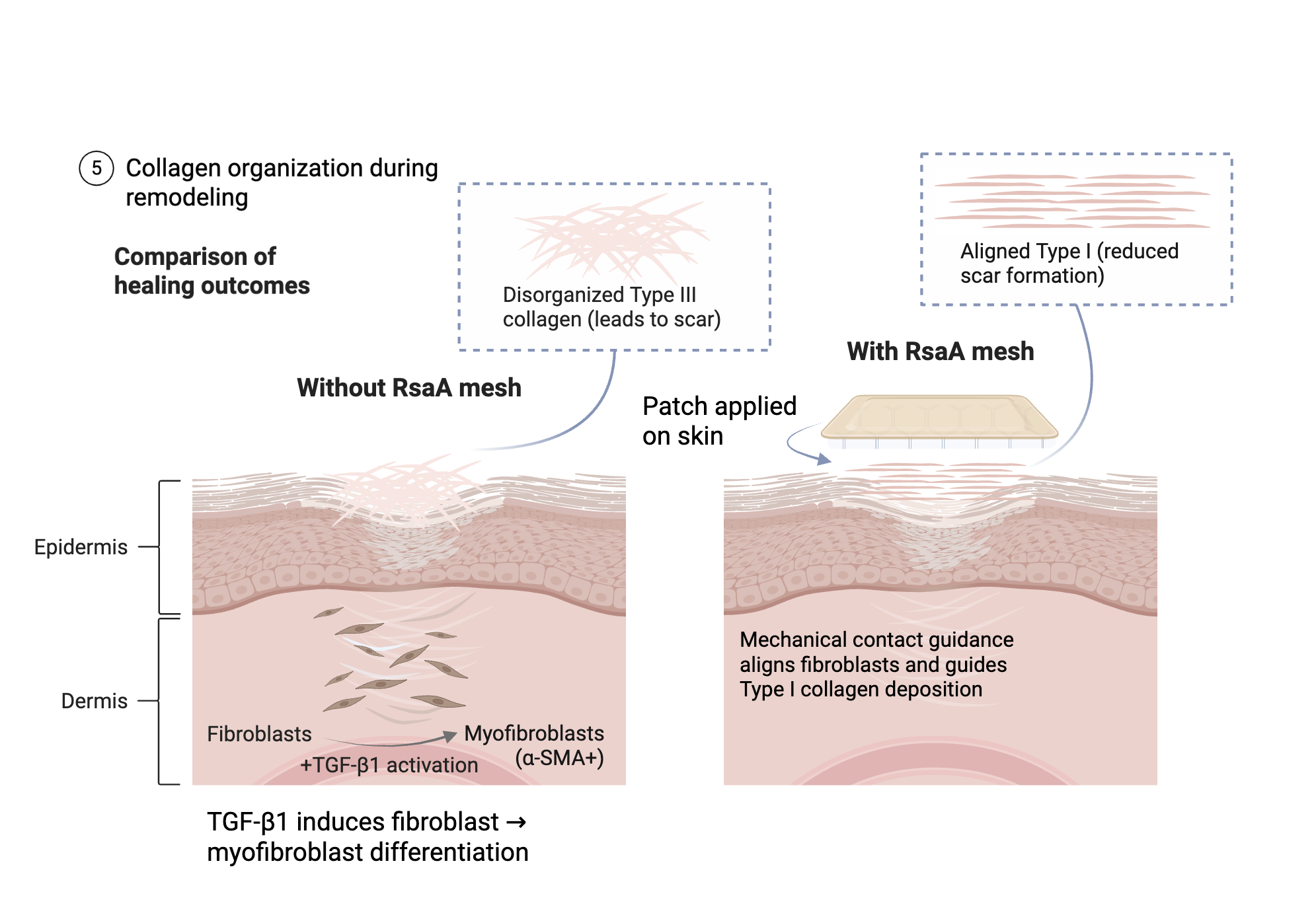 |
|---|
Systems level
Biological environment
Second-degree burns trigger a complex wound-healing cascade, in which TGF-β1 signaling promotes the fibroblast-to-myofibroblast differentiation and increases deposition of disorganized type III collagen. During the remodeling phase, type III collagen is gradually replaced by type I collagen, but if the local environment does not provide proper structural cues, this process can result in hypertrophic scar formation (Fleischmajer et al., n.d.). Our proposed system integrates into this healing process by introducing a temporary, two-dimensional mesh composed of RsaA S-layer protein lattices during the active remodeling phase. By mimicking key structural features of the extracellular matrix, the lattice is intended to provide contact guidance that helps orient cells and organize collagen deposition as the tissue matures (Hinz et al., 2007).
Systems-level effect
At the system level, our approach acts on multiple stages of wound healing simultaneously. It influences cell behavior, guides collagen fiber organization, and supports more ordered tissue structure during wound healing. Rather than replacing the natural healing process, the system is designed to shift the biological environment toward more organized collagen fiber alignment and reduced scar development.
Individual variability
While the RsaA protein lattice structure is designed as a broadly applicable, one-size-fits-all mesh, we recognize that skin structure and wound-healing dynamics can vary across different skin types and individuals. Factors such as tissue tension, skin thickness, and patient-specific healing responses may influence the effectiveness of purely mechanical guidance from pressure applied by the patch. However, because contact guidance is a foundational process where cells align their physical shape and direct their movement in response to the physical geometry or texture of their surrounding environment, in this case hexagonal lattice structures, we expect the lattice to provide baseline organizational benefits across diverse conditions. Future iterations of our system could incorporate tunable structural or biochemical features to better adapt to specific wound environments and further optimize healing outcomes.
Device level
The device consists of an engineered plasmid containing the rsaA gene and a GFP reporter, which allows visual confirmation of protein expression. This plasmid is introduced into the non-pathogenic E. coli chassis through transformation, where it directs the production of RsaA S-layer proteins. These proteins self-assemble into two-dimensional hexagonal lattices that form the basis of the proposed mesh (Charrier et al., 2019). This hexagonal lattice structure is particularly advantageous, as it mimics key structural features of the extracellular matrix, providing a repeating, nanoscale structure that supports uniform collagen fiber alignment. After production, the RsaA proteins would be harvested and incorporated into an external patch for application to the wound surface (Figure 2). To improve export of the protein, the secretion genes rsaD and rsaE may also be included, as these genes support secretion of the S-layer protein (Awram & Smith, 1998).
The device is intended to function during the proliferative and early remodeling phases of wound healing, typically occurring from several days to a few weeks after injury, when collagen organization is still responsive to external cues (Donnelly et al., 2013). In this system, the two-dimensional mesh provides nanoscale mechanical guidance that influences fibroblast alignment and helps direct collagen deposition (Figure 1). By shaping the local physical environment, the device is designed to support more organized tissue remodeling and reduce the likelihood of disorganized scar formation (Ford et al., n.d.).
Parts level
The functional system is built from genetic components from Caulobacter crescentus and expressed in E. coli to produce RsaA S-layer proteins. The main genetic component is the rsaA gene, which encodes for the RsaA proteins that can self-assemble into a two-dimensional hexagonal lattice (Charrier et al., 2019). In addition, the RsaA protein contains a C-terminal secretion signal, which functions as a tag that helps direct the protein out of the cell (Ford et al., n.d)
To improve secretion, the rsaA gene is paired with the accessory genes rsaD and rsaE, which are part of a type I secretion system. rsaD encodes a transporter protein within the inner membrane that uses ATP (cellular energy) to facilitate the movement of RsaA proteins (Awram & Smith, 1998). rsaE encodes a membrane fusion protein that helps connect the transport machinery across the periplasm, the space between the inner and outer membranes. Together, rsaD and rsaE form a pathway that allows the RsaA proteins to move directly from the cytoplasm to the outside of the cell, without stopping in the periplasm. These genes are likely organized as a polycistronic operon, meaning they can be expressed together in a coordinated way (Ford et al., n.d.).
Safety
The proposed system is designed to minimize risk by ensuring that E. coli is never applied directly to the skin. Instead, the S-layer proteins produced by non-pathogenic E. coli carrying the engineered rsaA plasmid would be harvested and incorporated into an external patch. In addition, the harvested RsaA proteins would undergo purification to remove bacterial byproducts, such as endotoxins, before incorporation into the patch. This ensures that only the structural protein lattice is applied to the wound, minimizing the risk of inflammatory or immune responses associated with residual E. coli components. This patch would then be applied to the surface of an actively healing burn wound. No live bacteria will be introduced into the wound, and the protein lattice will provide structural guidance to the healing tissue (Donnelly et al., 2013). In addition, the mesh is intended to be temporary and self-degrading, leaving no residue after wound healing is complete (Yehya, 2023). Rather than altering the natural healing process, this project aims to support it by guiding collagen organization and fiber alignment to reduce the likelihood of hypertrophic scar formation (Figure 2).
Discussions
This project aims to prevent the unorganized alignment of collagen fibers during the remodeling phase of wound healing, during which type III collagen is gradually replaced by type I collagen, through the use of a two-dimensional mesh. The mesh is intended to guide fiber orientation by providing nanoscale mechanical cues that promote contact guidance (Donnelly et al., 2013). By also mimicking structural features of the extracellular matrix, a protein network naturally occurring in human connective tissue, the system may support organized cellular behavior during healing (Chung et al., 2010). Preventing disorganized collagen deposition could reduce hypertrophic scar formation, which can negatively affect mobility, comfort, and quality of life (Epidemiology of Burn Scars, n.d.). In addition, this patch system could potentially be used alongside other approaches to scar prevention.
However, the mesh may also present challenges if it interacts with the skin or wound in unintended ways, such as causing irritation or disrupting normal healing. Because the system relies on mechanical guidance, it may be difficult to control the exact amount of pressure applied by the patch and the length of time that this pressure is maintained (Hinz et al., 2007). In addition, the degradation rate of the protein mesh will need to be tuned to match the wound healing timeline, so that mechanical guidance is maintained throughout the proliferative and early remodeling phases but does not persist longer than necessary. A future improvement to the design would be to optimize the patch material so that it delivers more consistent nanoscale cues at the appropriate stage of healing. Further testing of material properties could help refine the system and improve its ability to guide collagen organization more precisely. Future testing should also examine how the system performs on wounds located over pre-existing scar tissue, since differences in tissue structure and mechanical properties may affect patch effectiveness.
Next steps
The next step in this project is to confirm that the rsaA gene can be successfully cloned into a plasmid, transformed into a non-pathogenic strain of E. coli, and expressed under controlled lab conditions. To verify expression, our team would include a GFP reporter and compare fluorescence in induced and uninduced bacterial cultures. This would provide early visual evidence that the construct is working before moving to more advanced testing. After expression is confirmed, we would optimize basic culture conditions, such as incubation time and growth temperature, to improve gene expression. These tests would help identify the simplest conditions that produce the strongest and most consistent gene expression in a high school lab setting. A later step would be to examine whether the expressed rsaA can form the expected lattice structure and, in future work, be incorporated into a patch material. However, in our current phase, the main goal is to establish a working expression system and demonstrate that the construct can be produced in a lab setting. That would create a realistic foundation for future testing of the protein’s structural and wound-healing potential. If successful, this project could also be adapted for surgical wounds, chronic wounds, or other fibrotic conditions in different tissues.
Author contributions
SE conducted the literature review for the Background section. TP developed the Systems level section. ES and RQ wrote the Device level and Parts level sections. RQ also wrote the Safety and Discussion sections. AM wrote the Next steps section, reviewed and revised multiple sections of the manuscript, conducted a literature review on the gene choice for the project, identified the gene selected for the project, and created all figures and diagrams. SH wrote the Author contributions and Acknowledgments sections. LV and CD developed the video script, and LV assisted with video editing. All authors participated in the video production, contributed to the development of the project concept, conducted research, and reviewed and edited the manuscript. AM and RQ helped lead the project overall.
Acknowledgements
This project would not have been possible without the guidance and support of our mentor, Kelly Doering from KSBD LLC, who met with us weekly over several months and provided valuable feedback throughout the development of the project. We would also like to thank Dr. Natalie Kuldell, founder of BioBuilder, for her encouragement and interest in our project at the March 12 conference, which gave our team confidence to pursue publication. We are especially grateful to Ms. Sarah Hanna and Dr. Paola Favaretto, our club supervisors, whose support, advice, and oversight helped make the BioBuilder Club possible and contributed to the completion of this project.
References
Awram, P., & Smith, J. (1998, June 18). The Caulobacter crescentus Paracrystalline S-Layer Protein Is Secreted by an ABC Transporter (Type I) Secretion Apparatus. PubMed. Retrieved April 24, 2026, from https://pmc.ncbi.nlm.nih.gov/articles/PMC107805/
Charrier, M., Li, D., Mann, V. R., Yun, L., Jani, S., Rad, B., Cohen, B. E., Ashby, P. D., Ryan, K. R., & Ajo-Franklin, C. M. (2019, January 7). Engineering the S-layer of Caulobacter crescentus as a Foundation for Stable, High-Density, 2D Living Materials. PubMed. Retrieved April 25, 2026, from https://pmc.ncbi.nlm.nih.gov/articles/PMC6945806/
Chung, K., DeQuach, J. A., & Christman, K. L. (2010, July 12). Nanopatterned Interfaces for Controlling Cell Behavior. PubMed. Retrieved April 24, 2026, from https://pmc.ncbi.nlm.nih.gov/articles/PMC4221096/
Donnelly, P. E., Jones, C. M., Bandini, S. B., Singh, S., Schwartz, J., & Schwarzbauer, J. E. (2013, June 4). A Simple Nanoscale Interface Directs Alignment of a Confluent Cell Layer on Oxide and Polymer Surfaces. PubMed. Retrieved April 23, 2026, from https://pmc.ncbi.nlm.nih.gov/articles/PMC3735232/
Epidemiology of Burn Scars. (n.d.). Basics of Burn Care. Retrieved April 24, 2026, from https://basicsofburncare.org/epidemiology-of-burn-scars/
Fleischmajer, R., Perlish, J. S., Burgeson, R. E., Shaikh-Bahai, F., & Timpl, R. (n.d.). Type I and type III collagen interactions during fibrillogenesis. PubMed. Retrieved April 24, 2026, from https://pubmed.ncbi.nlm.nih.gov/2186689/
Ford, M. J., Nomellini, J. F., & Smit, J. (n.d.). S-Layer Anchoring and Localization of an S-Layer-Associated Protease in Caulobacter crescentus. Journal of Bacteriology, 189. https://doi.org/10.1128/JB.01690-06
Hinz, B., Wipff, P.-J., Rifkin, D. B., & Meister, J.-J. (2007, December 17). Myofibroblast contraction activates latent TGF-beta1 from the extracellular matrix – PubMed. PubMed. Retrieved April 23, 2026, from https://pubmed.ncbi.nlm.nih.gov/18086923/
Hypertrophic Scar: What Is It, Causes, Treatment. (2021, June 1). Cleveland Clinic. Retrieved April 23, 2026, from https://my.clevelandclinic.org/health/diseases/21466-hypertrophic-scar
Jeschke, M. G., van Baar, M. E., Choudhry, M. A., Chung, K. K., Gibran, N. S., & Logsetty, S. (2020, February 13). Burn Injury. PubMed. Retrieved April 23, 2026, from https://pmc.ncbi.nlm.nih.gov/articles/PMC7224101/
Scar Revision. (n.d.). Johns Hopkins Medicine. Retrieved April 23, 2026, from https://www.hopkinsmedicine.org/health/treatment-tests-and-therapies/scar-revision
Yehya, N. A. (2023, December 19). New bioengineered scaffold may help large deep burn wounds heal faster. UC Davis Health. Retrieved April 24, 2026, from https://health.ucdavis.edu/news/features/new-bioengineered-scaffold-may-help-large-deep-burn-wounds-heal-faster/2023/12