
Ismail Farid Allauddin Khan Ghori, Parisha Goyal, Saanvi Moota, Victoria Semanduyeva, Jahnavi Singh, and Vivek Venigalla, Denmark High School, Alpharetta, Georgia, United States of America
Reviewed on 9 May 2026; Accepted on 22 June 2026; Published on 26 October 2026
With help from the 2026 BioTreks Production Team.
Diabetes, a global health challenge, is currently one of the leading causes of death, as stated by the American Diabetes Association. One of its most serious complications is a chronic condition, diabetic retinopathy, which damages the retina and can result in irreversible blindness if left unaddressed. Diabetic retinopathy is highly prevalent, with an estimated number of affected Americans being 16 million by 2050. Despite the great concern, many cases go undiagnosed because conventional clinical eye examinations require specialized equipment and trained specialists. The design we plan to implement uses biochemical detection and AI-based image analysis to improve detection rates compared to traditional clinical eye screening. We implemented an AI image recognition model trained using machine learning that detects diabetic retinopathy. We also designed a lateral flow assay (LFA) that uses the color produced when gold nanoparticles bind to proteins associated with diabetes and retinal damage. The LFA is integrated into a circuit that sends an electrical current through the assay in order to measure the resistance of the LFA. Our key result is that the AI model successfully learned to distinguish between healthy and diseased retinas using real, open-source data from retinal image banks, achieving 96% accuracy and 0.03 loss. We have also successfully tested our electrochemical lateral flow assay (eLFA) concept using a COVID test, as a proof of concept, with a 41.7% change in electrical resistance, results that cement that there is a significant change in resistance during testing. Acting as a proof of concept, this dual-modal system allows us to combine qualitative and quantitative results to enhance screening accuracy. By combining image recognition and a rapid, noninvasive biochemical test, diabetic retinopathy can be detected earlier, faster, and more affordably, before vision loss becomes irreversible.
Keywords: Diabetic retinopathy, lateral flow assay, image recognition
Authors are listed in alphabetical order. Mary Cartenuto mentored the group. Please direct all correspondence to mailto:mcartenuto@forsyth.k12.ga.us.


Background
Over 40 million people, or 12% of the entire population of the United States, are living with diabetes (American Diabetes Association, 2023). Diabetes remains one of the most significant global health crises, leading to various life-altering complications (American Diabetes Association, 2023). Among these, diabetic retinopathy (DR) is a primary concern as a leading cause of preventable blindness worldwide. While diabetes was once considered a disease of the affluent, it has reached epidemic proportions in developing countries, with the global number of DR cases projected to grow from 126.6 million in 2010 to 191 million by 2030 (Mayo Clinic, 2023). DR occurs when chronic hyperglycemia causes microvascular damage to the retina, often progressing silently without noticeable symptoms until irreversible vision loss has occurred, or vision-threatening diabetic retinopathy, which is a condition expected to affect 56.3 million people by 2030 (Shukla & Tripathy, 2023).
The primary obstacle in diagnosing DR is that there are limited methods of early detection. Standard screening typically requires expensive photography and specialized ophthalmologists, which are often inaccessible in low-income or rural areas (Diabetic Retinopathy | National Eye Institute, 2025). Because the disease is asymptomatic in its early stages, many patients remain undiagnosed until the condition reaches an advanced, untreatable state. All these barriers also contribute to a crisis of compliance rates for eye examinations falling below 40% in some areas (Mya Polkamp et al., 2025). This highlights the critical need for a diagnostic tool that is cost-effective for widespread use, highly accurate to prevent false negatives, and quantitative, allowing for the measurement of disease severity.
To address this problem, our team proposes a multi-method diagnosis strategy for early detection mechanisms, an AI image recognition model combined with an electric LFA (Al-Shabrawey et al., 2015). Unlike invasive laboratory blood tests or expensive hospital-grade fundus cameras, which can cost up to $25,000, the LFA and the AI model are less expensive options, making them vital tools for diagnostics in underserved regions (Congdon et al., 2012).
Electric Lateral Flow Assay Design
Systems level
Our design is intended to serve as a diagnostic tool that combines a biochemical test for detection, electrical resistance for sensing, and artificial intelligence to detect and measure the severity of DR. This system utilizes a multiple-method strategy for detection with two different, but interconnected components: an Arduino-based electrical resistance sensor that builds off of a sandwich LFA design and an AI image analysis model. The eLFA is designed for at-home use to detect changes in electrical resistance, while the AI model acts as a secondary tool used by clinicians to verify detection.
The eLFA system begins by taking a non-invasive, biological input in the form of tear fluid. To ensure a consistent volume, the fluid is collected using a microcapillary tube, designed to hold a fixed amount. This collection method prevents the skewing of electrical resistance readings and ensures proper screening data (Comito et al., 2025). This fluid is placed at one end of the LFA, where capillary action transports it across a nitrocellulose membrane (Lateral Flow Immunoassay Protocol, 2023). As the fluid travels, Vascular Endothelial Growth Factor (VEGF) and glycated albumin (GA), the antibodies of the biomarkers associated with the detection of retinopathy, are conjugated to gold nanoparticles (Ang et al., 2019). These gold nanoparticle-conjugated antibodies are needed to produce the output, a red line that visually indicates the presence of retinopathy at the test lines (Secondary Antibodies – Jackson ImmunoResearch, 2017). The system works similarly to the traditional COVID or pregnancy tests, from which the basis of the eLFA was derived.
Unlike a traditional LFA, the main differentiating factor that is present in this project is an electrical sensor. An Arduino supplies 3.3 V through a circuit called a Wheatstone bridge (See Device level for more details), comprising two branches: one with known resistors, and another containing a variable resistor and the LFA. As the gold nanoparticles accumulate, the electrical resistance of the membrane changes (See Device level for more details) (Abarintos et al., 2025). An Arduino Uno microcontroller converts analog voltage signals from both branches to digital (Digital Pins, 2024), which is then used to calculate resistance(See Device level for more details). Once the tear fluid sample is applied and travels across the membrane, the Arduino compares the voltage readings before and after the binding occurs, and calculates the percent change in resistance at each electrode. This value is then revealed to the user, providing a quantitative measure of the biomarker concentration that enhances the functionality of the visual line display. These quantitative resistance values are utilized to establish biomarker concentration levels rather than provide a guaranteed diagnosis. The AI model evaluates these concentration categories to verify findings and improve screening reliability.
Device level
| Figure 1. Diagram of lateral flow assay and antigen progression through the assay. A tear sample moves from the sample pad through the conjugate pad via capillary action, allowing target biomarkers to bind with gold-nanoparticle conjugated antibodies. Once the fluid flows down the strip, the biomarkers become trapped by the antibodies at Test Lines 1 and 2, concentrating gold nanoparticles. Alligator clips apply voltage across the system to measure changes in electrical resistance. | 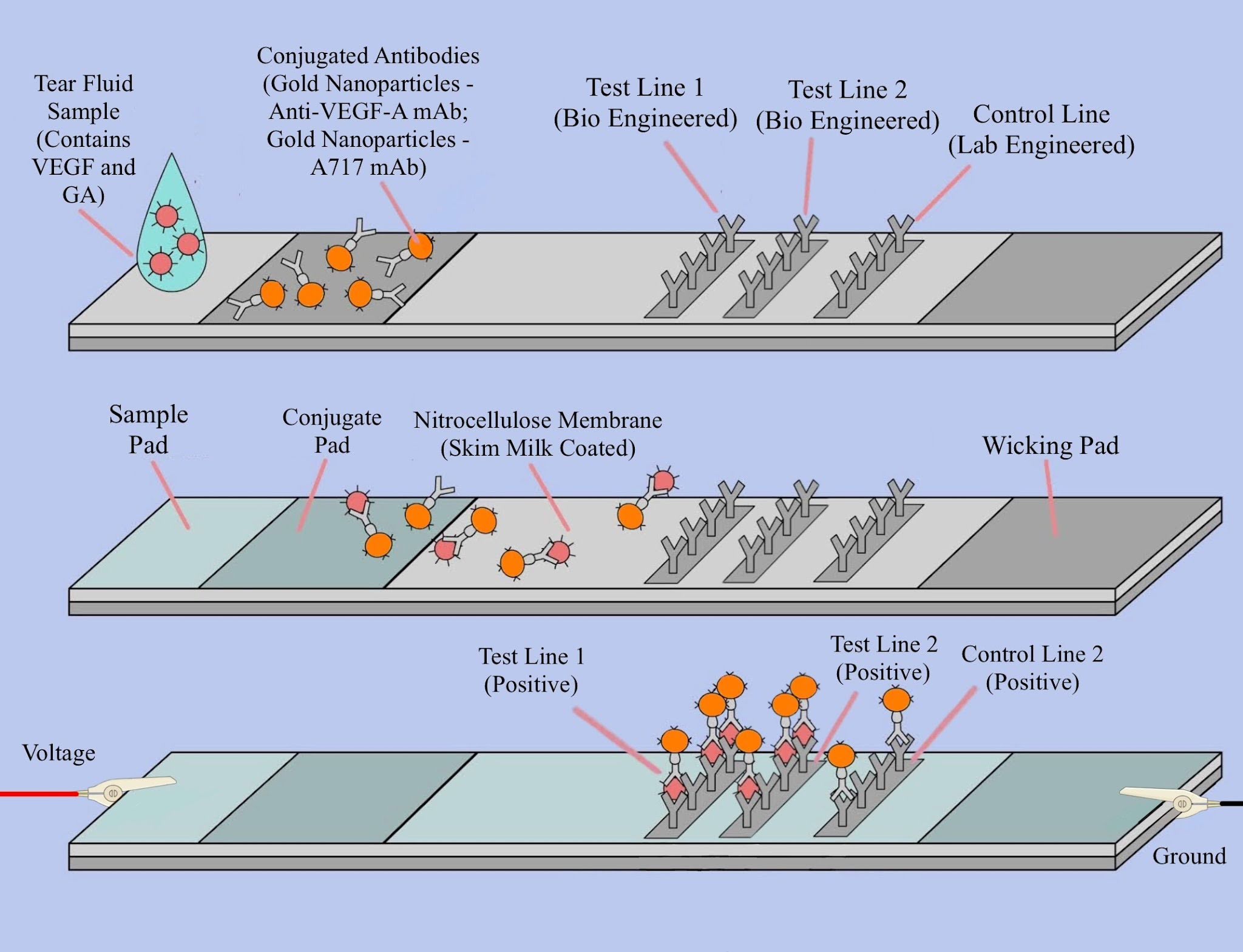 |
|---|
Overview
The LFA’s shell contains a sample pad, conjugate pad, nitrocellulose membrane, and absorbent pad (see Figure 1). The sample pad is where the sample fluid, in this case, the tear sample from a patient, is dropped onto. It contains surfactants and buffers that help maintain a stable environment that prevents analytes from denaturing and also ensures proper binding at the conjugate pad (Viro Research, 2019).
The conjugate pad contains the gold conjugated antibodies (see Parts level for conjugated antibodies information) that will bind to the analytes as the tear fluid flows through the pad. These antibodies are specific to both VEGF and GA. These conjugated antibodies are bonded to gold nanoparticles, which create a red color when present in high concentrations (Bucolo et al., 2021).
The detection zone for the test is a porous nitrocellulose membrane, where immobilized bioengineered capture antibodies are fixed at specific locations. Their role is to bind to the analyte–AuNP-conjugated antibody complexes and exhibit red lines. In this design, three lines are printed onto the membrane as illustrated in Figure 1: Test Line 1 (TL1) contains a bioengineered antibody to capture the VEGF-gold-conjugated-anti-VEGF complex, Test Line 2 (TL2) contains a bioengineered antibody to capture the GA-gold-conjugated-anti-GA complex, and the Control Line (CL) contains antibodies that bind to each of the complexes regardless of analytes’ presence, confirming flow of the tear fluid. The density of conductive gold nanoparticle conjugated antibodies-antigen complexes captured at TL1 and TL2 is the primary variable that changes, affecting the strip’s resistance. This is attributed to the attached gold nanoparticles, which exhibit slight electrical conductivity. Their placement and accumulation can affect the resistivity of the assay at specific locations (Koczula & Gallotta, 2016).
At the end of the test, the absorbent pad wicks out excess reagents and prevents any backflow of the liquid. This allows for a unidirectional flow of the tear fluid through the assay, ensuring that the antibodies bind effectively to the test and control lines for a stable resistance reading (Koczula & Gallotta, 2016).
Materials and Additional Factors
The sample pad is composed of glass fiber. According to Ahlstrom’s guidance on material selection, glass fiber materials have a more porous surface, which is suitable for fluids that do not require filtration, such as viscous and limited-volume samples like saliva and nasal fluids. Tear fluid fits this profile: tears exhibit viscosity levels of 0.91 mPa, which makes tears similar in viscosity to saliva (Rantonen & Meurman, 1998; Graf et al., 2025). Also, tear fluids do not contain any cells or large particulates that require filtration. Therefore, this material will allow the tear fluid to flow downstream in the assay without any resistance or analyte impedance that can compromise the assay’s results.
In order to maintain an environment that does not damage or denature the biomarkers, the sample pad must be prepped with surfactants and buffers to match crucial parameters such as pH to keep the proteins from denaturing or being damaged. Tear fluid, on average, has a pH from 6.5 to 7.6 with a mean of 7.0 (Abelson et al., 1981). Standard tear-based diagnostics utilize phosphate-buffered saline, a widely used buffer to maintain pH and prevent osmosis and denaturation of any particulates in biological fluids (Comito et al., 2025; ScienceDirect, 2025). Lateral flow assays also utilize detergents and emulsifiers, such as Tween-20 and CHAPS, to reduce the surface tension of the pad and to prevent irregular flow patterns (CHAPS Detergent, 2019). The typical concentrations of these two detergents are 0.25% and 0.5% (Adornetto et al., 2025). These detergents ensure the liquid sample flows in a smooth, straight line down the strip. By keeping the fluid moving evenly, the PBS buffer is able to prevent an alkaline surface environment from forming. Avoiding an alkaline shift is important because an elevated pH can alter the structure of the test line antibodies, skewing the resistance readings (Liang, 2019; Conjugate Pad Considerations for Lateral Flow Assays – NanoComposix, 2026). However, even with a consistent flow with the reduced surface tension, glass fibers contain high activation sites that can bind to the conjugated components. To prevent any false negatives, 1% bovine serum albumin is spread throughout the pad, which covers these high-activation sites to maintain flow-through without any conjugate being trapped in the glass fibers (“Blue-White Screening & Protocols for Colony Selection,” 2025).
The conjugate pad is also composed of glass fiber, a regularly used choice for the parameters of tear fluid (Conjugate Pad Considerations for Lateral Flow Assays – NanoComposix, 2026). Among Ahlstrom’s offerings, glass fiber offers a wicking rate that is suitable for rapid test results, along with a neutral pH to allow for a stable environment for the conjugate even without any pretreatment (Ahlstrom IVD Materials.pdf, 2026). The wicking rate is crucial as it governs how quickly the conjugate can bind with the antibodies and enter the nitrocellulose membrane. With a consistent wicking rate, the conjugate pad ensures the front of the stream does not contain any unbounded conjugated antibodies, preventing false negatives or weakened signals (Guide to Lateral Flow Immunoassays Innova Biosciences Guide, n.d.).
The conjugate pad contains two gold-conjugated antibodies in order to bind to the analytes, VEGF and GA. Bevacizumab (Avastin) is the monoclonal antibody targeting VEGF, while the Monoclonal Antibody Clone A717 (A717 mAb) targets GA. These two antibodies are conjugated to gold nanoparticles, which are crucial for creating a visual signal when found together in closed clusters (Koczula & Gallotta, 2016). Since this diagnostic is a sandwich immunoassay, the typical gold nanoparticle size is 40 nm at 10 OD (optical density) (Child, 2023). These gold nanoparticles are bound to the antibodies through physical passive adsorption, as it is the most viable for biological compounds and also offers an advantage in scaling during production (Child, 2023). This process works by simply mixing the gold nanoparticles with the antibodies, taking advantage of intermolecular forces, such as Van der Waals and ionic forces, to facilitate the spontaneous binding and formation of the conjugated antibodies. Spare antibodies are removed through filtration after the conjugation is complete(Passive Nanoparticle Conjugation for Lateral Flow Assays – NanoComposix, 2025). When transferring the conjugated antibodies onto the glass fiber, the conjugated antibodies are dispensed at a rate of 10 µL/cm of glass fiber. The pad is then dried in a forced-air convection oven at 37°C for 1 h (Conjugate Pad Considerations for Lateral Flow Assays – NanoComposix, 2026). When the antibodies are dried on the pad, they tend to denature or stick to the conjugate pad. To prevent this, this design implements two sugars: Sucrose and Trehalose. These two sugars act as a ‘replacement’ for water, stabilizing the antibody's structure while also ensuring it does not stick to the conjugate pad. Unsticking the antibodies can ensure the analyte in the tear fluid can bind with the antibodies and travel through the nitrocellulose membrane(Fleet Bioprocessing Ltd, 2026). Selecting the right concentration of these sugars depends on the OD level of the gold nanoparticles bonded with the conjugate antibodies; for the design’s OD level of 10, the recommended concentrations for sucrose and trehalose are 10% and 5%, respectively(Conjugate Pad Considerations for Lateral Flow Assays – NanoComposix, 2026).
The nitrocellulose membrane is made of cellulose ester of nitric ester (Bajpai, 2018). This biological membrane allows for sample liquid to flow at predetermined rates based on its pore size, thickness, and chemical treatment (nanoComposix, n.d.). In this diagnostic tool, the membrane also houses the capture antibodies that capture VEGF and GA, which are conjugated with the gold nanoparticles. To obtain accurate and consistent results, many commercial lateral flow assays utilize a membrane that has a slow run-time (the time it takes for the liquid front to traverse the entire length of the membrane. A larger run-time can improve analyte binding efficiency by allowing more interaction time between targets and capture antibodies (nanoComposix, n.d.). To create a membrane with a longer run-time, the membrane must have a smaller pore size and density. Our design is modeled after the Millipore HF180, which has a run-time of 180 s (“Blue-White Screening & Protocols for Colony Selection,” 2025). On the nitrocellulose membrane, capture antibodies corresponding to TL1, TL2, and CL are printed through hydrophilic interactions using either a pneumatic contact dispenser or a non-contact dispenser. Then the antibodies are cured under hydrophobic forces after the water is driven off under forced air at 40°C (Lateral Flow Immunoassays – Jackson ImmunoResearch, 2017). It is important to note that the thickness of the membrane plays a crucial role in the electrical resistance of the LFA over time.
The absorbent pad is composed of a blend of cotton and glass fibers. Ahlstrom’s documentation shows that these materials offer consistent absorbance rates, which allows for the nitrocellulose membrane to clear out and enables faster result readouts without sample evaporation delays (Ahlstrom IVD Materials.pdf, 2026). This specific material was chosen due to the absorbent pad’s correlation with the wicking speed of the conjugate pad. Different wicking speeds can affect the flow rate throughout the entire strip; inconsistent rates from the conjugate and absorbent pad can tip the scale of how long the conjugate can bind to the test/control lines in the membrane. A higher wicking rate at the conjugate pad relative to the absorbent pad’s absorbance rate results in an oversaturation of conjugate, leading to false positives. A lower wicking rate, conversely, creates a shortage of conjugate as the absorbent pad can absorb more conjugate than what is required to create visible lines (Kainz et al., 2021). To counteract any of these outcomes, the absorbent pad must maintain an absorption rate that is consistent throughout the duration of the test. The mixture indicated in the beginning achieves this goal, as it contains a large bed volume of cellulosic cotton, which contributes to the consistent absorption rate, and glass fiber, which contributes to dimensional stability (“Blue-White Screening & Protocols for Colony Selection,” 2025).
Assembly
| Figure 2. Side-view diagram of the electrical lateral flow assay showing overlaps of the components. The sample pad contains the tear fluid that migrates through the conjugate pad containing nanoparticle-linked antibodies. Then, it moves across the nitrocellulose membrane with Test Line 1 (TL1), Test Line 2 (TL2), and a Control Line (CL), and finally into the absorbent wick. All components lie on the backing card for consistent capillary flow. | 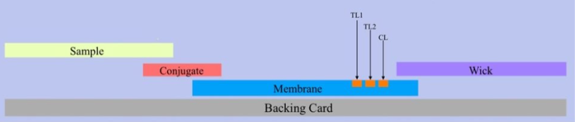 |
|---|
The strip assembly starts with a backing card that is electrically inert and has enough adhesion to hold the assembly together (Lateral Flow Backing Cards | IVD Backing Cards Manufacturer, 2024). Each component must be attached such that the nitrocellulose membrane is on the lowest level, with the conjugate and wick pad overlapping on the opposite ends of the membrane (see Figure 2). The sample pad is overlaid on the other end of the conjugate pad in Figure 2 as well, such that the sample pad and wick pad comprise the entire length of the test. These components should only have an overlap of ~2 mm to ensure there is enough space for the liquid to traverse through the various parts of the assay (Lateral Flow Immunoassay Protocol, 2023). The entire strip assembly is then mounted on an inert rigid backing made of polystyrene for mechanical stability. This ensures that the strip maintains a fixed position within the electrical measurement housing and allows consistent electrical contact during measurements (Useon, 2025). Conductive contact pads made of copper at both ends of the strip allow the Wheatstone bridge electrodes to make electrical contact. The shell is also coated in aluminum foil, transforming the shell into a Faraday Cage, preventing any electrical or magnetic interference for precise electrical resistance measurements (Koczula & Gallotta, 2016).
Electrical Wheatstone Bridge Integration
In order to accurately measure and detect minute changes in the electrical resistance of the lateral flow assay, the Wheatstone bridge employs two resistive branches. One branch contains a resistor of known electrical resistance and a variable resistor (Wikipedia, 2020). The other branch contains another resistor and the lateral flow assay (which is connected through the assay shell’s leads). These branches are clearly depicted in Figure 3, including the Arduino set-up. The Arduino supplies a stable 3.3 V DC Voltage through both resistive branches and monitors the differential voltage between the two midpoints through two analog pins (Digital pins).
| Figure 3. Circuit Diagram of the Wheatstone bridge with the lateral flow assay. The setup includes an ammeter, resistors, a potentiometer, and the lateral flow assay component, forming a circuit for measuring the unknown electrical resistance due to the LFA, specifically due to the biomarker interactions. | 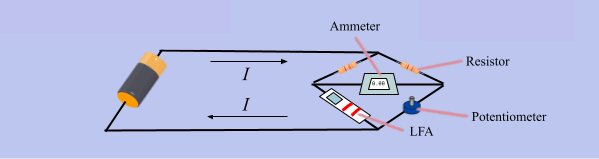 |
|---|
When the variable resistor is tuned to minimize this differential, the bridge approaches a balanced state (Wikipedia, 2020). The Arduino indicates the bridge is balanced through a console readout and an LED indicator. The LED blinking frequency increases with greater bridge imbalance and turns off when the bridge reaches a balanced state. Once the bridge is balanced, we use the formula:
(Wikipedia, 2020)
Where:
= a known resistance
= resistance of the variable resistor
= a known resistance
= Lateral Flow Assay’s electrical resistance(unknown)
Solving this equation allows us to determine the unknown resistance of the LFA:
Parts level
The presence of DR is what is being tested through an elaborately designed E-LFA. Within the E-LFA, both the Vascular Endothelial Growth Factor (VEGF) and Glycated Albumin (GA) are the target biomarkers for DR. The antibodies that are used to detect these are monoclonal antibodies. Bevacizumab (Avastin), a monoclonal antibody, is used for VEGF, while Monoclonal Antibody Clone A717 (A717 mAb) is used for GA. Since Avastin is a monoclonal antibody, it targets only VEGF (NCI Drug Dictionary, 2026). The same goes for the A717 monoclonal antibody; its binding affinity is preferentially solely for glycated albumin (Cohen & Hud, 1989). VEGF and GA are water-soluble, leading to a uniform solution containing the proteins and water, and thus can be used in a buffer solution, etc (Belinskaia et al., 2020; Florence T.H. Wu et al., 2009). The VEGF gene’s expression is regulated by hypoxia-inducible factor (HIF)-1α, which is activated under hypoxic conditions, a characteristic phenomenon of DR (Li et al., 2020). HIF-1α dimerizes with the unregulated HIF-1β to form a transcription (Ho & Kuo, 2007). Hypoxia is a characteristic phenomenon of DR (Li et al., 2020). Thus, the production of VEGF is directly correlated to DR. A person with non-proliferative diabetic retinopathy and proliferative diabetic retinopathy has significantly higher VEGF levels in tear fluid than those without DR (114.4 SD 52.5 pg/mL and 150.8 SD 49.7 pg/mL, respectively; 40.4 SD 26.5 pg/mL, p < 0.001) (Ang et al., 2019).
As mentioned in the device level, the antibodies are conjugated with the gold nanoparticles through physical passive adsorption. However, this process is most efficient when the pH is close to the isoelectric point of both antibodies (Passive Nanoparticle Conjugation for Lateral Flow Assays – NanoComposix, 2025). This ensures the antibodies have close to or 0 charge on their surface, allowing the gold nanoparticles to bind through intermolecular forces with relative ease. For Avastin and A717 mAb, the isoelectric points are 8.3 and 8.2, respectively (Kaja, 2011; Goyon et al., 2017). Since this complication can be caused by many other factors, to determine that the patient is guaranteed to have diabetes to cause to DR, we introduce another protein to detect: glycated albumin (GA). GA levels are significantly higher in type 1 diabetes than in type 2 diabetes (3.32 0.36 vs. 2.89 0.44, p<0.001) (Yoshiuchi et al., 2008). The mean values of GA in patients, both men and women, are approximately 40% higher than those of the controls (patients without type 2 diabetes) (Ai et al., 2009). Thus, GA is a reliable marker protein to determine if patients and test users have diabetes. Both of these proteins are present in the tear fluid of individuals with and without DR (Gospodarczyk et al., 2025; Ang et al., 2019). Since both of these proteins are found in tears, patients and testers can use tear fluid to detect DR.
This new solution, containing the buffer solution and the proteins from tear fluid, will flow through the nitrocellulose membrane with capillary action (Tang et al., 2022). The antibodies Avastin and A717 mAb will be conjugated with gold nanoparticles (AuNPs). This is crucial since gold nanoparticles are able to reflect a color, most often red. This reflection of color is due to vibrations labelled as surface plasmon resonance. The resonance of these particles is dependent on their structure and size, which will then induce different colors (AZoNano, 2023). However, the standard red color is induced by a particle the size of approximately 20 nm in size. AuNPs are also not water-soluble, but they can be stabilized using ligands, surfactants, and more. Gold nanoparticles also do not oxidize or corrode easily in biochemical environments. The solution will flow through the antibody and through the AuNP-conjugated antibodies Avastin and A717, which will be consolidated towards the beginning of the lateral flow assay, directly after the sample pad. The proteins in the buffer solution, VEGF and GA, will bind their respective monoclonal antibodies that have been conjugated with AuNPs. Then, a new complex is formed: AuNP-antibody-protein complexes.
These complexes will continue to flow due to the capillary action previously mentioned. Then, these new complexes will bind to pre-permanently-ingrained bioengineered antibodies, which are specifically engineered to adhere to the AuNP-antibody-protein complexes (Frenia et al., 2025). These are the two test lines. Then, complexes that are not AuNP-antibody-protein complexes, but are just AuNP-antibody complexes (i.e., complexes that have not bonded to any tear fluid proteins) will bind to a bioengineered protein line, which is also pre-permanently ingrained. This functions as the control line.
One of the possible issues is non-specific binding to protein, where the protein and complexes may bind to the membrane itself. To address this complication, the nitrocellulose membrane will be covered in a surfactant, specifically non-fat dry milk (NFDM). NFDM is a protein-based blocker that will fill in unoccupied protein-binding spaces in the membrane (Western Blot Blocking: Best Practices | Abcam, 2024). Skim milk is composed primarily of casein and whey proteins. This is important because the NFDM powder is derived from skim milk (Rathod et al., 2023). Caseins are insoluble, and whey proteins are soluble. 80% of the NFDM is composed of casein in its protein content (Madureira et al., 2007). Casein effectively prevents the non-specific binding issue of the membrane by saturating the available binding sites. This helps to lower the chances of the antibody and complexes binding elsewhere. Casein is a stabilizing protein, meaning it will help in preventing the complexes from denaturing. Whey protein affects the interactions and functions of proteins, helping with non-specific binding this way (Khatun et al., 2022).
Image Recognition Model
System Level
The AI diagnostic model serves as a non-invasive screening tool for DR and as a second opinion for optometrists. While the eLFA diagnoses DR through the presence of key biochemical signatures present in tear fluid, the image recognition utilizes retinal images to detect structural changes associated with DR, specifically vascular damage (Boyd, 2019).
This model takes in a retinal fundus image as input and returns a binary classification of 0 (DR Negative) or 1 (DR Positive). The model was trained using open-source retinal image banks (American Society of Retina Specialists, 2025), where we selected 124 labeled images of both healthy and diabetic retinas. The training teaches the model to distinguish pixel-level patterns among images that can suggest retinal damage, such as weakened blood vessels, hemorrhage, edema, etc. This trained model can then be used to quickly diagnose DR and support clinicians by providing additional insight.
Model Architecture
The model is built with Python utilizing the libraries TensorFlow, Keras, and NumPy, following a feedforward neural network design. Input images are fed into preprocessing, where they are converted to grayscale with the help of OpenCV and resized to a 128×128 pixel image. Grayscaling the image reduces the dimensionality of the input without sacrificing structural features related to DR. The resizing ensures that the image recognition model does not overfit to the image resolution difference between datasets. The pixel values are then normalized from a scale of 0-255 to a scale of 0-1, ensuring numerical stability while training. These pixel values are then flattened into a matrix, which is fed into the network.
| Figure 4. Model architecture of the image recognition algorithm used for retinal analysis. Data flows from the input layer of 128 neurons, through a hidden layer of 12 neurons, to a single output neuron. These pathways help the model learn patterns in retinal fundus images. | 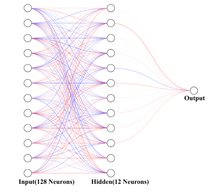 |
|---|
The model’s architecture is shown in Figure 4, illustrating the different layers used for classification in the model. The model contains one hidden layer and an output layer. The hidden layer contains 128 neurons, which perform ReLU activation to learn nonlinear features from the input. The output layer contains one neuron, which performs a sigmoid activation, mapping the ReLU activation’s output from each hidden-layer neuron into a probability between 0 and 1. Values above 0.5 are classified as positive, and the rest as negative. This model is compiled with the Adam Optimizer and binary cross-entropy loss, which are standard for binary classification models. The dataset undergoes an 80/20 split, with the former for training and the latter for testing. The model trains over the training dataset for 15 epochs, with the test dataset being used as a validation to adjust the neurons. Training accuracy and loss are monitored through each epoch in order to ensure there is no overfitting.
The trained model can be deployed via cloud service providers such as AWS or Azure, or locally through TensorFlow.js or Flask. Combined, the eLFA and image recognition model form a two-channel diagnostic test where both components complement each other. The eLFA provides rapid, biochemical data that can be quantified through tear samples, accounting for molecular changes in retinal damage before structural damage becomes apparent. The AI model functions independently, but not separately from the eLFA. This model analyzes retinal fundus images for physical markers of deterioration. Where one method may produce an ambiguous result, the other provides contrasting information, reducing the likelihood of false positives and false negatives. The dual-modal approach is particularly valuable in low-resource environments or primary care centers where a specialist’s second opinion can complement the results from the technologies, allowing the combined output to support clinical decisions.
Safety
The electronic lateral flow assay will be designed with safety considerations at every stage of development and use. Construction will be performed under appropriate laboratory safety conditions and standard PPE. Electronic components will operate under low-voltage conditions with proper insulation to minimize electrical hazards.
In the case of biological hazards, the safety level for this lab research would be BSL-1. This is because protein VEGF, GA, their respective antibodies, and other bioengineered antibodies would not be considered moderate-risk agents. However, labs should incorporate air filtration using HEPA filtration to remove a significant amount of particles from the air (Zacharias et al., 2021). Equipment can be cleaned using Presept 0.45%, if possible (Weidmann, 2023). Otherwise, researchers can use common 70% isopropyl, bleach, etc. A deeper clean for more heat-resistant material would be an autoclave. These methods will guarantee safety for those researching using these proteins, as they are not as dangerous as BSL-3 or BSL-4.
During development phases, the system will be designed so that it is easy to diagnose problems and issues, with components clearly spread out.
Discussions
The primary strength of this project lies in its multi-method diagnosis strategy, combining an electrical lateral flow assay and an AI-based image analysis. Unlike expensive hospital-grade cameras or invasive laboratory blood tests, both the LFA and the AI model are designed to be an inexpensive option, making them a great tool for diagnostics in resource-limited settings.
While traditional LFAs are often binary (positive/negative), the integration of a Wheatstone bridge to measure electrical resistance allows for a quantitative reading. This provides data on the concentration of VEGF and GA, potentially allowing clinicians to track the severity of the disease rather than just its presence. By leveraging neural networks, we tested our model architecture by training it on a dataset of 124 retinal images sourced from retinal banks. Through validation on our datasets, the model has achieved 96% diagnostic accuracy and a loss of 0.03, as depicted in Figure 5.
| Figure 5. Accuracy and Loss vs Epochs graphs of the AI Model. The left graph shows model accuracy increasing rapidly during early epochs and stabilizing near 0.9-1.0 accuracy values. The right graph shows a decrease in loss values approaching zero. These graphs confirm the model is reliable for classifying images. | 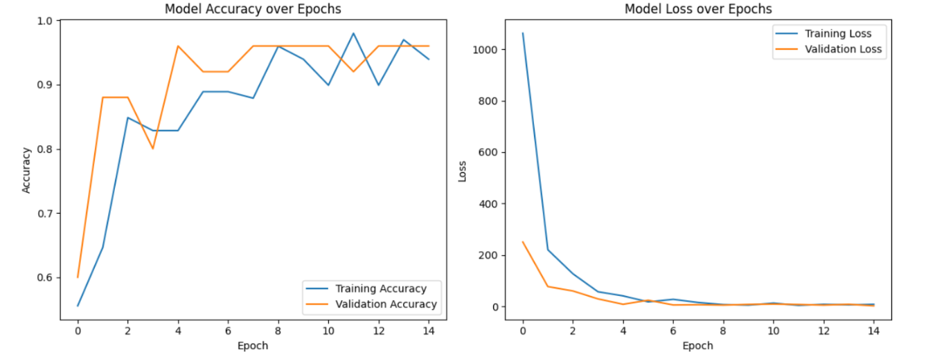 |
|---|
The eLFA takes inspiration from the widespread accessibility of COVID-19 rapid tests. We have theoretically modified the standard LFA by integrating biological receptors with specialized electrical components. Unlike traditional "yes/no" tests, this bio-electrical system utilizes a Wheatstone bridge–an electrical circuit used to measure an unknown electrical resistance by balancing two voltage dividers (legs) of a bridge circuit–circuit to translate the biological binding of analytes, such as VEGF and GA, into measurable changes in electrical resistance. This allows the device to move beyond qualitative detection and provide a quantitative measurement of protein concentration, potentially allowing clinicians to track the severity of the disease and its vascular progression rather than just its presence. To test the proof of concept, we utilized the COVID-19 test without the antibodies, where we connected the test to the Wheatstone bridge circuit, comparing the results of a buffer solution to a test with a buffer solution, replicating the way the test would work without actually testing for DR. However, with the full experiment, the added antibodies would be on the test lines, not the COVID-determining antibodies, and instead of the buffer solution, tears would be used. For our testing, we purchased the Flowflex COVID-19 Antigen At Home test. We cracked open the casing to access the LFA and used copper tape to connect the electrode on both ends of the LFA. Then, we wired a Wheatstone bridge using an Arduino and a breadboard, following our design depicted in Figure 6.
| Figure 6. Experimental setup of the eLFA concept test. The setup includes an Arduino microcontroller connected to a breadboard circuit, a multimeter for voltage and resistance measurements, and a battery pack supplying power to the system. The jumper wires link the parts of the circuit together. | 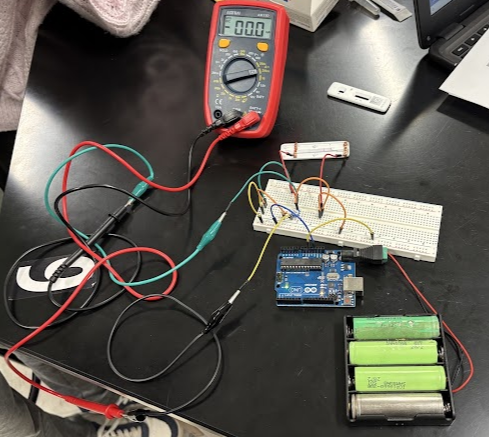 |
|---|
When we ran the test without the buffer solution, the LFA resistance when the circuit zeroed out was 1.27 kohms, and the resistance of the LFA with the buffer solution was 1.8 kohms, resulting in a significant change in resistance of 41.7%. It is important to note that the code is measuring the bridge differential voltage directly, not solving for resistance, which we calculated by hand.
Because this project is currently a proof of concept, the primary challenge remains the physical transition from individual parts to an integrated system. Specifically, while the AI and the electrical circuit have been tested independently, the actual physical integration of antibodies onto the LFA strip is still in the testing phase. The testing phase of the integration is important for identifying reactivity between the two test lines with the VEGF and GA antibodies before the device can be considered for clinical use. Since there are no current rigorous validation studies involving the use of tear fluids in an immunoassay diagnostic, many of our parameters concerning the materials/ chemicals used in the pads will have to be tested to ensure they do not interfere with assay performance.
Another significant technical issue remains in characterizing the biochemical interaction between the two primary antibodies. Our design utilizes separate test lines for VEGF and GA to prevent potential cross-reactivity. However, a phenomenon known as steric hindrance can interfere with the binding sites of VEGF and GA, isolating them and compromising the integrity of the electrical resistance data (Matos, 2020). The control line where the AuNP-antibody complexes are unbound by the biomarkers will accumulate and may cause issues in the future. This control line will also create resistance and lead to a false measurement of the total resistance due to the gold-nanoparticle accumulation pattern. However, this issue may be able to be overcome by keeping a consistent amount of binding in the control line.
Next steps
Building on the initial proof of concept, our team will spend the upcoming semester transitioning to the physical development and construction phase. The project will focus on the actual synthesis and assembly of the lateral flow assay components. Our team will also do further research and develop a method for collecting tears without damaging the retina, determining the chemical and protein composition of the tears, or harming the proteins themselves in the tears. Then, for our eLFA, initial testing will be conducted to verify that our biological sensor correctly responds to the corresponding retinopathy markers. Once the team ensures the antibodies work as intended, the team will then focus on rigorous testing to establish ranges of electrical resistances that pertain to certain DR severities. During this stage, the team will also start the development of a product design, involving the casing of the LFA and a custom printed circuit boardto replace the Arduino-based setup with a more cost-effective electrical measurement system.
The image recognition model will be refined by training it on larger, more diverse datasets of retinal fundus images to not only detect the presence but also the severity of DR. The team plans to contact multiple eye clinics to obtain direct access to retinal banks for training the model in real time. This training process aims to increase the model’s sensitivity and specificity, ensuring the AI model provides a highly reliable diagnosis. The team will also focus on the deployment of the AI model so it can be used by eye clinics with relative ease.
Author contributions
VS contributed to the development of the discussion, background, and references sections, explaining the importance of early detection and the overall mechanisms of the multimethod diagnosis strategy, and integrating resources utilized. VV contributed to the development of the electrical component of the lateral flow assay, specifically the Wheatstone bridge, and constructed and tested the AI image recognition model. IA contributed to the initial development and understanding of the lateral flow assay, specifically research of the biomarkers and antibodies, the writing of the Parts level in its entirety, the biological aspect of the Safety section, creation of figure 1, figure 2, figure 3, and refinement of the paper. PG contributed to refining sections of the manuscript based on reviewer feedback, developing the next steps and references sections, and researching the proteins VEGF and GA to identify them as appropriate biomarkers for the lateral flow assay. JS contributed to the abstract and background sections, the creation of the AI model and a user-accessible HTML interface, and the development of the eLFA through the Arduino-based experimental setup. SM contributed to the protein research for the eLFA, real world applications of the eLFA and AI, and research on the feasibility of the eLFA and AI on a larger scale.
Acknowledgements
We would like to thank our mentor, Dr. Kissaou Tchedre, Ph.D for investing his time and knowledge in us and our ideas. We are deeply indebted to his guidance, willingness to assist, and encouragement to push beyond our boundaries. This project and team would not have been possible without the help of our teacher and mentor, Mrs. Mary Cartenuto, without whom this project would not have come to fruition. Only with her guidance and willingness to spend countless hours aiding us in our research and obtaining resources to test were we able to bring the project as far as we have.
References
Abarintos, V., Piper, A., & Merkoci, A. (2025). Electrochemical lateral flow assays: A new frontier for rapid and quantitative biosensing. Current Opinion in Electrochemistry, 54, 101750. https://www.sciencedirect.com/science/article/pii/S2451910325001097
Abelson, M. B., Udell, I. J., & Weston, J. H. (1981). Normal Human Tear pH by Direct Measurement. Archives of Ophthalmology, 99(2), 301–301. https://doi.org/10.1001/archopht.1981.03930010303017
Adornetto, G., La Manna, F., & Rapti, E. (2025). Lateral flow test arrangement suitable for detection of an analyte in saliva (European Patent Office Patent). https://patents.google.com/patent/EP3816626A1/en
Ahlstrom IVD Materials.pdf. (2026). Showpad.com. https://ahlstrom-munksjo.showpad.com/share/F8zviFcyUFYbYiQtOIzUt
Ai, M., Seiko Otokozawa, Schaefer, E. J., Asztalos, B. F., Nakajima, K., Shrader, P., Sekar Kathiresan, Meigs, J. B., Williams, G., & Nathan, D. M. (2009). Glycated albumin and direct low density lipoprotein cholesterol levels in type 2 diabetes mellitus. Clinica Chimica Acta, 406(1-2), 71–74. https://doi.org/10.1016/j.cca.2009.05.015
Al-Shabrawey, M., Zhang, W., & McDonald, D. (2015). Diabetic Retinopathy: Mechanism, Diagnosis, Prevention, and Treatment. BioMed Research International, 2015, 1–2. https://doi.org/10.1155/2015/854593
American Diabetes Association. (2023). Statistics about diabetes. Diabetes.org; American Diabetes Association. https://diabetes.org/about-diabetes/statistics/about-diabetes
American Society of Retina Specialists. (2025). Retina Image Bank – The American Society of Retina Specialists. Asrs.org. https://www.asrs.org/clinical/retina-image-bank
Ang, W. J., Zunaina, E., Norfadzillah, A. J., Raja-Norliza, R. O., Julieana, M., Ab-Hamid, S. A., & Mahaneem, M. (2019). Evaluation of vascular endothelial growth factor levels in tears and serum among diabetic patients. PLOS ONE, 14(8), e0221481. https://doi.org/10.1371/journal.pone.0221481
AZoNano. (2023, November 20). AZoNano. AZoNano. https://www.azonano.com/article.aspx?ArticleID=6619
Bajpai, P. (2018). Wood and Fiber Fundamentals. Biermann’s Handbook of Pulp and Paper, 19–74. https://doi.org/10.1016/b978-0-12-814240-0.00002-1
Belinskaia, D. A., Voronina, P. A., Shmurak, V. I., Vovk, M. A., Batalova, A. A., Jenkins, R. O., Goncharov, N. V., & Belinskaia, D. (2020, October 30). Serum Albumin. Encyclopedia.pub; Nikolay V. Goncharov. https://encyclopedia.pub/entry/2844
Blue-White Screening & Protocols for Colony Selection. (2025). Merck, 1(1). https://www.sigmaaldrich.com/MX/en/technical-documents/technical-article/genomics/cloning-and-expression/blue-white-screening
Boyd, K. (2019). What Is Diabetic Retinopathy? American Academy of Ophthalmology. https://www.aao.org/eye-health/diseases/what-is-diabetic-retinopathy
Bucolo, C., Barbieri, A., Ilaria Viganò, Marchesi, N., Bandello, F., Drago, F., Govoni, S., Zerbini, G., & Pascale, A. (2021). Short-and Long-Term Expression of VEGF: A Temporal Regulation of a Key Factor in Diabetic Retinopathy. Frontiers in Pharmacology, 12. https://doi.org/10.3389/fphar.2021.707909
CHAPS Detergent: Protocols and Frequently Asked Questions. (2019, May). Agscientific.com. https://agscientific.com/blog/chaps-detergent-faqs-and-protocols.html?srsltid=AfmBOopPYCk2dx-3VRAIVlbDDQ1plU797V-WG0w-cFz3UplsVO5V_U99
Child, C. (2023, August 2). LFA Design: Mastering the Art of Choosing Gold. Abingdon Health Plc. https://www.abingdonhealth.com/lfa-design-mastering-art-of-choosing-gold/
Cohen, M. P., & Hud, E. (1989). Production and characterization of monoclonal antibodies against human glycoalbumin. Journal of Immunological Methods, 117(1), 121–129. https://doi.org/10.1016/0022-1759(89)90126-9
Comito, R., Ciavarella, C., Astolfi, G., Conti, M., Emanuele Porru, Violante, F. S., & Piera Versura. (2025). Tear Sampling and Biomarker Discovery: A Robust Workflow for Routine Clinical Applications Using UHPLC-MS/MS and Schirmer Strips. International Journal of Molecular Sciences, 26(5), 2041–2041. https://doi.org/10.3390/ijms26052041
Congdon, N., Zheng, Y., & He, M. (2012). The worldwide epidemic of diabetic retinopathy. Indian Journal of Ophthalmology, 60(5), 428.
https://doi.org/10.4103/0301-4738.100542
Conjugate Pad Considerations for Lateral Flow Assays – nanoComposix. (2026). NanoComposix. https://nanocomposix.com/pages/conjugate-pad-selection-treatment-and-conjugate-drying-for-lateral-flow-assays
Diabetic Retinopathy | National Eye Institute. (2025, May 27). Nih.gov. https://www.nei.nih.gov/eye-health-information/eye-conditions-and-diseases/diabetic-retinopathy
Digital pins. (2024). Arduino.cc. https://docs.arduino.cc/learn/microcontrollers/digital-pins/
Fleet Bioprocessing Ltd. (2026, February 24). Detector Labels and Bioconjugation in Lateral Flow Assays. Fleet Bioprocessing. https://www.fleetbioprocessing.co.uk/post/detector-labels-and-bioconjugation
Frenia, K., Fu, Y., Beatty, M. A., Garwood, K. C., Kimmel, J., Veena Raiji, Pan, D., Bartlett, D., Labriola, L. T., & Xiao, K. (2025). Precision in Tear Fluid Biomarker Discovery: Quantitative Proteomic Profiling of Small-Volume, Individual Samples Using Capillary Tube Collection. Biomedicines, 13(2), 386–386. https://doi.org/10.3390/biomedicines13020386
Gospodarczyk, N., Martyka, A., Urszula Błaszczyk, Wiktoria Czuj, Piekarska, J., Wylęgała, E., & Nowińska, A. (2025). Tear Fluid Biomarkers in Diabetic Ocular Surface Disease: A Systematic Review. Journal of Clinical Medicine, 14(19), 6958–6958. https://doi.org/10.3390/jcm14196958
Goyon, A., Excoffier, M., Janin-Bussat, M.-C., Bobaly, B., Fekete, S., Guillarme, D., & Beck, A. (2017). Determination of isoelectric points and relative charge variants of 23 therapeutic monoclonal antibodies. Journal of Chromatography B, 1065-1066, 119–128. https://doi.org/10.1016/j.jchromb.2017.09.033
Graf, U., Schmidl, D., Garhöfer, G., & Schmetterer, L. (2025). Shear-Stress-Dependent Viscous Properties of Hyaluronic-Based Lubricants. Journal of Clinical Medicine, 14(24), 8753. https://doi.org/10.3390/jcm14248753
Guide to Lateral Flow Immunoassays Innova Biosciences Guide. (n.d.). https://fnkprddata.blob.core.windows.net/domestic/download/pdf/IBS_A_guide_to_lateral_flow_immunoassays.pdf
Ho, Q. T., & Kuo, C. J. (2007). Vascular endothelial growth factor: Biology and therapeutic applications. The International Journal of Biochemistry & Cell Biology, 39(7-8), 1349–1357. https://doi.org/10.1016/j.biocel.2007.04.010
Kainz, D. M., Breiner, B. J., Früh, S. M., Hutzenlaub, T., Zengerle, R., & Paust, N. (2021). Eliminating viscosity bias in lateral flow tests. Microsystems & Nanoengineering, 7(1). https://doi.org/10.1038/s41378-021-00296-5
Kaja, S. (2011, November). Effects of dilution and prolonged storage with preservative in a polyethylene container on Bevacizumab (AvastinTM) for topical delivery as a nasal spray in anti-hereditary hemorrhagic telangiectasia and related therapies. ResearchGate; PubMed. https://www.researchgate.net/publication/51842315_Effects_of_dilution_and_prolonged_storage_with_preservative_in_a_polyethylene_container_on_Bevacizumab_Avastin_for_topical_delivery_as_a_nasal_spray_in_anti-hereditary_hemorrhagic_telangiectasia_and_r
Khatun, S., Appidi, T., & Rengan, A. K. (2022). Casein nanoformulations – Potential biomaterials in theranostics. Food Bioscience, 50, 102200. https://doi.org/10.1016/j.fbio.2022.102200
Koczula, K. M., & Gallotta, A. (2016). Lateral flow assays. Essays in Biochemistry, 60(1), 111–120. https://doi.org/10.1042/ebc20150012
Lateral Flow Backing Cards | IVD Backing Cards Manufacturer. (2024, October 28). Hi-Tech Products Die Cutting and Converting. https://hi-tech-products.com/lateral-flow-backing-cards/
Lateral Flow Immunoassay Protocol. (2023, April 10). Creative-Diagnostics.com. https://www.creative-diagnostics.com/lateral-flow-immunoassay-protocol.htm
Lateral Flow Immunoassays – Jackson ImmunoResearch. (2017). Jacksonimmuno.com. https://www.jacksonimmuno.com/technical/products/applications/elisa/lateral-flow/immunoassays-introduction#application
Li, H., Yuan, Y., Fu, Y., Wang, Y., & Gao, X.-Y. (2020). Hypoxia-inducible factor-1α: A promising therapeutic target for vasculopathy in diabetic retinopathy. 159, 104924–104924. https://doi.org/10.1016/j.phrs.2020.104924
Liang, S. (n.d.). Application Highlight Surfactants Selection Guide for Lateral Flow Assays. Retrieved May 1, 2026, from https://api.sartorius.com/document-hub/dam/download/165210/Lateral-Flow-Assays-Surfactants-Selection-Guide-en-Sartorius.pdf
Madureira, A. R., Pereira, C. I., Gomes, A. M. P., Pintado, M. E., & Xavier Malcata, F. (2007). Bovine whey proteins – Overview on their main biological properties. Food Research International, 40(10), 1197–1211. https://doi.org/10.1016/j.foodres.2007.07.005
Matos, D. M. (2020). Steric hindrance: A practical (and frequently forgotten) problem in flow cytometry. Cytometry Part B: Clinical Cytometry. https://doi.org/10.1002/cyto.b.21959
Mayo Clinic. (2023). Diabetic Retinopathy – Symptoms and Causes. Mayo Clinic; Mayo Clinic. https://www.mayoclinic.org/diseases-conditions/diabetic-retinopathy/symptoms-causes/syc-20371611
Mya Polkamp, Nhan, Wilson, Hardikar, H. P., Kunte, P. S., Cameron, M. A., Hardikar, A. A., & Joglekar, M. V. (2025). Tear-fluid-derived biomarkers of ocular complications in diabetes: a systematic review and meta-analysis. BMC Medicine, 23(1). https://doi.org/10.1186/s12916-025-03855-z
nanoComposix. (n.d.). Nitrocellulose Membrane Selection and Striping for Lateral Flow Assays. NanoComposix. https://nanocomposix.com/pages/nitrocellulose-membrane-selection-and-striping-for-lateral-flow-assays
NCI Drug Dictionary. (2026). Cancer.gov. https://www.cancer.gov/publications/dictionaries/cancer-drug/def/bevacizumab
Passive Nanoparticle Conjugation for Lateral Flow Assays – nanoComposix. (2025). NanoComposix. https://nanocomposix.com/pages/passive-physisorbed-nanoparticle-conjugation-for-lateral-flow-assays
Phosphate Buffered Saline – an overview | ScienceDirect Topics. (n.d.). Www.sciencedirect.com. https://www.sciencedirect.com/topics/pharmacology-toxicology-and-pharmaceutical-science/phosphate-buffered-saline
Rantonen, P. J. F., & Meurman, J. H. (1998). Viscosity of whole saliva. Acta Odontologica Scandinavica, 56(4), 210–214. https://doi.org/10.1080/00016359850142817
Rathod, G., Beckman, S., & Amamcharla, J. K. (2023). Production and functional evaluation of nonfat dry milk with whey proteins as fibrils. Journal of Dairy Science, 106(12), 8479–8492. https://doi.org/10.3168/jds.2023-23599
Secondary Antibodies – Jackson ImmunoResearch. (2017). Jacksonimmuno.com. https://www.jacksonimmuno.com/technical/products/applications/elisa/lateral-flow/immunoassays
Shukla, U. V., & Tripathy, K. (2023). Diabetic Retinopathy. PubMed; StatPearls Publishing. https://www.ncbi.nlm.nih.gov/books/NBKNBK560805/
Tang, R., Xie, M. Y., Li, M., Cao, L., Feng, S., Li, Z., & Xu, F. (2022). Nitrocellulose Membrane for Paper-based Biosensor. Applied Materials Today, 26, 101305. https://doi.org/10.1016/j.apmt.2021.101305
useon. (2025, April 10). Polystyrene (PS): Properties, Uses and Processing – USEON. USEON. https://www.useon.com/polystyrene/
Viro Research. (2019, September 17). Manufacturing of Lateral Flow Assays (Part 1) – Viro Research. Viro Research. https://viroresearch.com/mfg-lateral-flow-assays-1/
Weidmann, M. (2023). Efficiency of surface inactivation disinfectants for bacteria studied in an aquaculture microbiology laboratory. Journal of Biosafety and Biosecurity, 5(2), 84–86. https://doi.org/10.1016/j.jobb.2023.06.001
Western Blot Blocking: Best Practices | Abcam. (2024). Abcam.com. https://www.abcam.com/en-us/knowledge-center/western-blot/western-blot-blocking-methods
wikipedia. (2020, February 26). Wheatstone bridge. Wikipedia. https://en.wikipedia.org/wiki/Wheatstone_bridge
Yoshiuchi, K., Matsuhisa, M., Katakami, N., Nakatani, Y., Sakamoto, K., Matsuoka, T., Umayahara, Y., Kosugi, K., Kaneto, H., Yamasaki, Y., Hori, M. (2008). Glycated Albumin is a Better Indicator for Glucose Excursion than Glycated Hemoglobin in Type 1 and Type 2 Diabetes. Endocrine Journal, 55(3), 503–507. https://doi.org/10.1507/endocrj.k07e-089
Zacharias, N., Haag, A., Brang-Lamprecht, R., Gebel, J., Essert, S. M., Kistemann, T., Exner, M., Mutters, N. T., & Engelhart, S. (2021). Air filtration as a tool for the reduction of viral aerosols. Science of the Total Environment, 772, 144956. https://doi.org/10.1016/j.scitotenv.2021.144956
(2026). Usbio.net. https://www.usbio.net/biochemicals/P4379/Tween-20-Polysorbate-20-Polyoxyethylenesorbitan-Monolaurate