
Swetha Anand, Shreeansh Bommoju, Sai Jaggannagari, Hyowon Kim, Bhargavi Ladekar, Aleena Rasheed, Aarna Ravikanth, Nitya Shool, and Rachel Weitz, Alpharetta High School Biotechnology Club, Alpharetta, Georgia, United States of America
Reviewed on 9 May 2026; Accepted on 22 June 2026; Published on 26 October 2026
With help from the 2026 BioTreks Production Team.
Pancreatic cancer is one of the most lethal cancers due to diagnostic difficulties and poor prognosis. Current detection methods, such as imaging and biopsies, are invasive, costly, and inaccessible to at-risk populations. Early, affordable, and convenient diagnostic tools are urgently needed. Our project targets the KRAS G12D mutation, a key driver of pancreatic cancer, using a CRISPR-Cas12a detection system. Cas12a, guided by a custom crRNA, would bind selectively to mutant KRAS DNA. Upon binding, Cas12a would undergo activation and cleave nearby single-stranded DNA (ssDNA) reporters in a non-specific manner (collateral cleavage). This cleavage would separate the fluorophore from its quencher on the reporter, producing a measurable fluorescence signal that indicates the presence of the mutation. To enhance sensitivity, we propose combining REA-RPA (Restriction Endonuclease Assisted-Recombinase Polymerase Amplification) with NlaIII enzymes. NlaIII would create recognition sites that enable Cas12a to detect even low concentrations of the target DNA. Meanwhile, RPA primers and recombinase proteins would allow the assay to function without requiring a PAM sequence or temperature changes, making it more versatile and resilient. Successful development could enable early pancreatic cancer detection outside clinical settings, improving survival rates and reducing healthcare burdens. This project advances CRISPR diagnostics toward accessible, point-of-care cancer screening with broad public health impact.
Keywords: Pancreatic cancer, KRAS-G12D, early detection, CRISPR-Cas12a
Authors are listed in alphabetical order. Katherine Gell mentored the group. Please direct all correspondence to mailto:etheridgel@fultonschools.org.
Background
Pancreatic cancer is an aggressive disease in which malignant cells grow uncontrollably in the pancreas forming a tumor. Pancreatic cancer is one of the deadliest cancers, with a five-year survival rate of 13%, mainly because it is usually diagnosed at a late stage, due to low mutation frequency and size. One of the main causes for pancreatic cancer is the KRAS G12D mutation, a major driver of pancreatic cancer that can be detected in circulating tumor DNA (ctDNA) which are small fragments of tumor-derived DNA that can be found in the bloodstream (Luo, 2021). The KRAS G12D mutation is a point mutation, a guanine to adenine base pair change on codon 12 and a glycine to an aspartate amino acid change (Ardalan et al., 2025). Because the KRAS G12D mutation occurs early in pancreatic cancer development, it serves as a valuable biomarker for early diagnosis (Kanda et al., 2012). However, detecting this mutation is challenging due to its nature as a single base pair change and because of its low mutation frequency. To overcome this, the mutation would first be amplified using Recombinase Polymerase Amplification (RPA) primers, enabling Cas12a to detect it.
Detecting the KRAS G12D mutation through a blood‑based test creates a non-invasive approach to diagnosing pancreatic cancer earlier and safer than current detection methods and could facilitate more accurate early stage detection of pancreatic cancer (Jaber, 2024). Our team proposes a rapid, sensitive, and low‑cost detection assay using CRISPR‑Cas12a combined with REA‑RPA amplifiers. To achieve this, we propose designing a mutation-specific crRNA that would guide Cas12a to the mutated KRAS G12D DNA sequence. If the target mutation is present, Cas12a would bind to it and become activated, triggering its collateral cleavage activity (He et al., 2023). This means it would cut nearby single-stranded DNA (ssDNA) reporters, including a fluorescently labeled reporter, causing the fluorophore to separate from its quencher, to produce a detectable signal if the mutation is present. This system would enable precise identification of the mutation, ultimately allowing healthcare providers to diagnose pancreatic cancer at earlier stages.
Systems level
The proposed detection system would integrate CRISPR/Cas12a technology with Recombinase Polymerase Amplification (RPA) to enable rapid and specific identification of the KRAS G12D mutation from liquid biopsy samples. A key design feature of this system would be Cas12a activity, which would discriminate between mutant KRAS G12D and wild-type KRAS, despite their difference being only a single nucleotide (Amintas et al., 2024). This would be achieved by designing a mutation-specific crRNA with high complementarity to the mutant target, thereby minimizing false positives from the wild-type sequence. Cas12a activity would be guided by a mutation-specific CRISPR RNA (crRNA), which would be designed to selectively bind the single nucleotide substitution characteristic of the KRAS G12D mutation (Zhou et al., 2021). The crRNA sequence would be selected to ensure high complementarity to the mutant target while minimizing off-target interactions with background DNA sequences commonly present in plasma-derived circulating tumor DNA. (Figure 1)
| Figure 1. | 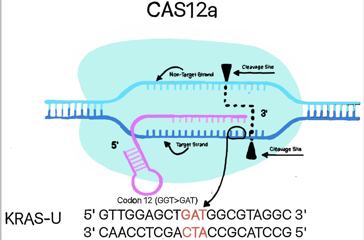 |
|---|
To amplify the target DNA region, RPA primers would enable isothermal amplification at moderate temperatures, offering a faster and simpler alternative to conventional PCR. Primer sequences would be carefully selected to maximize specificity for the KRAS G12D locus and to avoid amplification of non-target sequences, thereby reducing background noise in complex biological samples. And, to maintain high specificity, we propose using pre-existing primer sequences optimized for the KRAS G12D locus (He et al., 2025).
Together, the RPA amplification and CRISPR/Cas12a detection modules would form a cohesive system that processes the liquid biopsy sample through targeted amplification, mutation-specific recognition, and fluorescence-based signal generation. This integrated approach allows for sensitive, rapid, and minimally invasive detection of oncogenic mutations within a complex biological matrix, facilitating early cancer diagnosis.
Device level
The detection system consists of key functional components designed to enable rapid, sensitive identification of the KRAS G12D mutation.
Detection Experiment Setup: Four sample tubes would be prepared, each containing one of the following: mutant KRAS G12D DNA associated with pancreatic cancer, wild-type KRAS DNA, a no-template control (no DNA), and a non-target DNA control for cross-reactivity confirmation. These samples would be prepared in a laboratory setting. The Recombinase Polymerase Amplification (RPA) reagents would then be added to each tube, followed by incubation at 40°C for 40 minutes to facilitate DNA amplification. After incubation, the CRISPR complex, comprising Cas12a protein, mutation-specific crRNA, and a fluorescent quenched (FQ) reporter, would be introduced. The Cas12a protein and crRNA would be designed in silico, assembled, and supplied commercially. The FQ reporter would be labeled with a fluorophore such as FAM (6-carboxyfluorescein) for fluorescence detection. We predict that only the tube with mutant DNA would demonstrate fluorescence. The tube with the wild-type DNA would not fluoresce, demonstrating that the CRISPR complex would detect only the mutated KRAS G12D DNA. The blank tube would also not fluoresce, showing that cleavage would not occur without interaction between Cas12a, crRNA, and the DNA. The non-target tube would also not fluoresce, showing that the CRISPR complex would detect only KRAS G12D (Figure 2).
| Figure 2. | 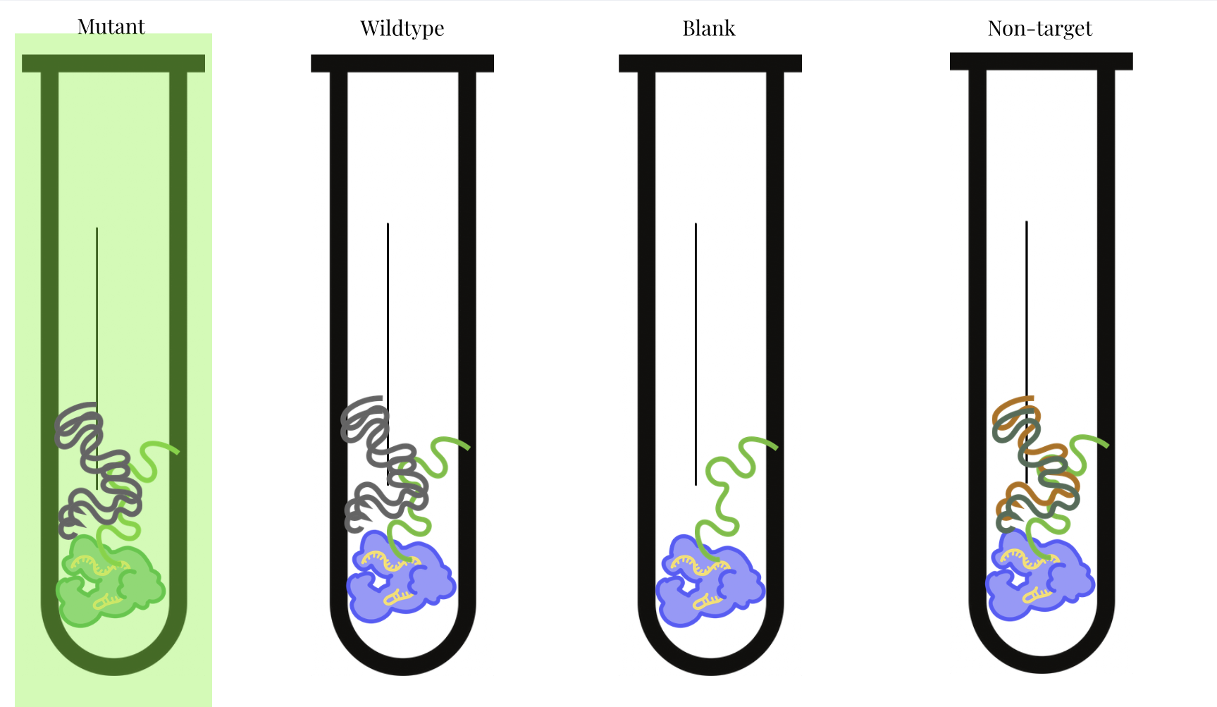 |
|---|
Blood/Plasma Sample Processing: Before amplification, ctDNA would need to be extracted and purified from the blood/plasma sample to isolate the target DNA and remove potential inhibitors (Ballehaninna & Chamberlain, 2011). This would ensure the sample is compatible with the RPA and CRISPR reactions.
Device Operation in Practice: The proposed device would collect a 20 mL liquid biopsy sample via a sterile syringe interface connected to the device. This volume would be chosen to ensure sufficient sample for ctDNA extraction and downstream processing, balancing sensitivity and practicality. The sample would be transferred into a reaction chamber where the RPA reagents would be automatically dispensed. Electric induction heating panels would maintain the reaction chamber at 40°C to ensure optimal amplification conditions. (Figure 3) After 40 minutes, the CRISPR complex would be automatically introduced into the reaction chamber. In the presence of the KRAS G12D mutation, activated Cas12a would cleave the FQ reporter, producing fluorescence. This fluorescence would be detected by an integrated optical sensor and displayed on a monitor as a clear visual signal, confirming the presence of the mutation (Zhao et al., 2022).
| Figure 3. | 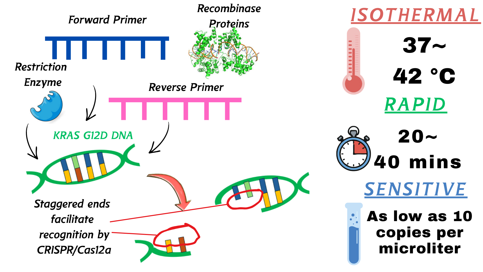 |
|---|
This proposed integrated system would combine automated sample processing, precise temperature control, mutation-specific CRISPR detection, and real-time fluorescence readout, enabling rapid, minimally invasive diagnostics for pancreatic cancer-associated KRAS mutations.
Parts level
The proposed assay design would incorporate a series of well-defined genetic components to enable sensitive and specific detection of the KRAS G12D mutation (Felsenstein et al., 2018). The core elements would include:
Synthetic KRAS DNA templates: Mutant (G12D) and wild-type sequences serve as controls and targets for amplification and detection.
REA-RPA primers: Primers are engineered to include NlaIII restriction sites, facilitating the generation of sticky ends post-amplification. This modification removes the protospacer adjacent motif (PAM) constraints typically required for Cas12a activity (He et al., 2025), thereby increasing convenience and versatility. Without this step, Cas12a would be unable to recognize and cleave the KRAS G12D target due to the lack of a suitable PAM sequence in the mutation site. The recombinase enzyme wraps around the primer, and causes the target DNA to unwind when it finds that its sequence matches the primer. The restriction endonuclease makes staggered cuts in the amplification products, resulting in sticky ends that the CRISPR can recognize.
CRISPR-Cas12a enzyme: Would function as the sequence-specific nuclease activated upon recognition of the mutation site by its guide RNA (He et al., 2023).
Mutation-specific CRISPR RNA (crRNA): Would be designed to discriminate the single nucleotide polymorphism characteristic of the G12D mutation with high specificity, guiding Cas12a to the mutant DNA target (Amintas et al., 2024).
Fluorescent single-stranded DNA (ssDNA) reporter: Would be labeled with a fluorophore and quencher, this reporter is cleaved nonspecifically by Cas12a upon activation, resulting in fluorescence emission that serves as a real-time readout (Zhao et al., 2022).
Circulating tumor DNA (ctDNA) from plasma samples: Utilized as a minimally invasive clinical sample source for assay validation. ctDNA can be present at very low abundance, especially in early-stage cancer, which is why assay sensitivity and amplification steps like REA-RPA would be critical for reliable detection (Jaber, 2024).
The proposed workflow involves initial amplification of target DNA via REA-RPA using the engineered primers to produce Cas12a-compatible substrates. Subsequently, the Cas12a enzyme and mutation-specific crRNA would be introduced along with the fluorescent reporter. In the presence of mutant DNA, Cas12a would be activated, cleaving the reporter and generating a measurable fluorescence signal indicative of mutation presence (Figure 4). This assembly of genetic parts would enable rapid, precise detection of KRAS mutations in a streamlined diagnostic assay.
| Figure 4. | 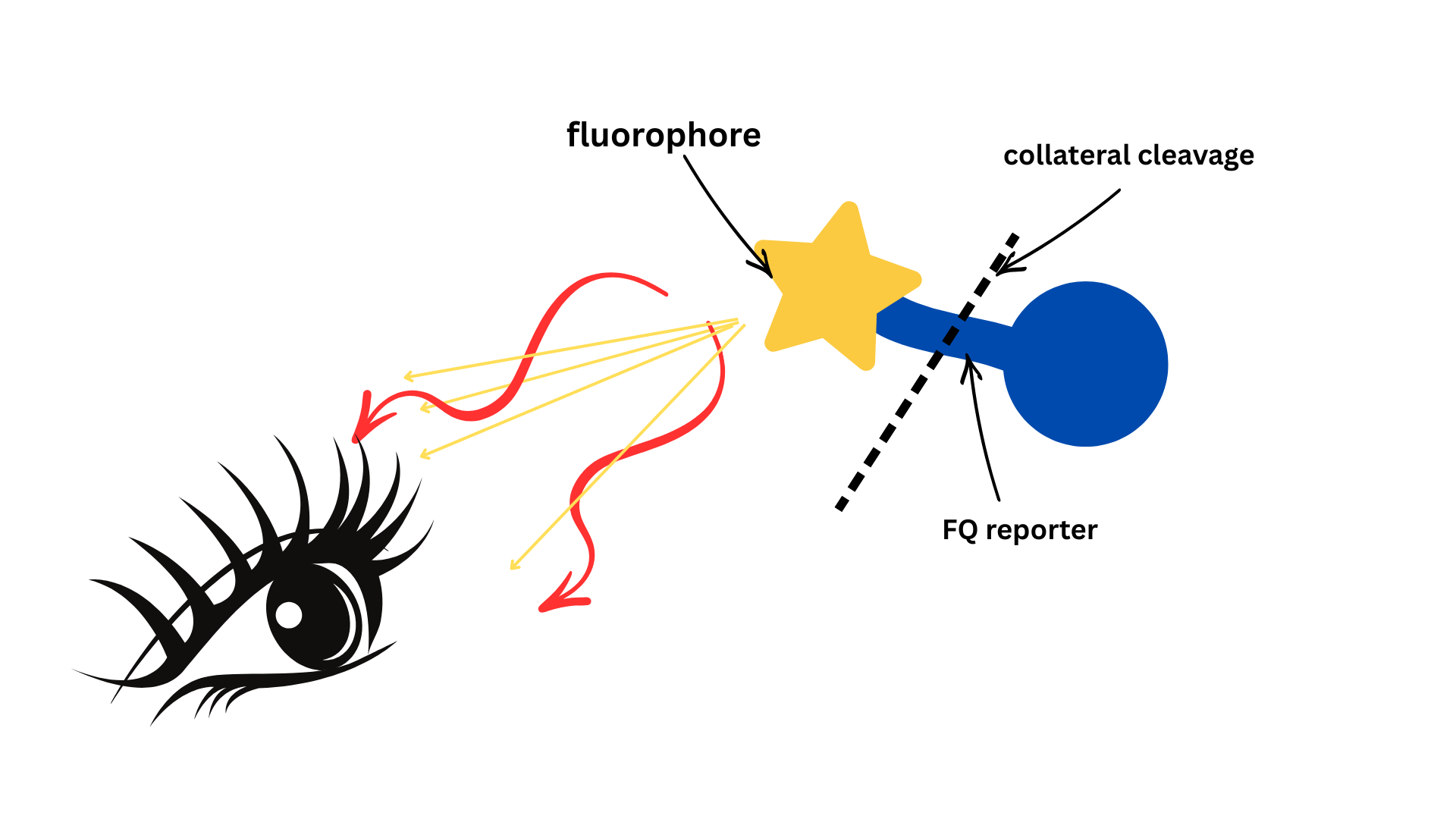 |
|---|
Safety
The proposed experiment would test for synthetic mutated KRAS DNA in blood samples under in vitro conditions. The system would use synthetic DNA and RNA components, which cannot replicate or integrate into the human genome, eliminating any risk of tumor formation or carcinogenic effects. After Cas12a would be added to the mutated DNA-containing blood samples, the test tubes would be sealed to prevent contamination. The tubes would be constructed from durable materials to minimize the risk of breakage and potential injury. Each blood sample would be handled according to biosafety level 1 (BSL-1) standards, as synthetic nucleic acids pose no known biological hazards. Researchers would wear standard personal protective equipment (PPE), including gloves and lab coats, to ensure safe handling. All biological samples and materials would be disposed of in accordance with biohazard waste protocols. This proposed experiment would have minimal risk due to the use of synthetic components and controlled laboratory conditions.
Risk Mitigation for Diagnostic Performance: As a cancer diagnostic tool, the proposed system would need to account for potential risks associated with false positives and false negatives. A false negative could delay medical intervention, while a false positive might lead to unnecessary stress or additional testing. To minimize these risks, the assay would incorporate: High-specificity crRNA design to reduce off-target interactions. Multiple controls (mutant, wild-type, no-template, and non-target) to validate results. Automated and standardized workflows to ensure consistency and accuracy.
Discussions
The fluorescence predicted in the mutant KRAS sample suggests that the CRISPR-Cas12a and REA-RPA system would be able to detect the KRAS G12D mutation. The predicted lack of fluorescence in the wild type and blank samples suggests that the system would be specific, but any signal in the non-target sample may indicate off-target activity. To distinguish true KRAS G12D detection from off-target effects or contamination, repeated trials, proper negative controls, and a defined fluorescence threshold would be used to confirm true positive signals. One challenge is that circulating tumor DNA is present in low amounts in real blood samples, which could make detection harder even with amplification (Jaber, 2024). There is also a chance that plasma related factors such as nucleases, reaction inhibitors, or challenges in sample preparation could affect how well the reaction works. Compared to previously published CRISPR-based KRAS detection methods, such as Cas13a based systems and Cas12a based diagnostic approaches that often require more complex amplification steps (Amintas et al., 2024; Zhou et al., 2021), our proposed REA-RPA and Cas12a based design aims to improve simplicity and compatibility while maintaining mutation specificity. In the future, we would look to improve the crRNA and primer design to increase accuracy and test the system with real patient samples. We would also look to make the test more portable so it can be used outside of a lab for early testing.
Next steps
In order to advance our project, we identified a need for refining the specificity of CRISPR-Cas12a recognition to reduce any off-target effects and to validate the system with clinical samples.
Performance Metrics for Success: We would define success using the following performance metrics: Limit of detection (LOD): The lowest concentration of KRAS G12D detectable in blood samples. Sensitivity: The ability to correctly identify samples with the KRAS G12D mutation. Specificity: The ability to correctly identify samples without the mutation. False-positive rate: The proportion of wild-type samples incorrectly identified as positive. Comparison with wild-type KRAS controls to ensure accurate discrimination.
First, we would focus on testing the amplification and detection system directly on patient-derived samples to assess real-world performance. Because our project emphasizes early detection of pancreatic cancer, we would aim to optimize the system to detect the KRAS G12D mutation at the lowest possible concentrations. Achieving this would enhance the clinical utility of the device by enabling earlier alerts for patients with pancreatic cancer, even when KRAS G12D concentrations are low in their blood. This step would evaluate how the assay performs under biological conditions, ensuring that the detection mechanism remains accurate, reliable, and clinically meaningful when exposed to human-derived material.
Sample Collection Considerations: Additionally, we would explore ways to collect samples from sites that may yield higher concentrations of pancreatic ductal adenocarcinoma (PDAC) biosignatures, as this could reduce detection challenges and the likelihood of false positives. While the pancreas is located deep within the abdominal cavity, making direct sampling impractical for routine screening, we would investigate whether alternative minimally invasive sample types, such as portal vein blood or lymph node aspirates, which could provide higher biomarker concentrations (Gurreri et al., 2023). These sample types would still align with our goal of a minimally invasive, point-of-care diagnostic while potentially improving detection sensitivity. Ultimately, the continued refinement of this CRISPR-based detection platform holds significant promise for transforming early pancreatic cancer screening into a rapid, accessible, and widely deployable diagnostic tool.
Author contributions
SA conducted research on reporter systems, specifically fluorescent reporters and contributed to the fluorometric detection mechanism. SB focused on CRISPR-Cas12a mechanisms and contributed to the understanding of fluorescence capabilities and system viability. SJ investigated UV usage in detection results, the function of CRISPR methods in the context of our device, and the limitations to our project to further improve the design. HK fluorescent proteins and also researched the idea to use amplification strategies including REA – RPA along with specific primers to make the detection quick and simple, not requiring a PAM sequence. BL contributed to the device design functions in point of care context, readout systems and limitations in current detection methods. AR conducted research on detection readout options including gold nanoparticles and fluorescence options and how each would accompany our device in detection. ATR researched the different mechanisms available and which one would be best suited for point of care detection, as well as researching which mutation is most prevalent and determining the overall viability of the assay. NS contributed towards the clinical implications as well as the background of pancreatic cancer and difficulties that must be overcome by the device. RW analyzed Cas12a and the design of the system as a whole as well as researching specifically on the specificity of the device.
Acknowledgements
The Alpharetta High School Biotechnology Club would like to acknowledge the support of Katherine Gell from Vanderbilt University and Eric Lu from Massachusetts Institute of Technology as well as our advisor, Letitiah Etheridge.
References
Amintas, S., Cullot, G., Boubaddi, M., Rébillard, J., Karembe, L., Turcq, B., Prouzet-Mauléon, V., Bedel, A., Moreau-Gaudry, F., Cappellen, D., & Dabernat, S. (2024). Integrating allele-specific PCR with CRISPR-Cas13a for sensitive KRAS mutation detection in pancreatic cancer. Journal of Biological Engineering, 18(1), Article 53. https://doi.org/10.1186/s13036-024-00450-3
Ardalan, B., Ciner, A., Baca, Y., Hinton, A., Darabi, S., Kasi, A., Lou, E., Azqueta, J. I., Xiu, J., Datta, J., Shields, A. F., Aguirre, A., Singh, H., Shroff, R. T., Pishvaian, M. J., & Goel, S. (2025). Distinct molecular and clinical features of specific variants of KRAS codon 12 in pancreatic adenocarcinoma. Clinical Cancer Research, 31(6), 1082–1090. https://doi.org/10.1158/1078-0432.ccr-24-3149
Ballehaninna, U. K., & Chamberlain, R. S. (2011). Serum CA 19-9 as a biomarker for pancreatic cancer—A comprehensive review. Indian Journal of Surgical Oncology, 2(2), 88–100. https://doi.org/10.1007/s13193-011-0042-1
Felsenstein, M., Hruban, R. H., & Wood, L. D. (2018). New developments in the molecular mechanisms of pancreatic tumorigenesis. Advances in Anatomic Pathology, 25(2), 131–142. https://doi.org/10.1097/PAP.0000000000000172
Gurreri, E., Genovese, G., Perelli, L., Agostini, A., Piro, G., Carbone, C., & Tortora, G. (2023). KRAS-dependency in pancreatic ductal adenocarcinoma: Mechanisms of escaping in resistance to KRAS inhibitors and perspectives of therapy. International Journal of Molecular Sciences, 24(11), 9313. https://doi.org/10.3390/ijms24119313
He, X., Deng, L., Zhou, S., Gu, T., Li, X., Zhu, S., Luo, X., Huo, D., & Hou, C. (2025). Breaking the PAM restriction: A universal double stranded DNA detection method based on the sticky end-mediated CRISPR/Cas12a coupled RPA and its application to KRAS G12C single base mutations. Analytical Chemistry, 97(48), 26886–26896. https://doi.org/10.1021/acs.analchem.5c05908
He, Y., Yan, W., Long, L., Dong, L., Ma, Y., Li, C., Xie, Y., Liu, N., Xing, Z., Xia, W., & Li, F. (2023). The CRISPR/Cas system: A customizable toolbox for molecular detection. Genes, 14(4), 850. https://doi.org/10.3390/genes14040850
Jaber, N. (2024, May 10). MicroRNA-based liquid biopsy detects early pancreatic cancer. National Cancer Institute. https://www.cancer.gov/news-events/cancer-currents-blog/2024/liquid-biopsy-detects-pancreatic-cancer
Kanda, M., Matthaei, H., Wu, J., Hong, S., Yu, J., Borges, M., Hruban, R. H., Maitra, A., Kinzler, K., Vogelstein, B., & Goggins, M. (2012). Presence of somatic mutations in most early-stage pancreatic intraepithelial neoplasia. Gastroenterology, 142(4), 730–733.e9. https://doi.org/10.1053/j.gastro.2011.12.042
Luo, J. (2021). KRAS mutation in pancreatic cancer. Seminars in Oncology, 48(1), 10–18. https://doi.org/10.1053/j.seminoncol.2021.02.003
Zhao, L., Qiu, M., Li, X., Yang, J., & Li, J. (2022). CRISPR-Cas13a system: A novel tool for molecular diagnostics. Frontiers in Microbiology, 13. https://doi.org/10.3389/fmicb.2022.1060947
Zhou, H., Tsou, J.-H., Leng, Q., & Jiang, F. (2021). Sensitive detection of KRAS mutations by clustered regularly interspaced short palindromic repeats. Diagnostics, 11(1), 125. https://doi.org/10.3390/diagnostics11010125