
Anish Anumakonda, Chethas Chittipiralla, Andrew Jiang, Sanel Lathiya, Sarah Manoj, Nila Prabhu, Smyan Reddy, Rochan Saireddy, and Raghavan Swaminathan, Lambert High School, Suwanee, Georgia, United States of America
Reviewed on 9 May 2026; Accepted on 22 June 2026; Published on 26 October 2026
With help from the 2026 BioTreks Production Team.
Mosquito-borne diseases result in 390 million infections annually, disproportionately affecting underprivileged regions. Existing treatments are often expensive and inaccessible in low and middle-income regions, leaving large populations without reliable protection. Wolbachia pipientis is a maternally inherited endosymbiont that reduces mosquito-vector competence by blocking virus replication and inducing cytoplasmic incompatibility. We engineer a Wolbachia-compatible expression plasmid to produce RNA targeting NS5 (Non-Structural 5) polymerase mRNA to form a dsRNA complex. This triggers Dicer-2-mediated cleavage of viral transcripts across three flaviviruses, with Saccharomyces cerevisiae serving as the proof-of-concept host for validating expression and binding of probe and target strands. First, we aligned NS5 polymerase coding sequences of Zika, Dengue, and Yellow Fever viruses to identify conserved regions across all three species, with candidate regions being evaluated using ViennaRNA’s Web Services and Python API to minimize secondary structure formation and maximize binding probability. Our selected NS5 target sequence is used to produce a probe mRNA transcript in S. cerevisiae (strain INVSc1). Upon annealing to the viral mRNA target, the resulting dsRNA duplex serves as a Dicer-2 substrate, initiating RISC-mediated degradation of viral transcripts in a separate host organism complete with RNA interference (RNAi) pathways after validation of gene expression is finished in S. cerevisiae, as S. cerevisiae lacks RNAi machinery. We have engineered a superplasmid containing all three NS5 sequences and our probe strand that will be ordered from VectorBuilder. In the following months, lab work will include gene expression and validation of binding, which will involve RT-qPCR and northern blotting, to assess viral mRNA degradation in S. cerevisiae. After successful expression validation, transfer to a suitable RNAi host (W. pipientis) can potentially be performed and protein functionality can be assessed with western blotting and mass spectrometry. The integration of Wolbachia-based biocontrol with siRNA-mediated viral suppression offers a sustainable strategy for flavivirus suppression in the face of rising insecticide resistance and climate change.
Keywords: NS5 Degradation, mosquito-borne diseases, RNA interference, probe strand, RNAi
Authors are listed in alphabetical order. Alyssa Morrow mentored the group. Please direct all correspondence to mailto:f19723@forsythk12.org.


Background
In 2025 alone, the World Mosquito Program reported 3.6 million Dengue cases and over 1,900 deaths attributable to mosquito-borne diseases across dozens of countries (Jackson, 2025). Mosquito-borne diseases disproportionately affect poorer countries in tropical areas, such as countries in sub-Saharan Africa and South America (World Health Organization, 2024). Figure 1 shows the Center for Disease Control’s estimate of the Dengue threat levels across the world in 2026, with a majority of the regions mentioned demonstrating low levels of development (CDC, 2026). This image does not imply endorsement by CDC, ATSDR, HHS or the United States Government and is available on the CDC’s website free of charge.
| Figure 1: Risk of contracting Dengue around the world. This image shows the risk of contracting Dengue at a national scale of analysis. Countries where risk is higher are shaded darker. These regions are typically in less-developed regions such as East Africa, South America, and the Indian subcontinent. Source: U.S. Centers for Disease Prevention and Control (2026) | 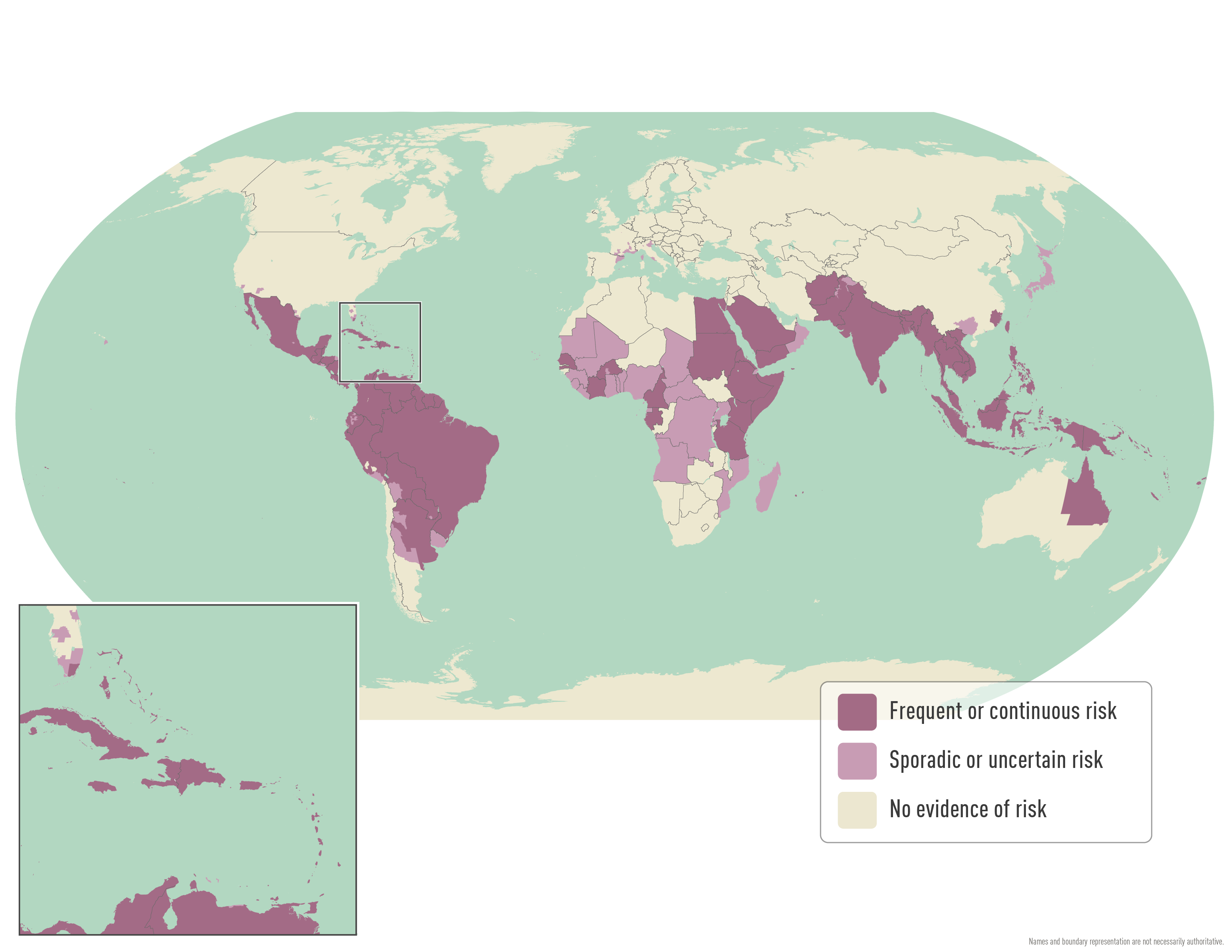 |
|---|
No approved antiviral treatment exists for Dengue; current clinical management is limited to supportive care and analgesics, leaving severe cases with no targeted therapeutic option (CDC, 2024). Especially in severe cases, the lack of proper treatment presents itself as a significant issue.
Much research has been ongoing about the use of W. pipientis to target mosquito-borne diseases. Currently, the World Mosquito Program engineers mosquitos with Wolbachia and attempts to release these mosquitos into the wild, as Wolbachia has been known to suppress mosquito-borne diseases such as Dengue and Zika when present in mosquitos (World Mosquito Program, 2011). However, surface-level vector control and symptom management fail to address viral replication directly. Hence, this design targets NS5 (Non-Structural 5) polymerase, the driver of flavivirus genome replication, across Dengue (DENV2), Zika (ZIKV), and Yellow Fever viruses. We set target sequences to require close identity across NS5 coding sequences, which we validated using Clustal Omega and Vienna RNA’s Folding Kinetics Web Services. Further binding metrics such as binding energy were evaluated using ViennaRNA’s Python API, the specifics of which will be addressed in the following sections. Our design targets the mRNA coding for viral NS5 Polymerase, which is highly conserved among the aforementioned flaviruses, is responsible for the replication of viral strands, and shares no significant homology with the human or S. cerevisiae transcriptome, minimizing the risk of off-target silencing (El Sahili et al., 2017). This is a critical safety consideration given that NS5 homologs are absent from vertebrate genomes. Using an episomal plasmid that is inserted into S. cerevisiae (proof-of-concept for W. pipientis, an endosymbiotic bacterium in mosquitoes), a probe strand that binds to viral NS5 mRNA will be expressed. This probe-target strand annealing produces a dsRNA complex recognized and cleaved by Dicer-2, initiating RISC assembly for targeted degradation of NS5 mRNA.
We predict that siRNA generated from this system will silence NS5 polymerase expression, directly halting genome replication of Dengue, Zika, and Yellow Fever viruses within the mosquito host. This system is designed to be hosted and expressed by W. pipientis, which is already known to suppress some viruses to a certain extent and provides its hosts–mosquitoes–with a comparative advantage against organisms without the endosymbiont. However, proof-of-concept validation is performed in S. cerevisiae, being an ideal model organism which supports the first step of RNA interference (RNAi): modeling expression and binding of target and probe genes as well as episomal plasmid maintenance, with Escherichia coli serving as the intermediate propagation host. Of course, yeast lacks insect-specific Dicer-2, R2D2, and AGO2-RISC, which require validation in host organisms complete with RNAi pathways. In this design, yeast is primarily used to validate expression of all genes prior to transformation into a potential host that comes with RNAi-specific proteins to implement in mosquitoes. Once lab validation of gene expression and binding has concluded, we plan on transforming our system into W. pipientis, thereby eliminating the limitation of yeast not having an inherent RNAi pathway.
Systems level
| Figure 2: Comprehensive diagram of our Wolbachia system. This diagram shows our expression and binding proof-of-concept that occurs in yeast (Steps 1, 2, and 3). Step 4 will be performed in Wolbachia following validation of previous steps. | 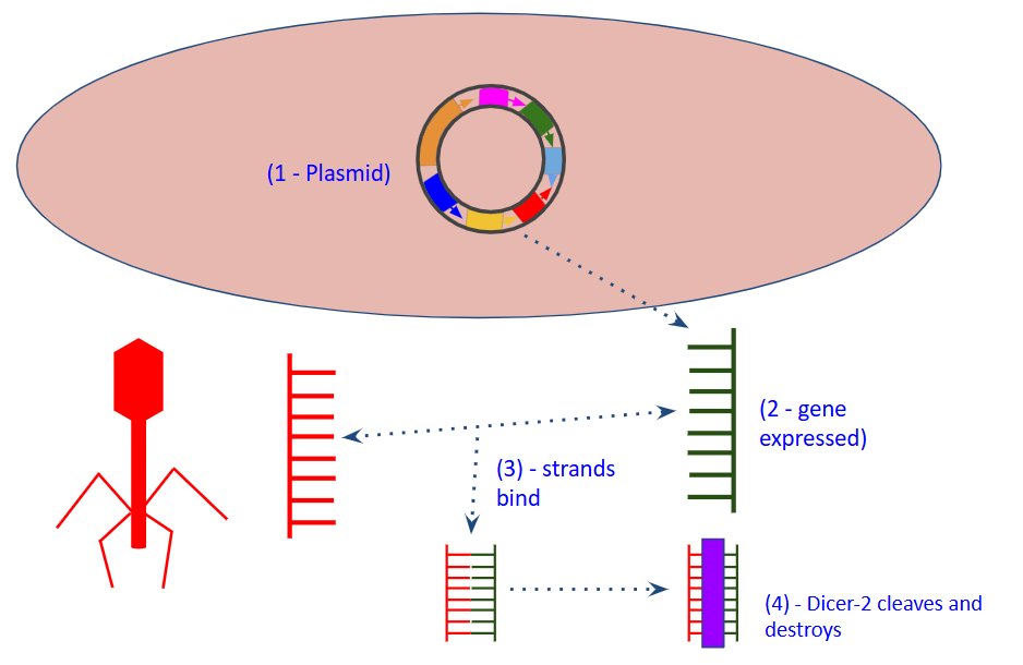 |
|---|
The system pictorially shown in Figure 2 is built around a yeast episomal shuttle plasmid (VectorBuilder pSC_Ex backbone), selected for its dual-origin replication, CUP1-inducible promoter, and URA3 selection marker compatible with S. cerevisiae. The plasmid encodes the antisense strand complementary to the conserved NS5 target sequence (5' → 3'), driven by the CUP1 Cu²⁺ – inducible promoter, and is propagated in E. coli via ampicillin selection before yeast transformation. Using RNA polymerase, the antisense of our double-stranded DNA (3’ to 5’) will be transcribed to create an mRNA strand (5’ → 3’) identical to the sense of our double-stranded DNA, thus being highly complementary to the viral NS5 mRNA when aligned antiparallel (3’ → 5’). This will be followed by the binding of the two highly complementary RNA strands, resulting in cleavage of the combined dsRNA strand by anticipating Dicer-2 and AGO2 proteins when extrapolated to an organism with these RNAi-specific proteins, as one limitation of S. cerevisiae is its lack of RNAi-specific proteins such as Dicer-2 and AGO2 (Duman-Scheel, 2019).
Device level
Plasmid assembly and propagation
Using VectorBuilder, we expanded upon a yeast episomal plasmid that contains a yeast inducible promoter that allows control over the expression of our probe gene. We plan on ordering this plasmid transformed into E. coli to propagate it using heat-shocking at 42°C and incubation at 37°C before transfer to S. cerevisiae. Direct transformation into yeast is feasible if there are stable plasmid yield and purity conditions. Once inside and propagated in yeast, we can perform selection to isolate the transformed bacteria.
RNA expression and protein function
Once our plasmid has been assembled and transformed into a yeast cell, using RNA polymerase, we will perform in vivo transcription of our custom gene of interest and model its interaction with viral NS5 mRNA. We hypothesize that—being highly complementary—the two strands will bind to create a double-stranded RNA. Of course, mere complementarity is insufficient to assess binding efficiency. Hence, we used ViennaRNA’s Web Services and Python API to assess secondary structure formation and binding thermodynamics. Following validation of gene expression and binding in the lab, endogenous Dicer-2 recognizes the dsRNA duplex and processively cleaves it into ~21 nt siRNA fragments, which are loaded into RISC to guide sequence-specific degradation of NS5 mRNA, all of which will be attempted in W. pipientis following expression of genes.
Parts level
Sequence analysis/selection
The first stage of the design involves determining the correct portion of the NS5 mRNA sequence to target. Analyzing the NS5 mRNA sequences of Yellow Fever (strain TN-96), Zika (strain MR-766), and Dengue virus Type-2 (strain Pecs/2003) from UniProt and GenBank, we identified four highly conserved regions, shown in Table 1. Their GenBank accession numbers are AF013417 (National Library of Medicine, 1998b), AF013415 (National Library of Medicine, 1998a), and AH013794 (National Library of Medicine, 2004) respectively.
| Conserved GenBank Sequence (cDNA Sense 5’ → 3’) | Conserved GenBank Sequence (cDNA Antisense 3’ → 5’) | Target mRNA Expressed by Virus (RNA 5’ → 3’) | Reversed Probe mRNA Expressed by Plasmid (RNA 3’ → 5’) | Probe mRNA Expressed by Plasmid (RNA 5’ → 3’) | Sequence in Plasmid (cDNA) |
|---|---|---|---|---|---|
| ATGGGAAAGGTTAGGAAAGAC D: 100% – 631-651 Z: 95% – 673-693 Y: 71% – 679-699 Avg: 89% | TACCCTTTCCAATCCTTTCTG | AUGGGAAAGGUUAGGAAAGAC | UACCCUUUCCAAUCCUUUCUG | GUCUUUCCUAACCUUUCCCAU | 5’ – GTCTTTCCTAACCTTTCCCAT – 3’ 3’ – CAGAAAGGATTGGAAAGGGTA – 5’ |
| GTTAGGAAAGACACACAGGAG D: 81% – 640-660 Z: 100% – 684-704 Y: 62% – 690-710 Avg: 81% | CAATCCTTTCTGTGTGTCCTC | GUUAGGAAAGACACACAGGAC | CAAUCCUUUCUGUGUGUCCUC | CUCCUGUGUGUCUUUCCUAAC | 5’ – CTCCTGTGTGTCTTTCCTAAC – 3’ 3’ – GAGGACACACAGAAAGGATTG – 5’ |
| GGGAGTGGACAAGTTGTCACT D: 81% – 377-397 Z: 100% – 421-441 Y: 62% – 427-447 Avg: 81% | CCCTCACCTGTTCAACAGTGA | GGGAGUGGACAAGUUGUCACU | CCCUCACCUGUUCAACAGUGA | AGUGACAACUUGUCCACUCCC | 5’ – AGTGACAACTTGTCCACTCCC – 3’ 3’ – TCACTGTTGAACAGGTGAGGG – 5’ |
| GGATGGAGCAATTGGGAAGAA D: 76% – 674-694 Z: 100% – 718-738 Y: 67% – 724-744 Avg: 81% | CCTACCTCGTTAACCCTTCTT | GGAUGGAGCAAUUGGGAAGAA | CCUACCUCGUUAACCCUUCUU | UUCUUCCCAAUUGCUCCAUCC | 5’ – TTCTTCCCAATTGCTCCATCC – 3’ 3’ – AAGAAGGGTTAACGAGGTAGG – 5’ |
Table 1: Sequence selection using NCBI databases and Clustal Omega. The table shows conserved cDNA sequences from GenBank converted to mRNA target strands, along with projected plasmid-expressed mRNA probe strands. Column 1 provides sequences with complementarity percentages and genomic coordinates (D – Dengue, Z – Zika, Y – Yellow fever, Avg – Average Complementarity). Columns 4 and 5 are mirror images of each other in order to synthesize an mRNA strand that, when aligned antiparallel to viral NS5 mRNA, will be complementary and ready to bind.
With Clustal Omega, we chose four 21 nucleotide regions in which commonalities were at a maximum. Strand 1 had an average complementarity rate of 89% across all three viruses, while Strands 2, 3, and 4 all had an average complementarity rate of 81% across all three viruses (Madeira et al., 2024). Using ViennaRNA’s Folding Kinetics Web Service, we modeled the folding path of each probe mRNA strand once it was transcribed from the corresponding DNA strand (Wuchty et al., 1999).
RNAsubopt analysis (Figure 3) identified Strand 1 as optimal (ViennaRNA, 2026a): it exhibited the fewest predicted secondary structures (n = 6) within an 8 kcal/mol window above the minimum free energy, indicating low conformational variance and high accessibility for mRNA base-pairing (Lorenz et al., 2011). With fewer possible structures, we have a probabilistically favorable system that can be implemented with the least possible variability. For context, Strand 2 exhibited 10 secondary structures within an 8 kcal/mol window (ViennaRNA, 2026b); Strand 3 exhibited 57 secondary structures within a 40 kcal/mol window (ViennaRNA, 2026c); and Strand 4 exhibited 7 secondary structures within a 40 kcal/mol window (ViennaRNA, 2026d).
| Figure 3: Statistics on strand efficiency from ViennaRNA’s Folding Kinetics Web Service. Graph showing predicted number of secondary structures and energy range above minimum free energy for each strand (Hofacker, 1994). | 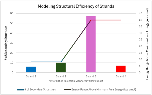 |
|---|
After ensuring proper RNA structure of our selected probe strand, we modeled the interactions between the target NS5 mRNA and our custom probe strand, the central focus of this proof-of-concept, by first analyzing the thermodynamics of the binding. Using ViennaRNA’s Python API Package, translating directly from their C-library documentation, we created a python script to model the binding structure and energy between the probe strand and each NS5 mRNA (Swaminathan et al., 2026), the code of which is shown in Figure 4. Specifically, we used the ViennaRNA python library and the RNA module from the main ViennaRNA Package (Bernhart et al., 2005). Running our code for each of the three interactions separately, we identified binding energies of -324.1 kcal/mol between Zika and the probe, -434.1 kcal/mol between Dengue and the probe, and -361.3 kcal/mol between Yellow Fever and the probe (Swaminathan et al., 2026). All code is stored in a public GitHub repository (see References). The negative energy values with high magnitudes indicate an extremely stable RNA complex that forms spontaneously as well as the change in the energy state of the strands before and after binding. A significant drop in energy is suggestive of a much more stable state than that before binding, increasing the probability of strand binding (Chaurasiya et al., 2010).
| Figure 4: Custom Python script modeling binding energy between two strands using ViennaRNA’s Python package. This code shows the custom Python script created to model binding energy between two strands. This specific input shows the sequence for Zika alongside the probe sequence and is one of the three such inputs modeling interaction. | 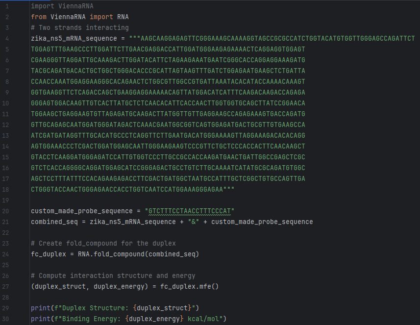 |
|---|
Using this python script, we determined that there exists a thermodynamically stabler state (release of energy) for hydrogen bonds to form. The interaction between the probe and target strand is shown in Figure 5, along with stable hydrogen bonds (Pettersen et al., 2020). Figure 5 was created with UCSF ChimeraX, developed by the Resource for Biocomputing, Visualization, and Informatics at the University of California, San Francisco, with support from NIH R01-GM129325 and the Office of Cyber Infrastructure and Computational Biology, National Institute of Allergy and Infectious Diseases. Both Figure 4 and Figure 5 indicate a stable dsRNA complex that is likely to form.
| Figure 5: ChimeraX model showing interaction between probe and target strands. This ChimeraX model shows the interaction between the probe strand (white) and the target strand (dark blue) to create a dsRNA complex. The specific target shown here is Zika. Hydrogen bonds connecting the two strands are shown as light green dashed lines. | 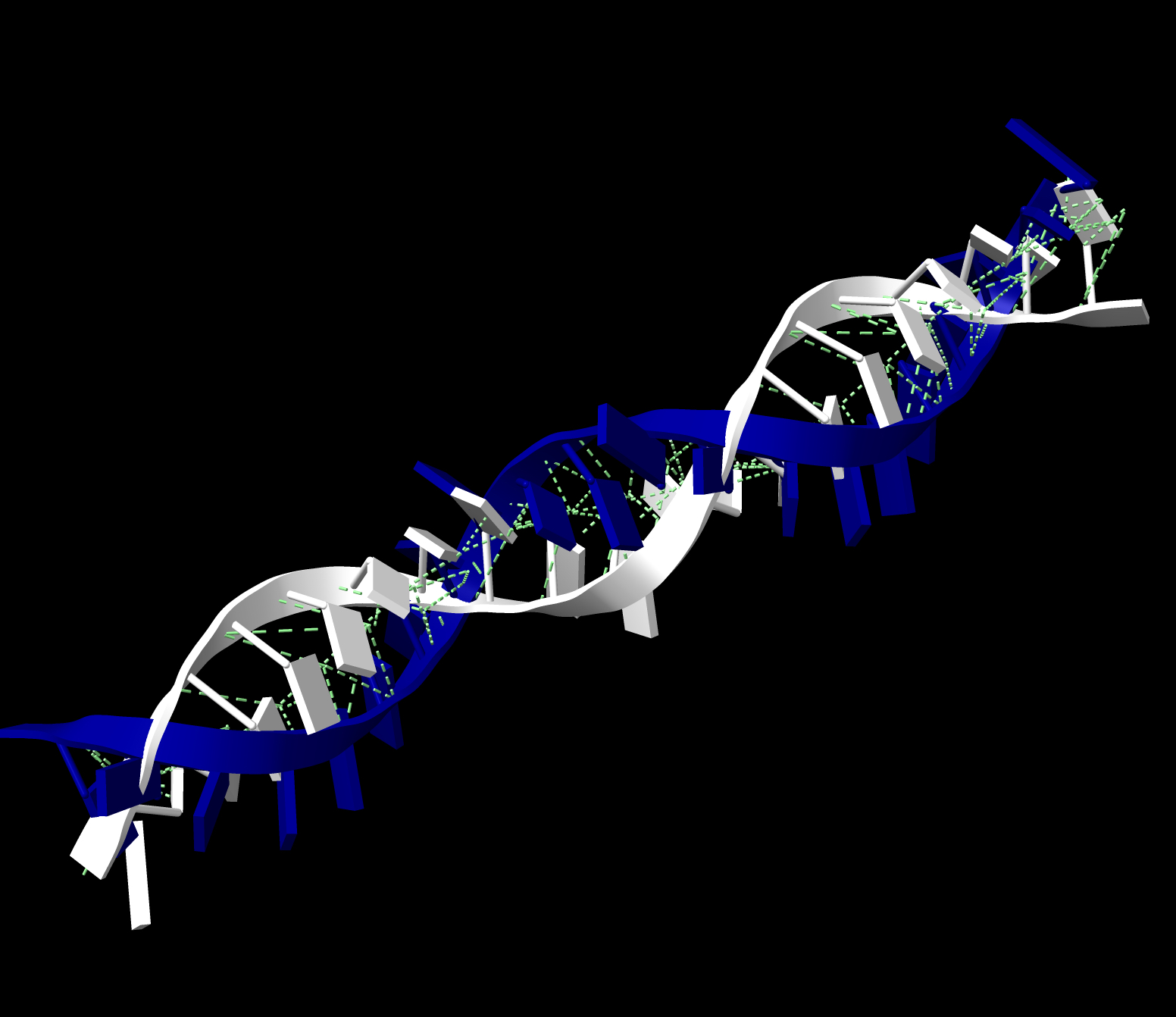 |
|---|
Plasmid design
As shown in the previous section, Strand 1 was identified as optimal across all tests including complementarity, secondary structure formation, energy range above MFE, and binding energy.
The next phase involves the design of our plasmid, which will be inserted into and propagated through E. coli before being transferred to S. cerevisiae. Our plasmid uses a yeast episomal plasmid from VectorBuilder (VectorBuilder, n.d.). The annotated plasmid map is shown in Figure 6.
| Figure 6: Episomal plasmid template from VectorBuilder. Image shows the yeast episomal base plasmid used for basic expression of the probe gene. | 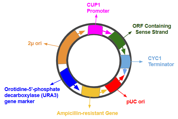 |
|---|
This plasmid contains a CUP1 promoter, which triggers a high-level transcription of our gene of interest when in the presence of highly soluble Cu(II)SO4 (VectorBuilder, n.d.). The ampicillin-resistant gene will ensure that our plasmid will be sustained when purifying our culture in E. coli. Next, the pUC origin of replication facilitates replication in E. coli, and following transfer to S. cerevisiae, the 2µ origin of replication permits maintenance in yeast, the host of our system. Furthermore, to isolate the specific yeast cells that have been transformed to host our plasmid, the orotidine-5'-phosphate decarboxylase (URA3) gene is expressed, which permits yeast transformant selection (VectorBuilder, n.d.). Finally, the CYC1 terminator will be employed to finish transcription and stabilize the expressed mRNA.
Safety
Our proposed system is designed with safety as a primary consideration during construction and testing. All recombinant DNA work will be conducted under BSL-1 conditions using non-pathogenic E. coli (DH5α) and S. cerevisiae strains, in compliance with NIH rDNA guidelines and institutional biosafety protocols. All experiments will be conducted under controlled lab settings to prevent accidents. Field deployment would require regulatory review under EPA FIFRA and WHO vector control guidelines, with controlled environmental release trials to assess ecological impact before any large-scale application.
Using Wolbachia to modify entire mosquito populations definitely has its risks, and careful measures need to be undertaken to limit ecological and environmental impact. For example, mutations in viral genomes and/or the probe strand sequences can cause unintended consequences and may result in viruses developing new forms of resistance (Duffy, 2018). Proper care must be taken when handling viral genomes to ensure that further mutations are not caused while testing that could potentially spread to other mosquitoes. Furthermore, while handling infected mosquitoes outside of BSL-1 conditions, proper care and containment is required to ensure that local epidemics are not unintentionally caused.
Discussions
Our team’s original plan was to create an RNAi system that could be implemented in W. pipientis. Due to its inaccessible nature in our position and the vast scope of our project, we started out by creating a proof-of-concept to first model the expression of our genes as well as the interaction that results between them. Creating a proof-of-concept and outlining our system proved effective, as we were able to perform quality research in identifying potential conserved strands to target and modeling efficiency. In that manner, we achieved our goal of outlining a viable solution to the problem that mosquito-borne diseases pose. We now plan on performing the second stage of this project: validating our proof-of-concept in the lab.
Using S. cerevisiae as a proof-of-concept host certainly has its limitations, the main one being the lack of inherent RNAi-specific proteins. The most common way to surpass this is to transform S. cerevisiae with an episomal plasmid containing the genes to express Dicer-2 and AGO2 (Chen et al., 2020). However, with such a vast scope, the first step was to validate mere expression of target NS5 genes and custom probe genes as well as their binding. This step is extremely crucial and dictates whether an RNAi system can be implemented in any organism to target mosquito-borne diseases. Research has been ongoing on inserting pure Wolbachia into mosquitoes, as their presence naturally defends mosquitoes against viruses like Zika and Dengue (Terradas et al., 2017). However, this project takes Wolbachia’s function a step further, analyzing how this intrinsic quality can be combined with the wonders of RNA interference to make mosquito-borne disease-targeting even more effective. Implementing our design in Wolbachia has the potential to target mosquito-borne diseases at a scale many times larger than what is currently being done.
Next Steps/Ongoing Labwork
Our goal was to use Wolbachia to target mosquito-borne diseases. However, the immediate experimental priority is validating probe strand expression via RT-qPCR and confirming dsRNA duplex formation through northern blotting in S. cerevisiae. Hence, we assembled all genes into one plasmid. To conserve resources, we built on the existing episomal plasmid that contains our custom insert to include the 3 NS5 genes of Yellow Fever, Zika, and Dengue, each with its own expression system. We plan to order this consolidated plasmid for sequential induction experiments, first confirming individual NS5 gene expression under GAL1, MET17, and AOX1, then co-inducing with the CUP1-driven probe strand and assessing binding efficiency by RT-qPCR and northern blotting.
This “superplasmid,” shown in Figure 7, contains all the interacting pieces of DNA that, in the real world, would come from different sources (i.e., the virus, the yeast), but are being consolidated when testing.
| Figure 7: Experimental superplasmid designed for in-lab expression of all genes. This superplasmid is designed using VectorBee software owned by VectorBuilder. We plan on ordering this plasmid from VectorBuilder transformed into E. coli and will express each system separately in the lab. | 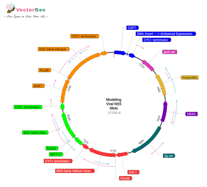 |
|---|
Being a 12,154-nucleotide-long plasmid, our experimental plasmid contains four systems in one (Swaminathan et al., 2026). The first system (blue) consists of our probe gene, accompanied by a CUP1 promoter, which is induced in the presence of copper ions, and a CYC1 terminator. The second system is composed of the NS5 gene for Yellow Fever (strain TN-96 – red), expression of which is initiated by the GAL1 promoter in the presence of galactose; a Kozak sequence for stability in translation; as well as a CYC1 terminator identical to the previous system. The other two systems of Zika (strain MR-766 – green) and Dengue (strain Pecs/2003 – orange) are identical to the Yellow Fever system: they each contain a yeast inducible promoter, a common Kozak sequence, their respective NS5 gene insert, and a CYC1 terminator. However, Zika’s system utilizes the MET17 promoter, induced in the presence of methionine, and Dengue’s uses the AOX1 promoter, induced in the presence of methanol.
In the lab, we plan on expressing these genes both individually and together. Once gene expression has been validated in the lab, we hope to copy our system into W. pipientis, a different host organism that can be engineered to support RNAi pathways while naturally targeting mosquito-borne diseases, which would further the effectiveness of targeting viruses. First, we will install RNAi machinery and cause W. pipientis to express Dicer-2 and AGO2 proteins under constitutive promoters. Following this, we will transform our plasmid containing the probe gene and express it under an inducible promoter. This will be followed by validation of Dicer-2 and AGO2 protein functionality using mass spectrometry and western blotting. Ultimately, we would like to collaborate with other researchers on finding a promising way to implement this RNAi system in W. pipientis, a naturally thriving bacterium in mosquitoes already working against mosquito-borne diseases. Using a self-sustaining bacterium such as Wolbachia reduces the need for multiple expensive clinical trials in the lab if our RNAi system can be implemented in it. However, further research is necessary as Wolbachia is extremely hard to culture outside of host cells and transforming Wolbachia poses a major challenge, but the combination of Wolbachia’s inherent antiviral qualities and a potential RNAi pathway can decrease mosquito-borne diseases by a much more significant amount (Sucupira et al., 2020), and we hope to continue researching on the RNAi capabilities of Wolbachia.
Author contributions
RaS: Ideation, led the manuscript writing, analyzed literature for manuscript, designed the plasmid using VectorBuilder and VectorBee, wrote code for computational analysis, used ViennaRNA for sequence selection, created visual graphs and diagrams to represent software results and plasmid, presented work in video; RoS: Created idea, assisted with sequence aligning using Clustal Omega, assisted with plasmid designing; SR: Contributed to manuscript writing, analyzed literature for manuscript, created visuals using UCSF ChimeraX; SL: Assisted with manuscript writing, editing, and revision, assisted with video creation; NP: Assisted with initial abstract writing and editing; SM: Assisted with ideation, editing, and revision of paper; AA, CC, AJ: Assisted with writing the safety portion of manuscript and editing.
Acknowledgements
We express our gratitude to BioBuilder’s partners, which include but are not limited to Ginkgo Bioworks, Carolina, BriteBound, New England BioLabs Inc., ucb, and the Ragon Institute. We are thankful to Dr. Alyssa Morrow for her expert guidance through the development of this project, Mrs. Catherine Sharer for her funding and continuous support, and the BioBuilder Educational Foundation for giving us the opportunity to present our research. Lastly, we would like to thank VectorBuilder Inc. for their continuous support in designing our plasmid for cloning, specifically Ms. Kathy Huang, Dr. Tautvydas Shuipys, and Dr. Micaela Ziraldo.
References
Bernhart, S. H., Hofacker, I. L., & Stadler, P. F. (2005). Local RNA base pairing probabilities in large sequences. Bioinformatics, 22(5), 614–615. https://doi.org/10.1093/bioinformatics/btk014
CDC. (2026, May 12). Areas with Risk of Dengue. Dengue; CDC. https://www.cdc.gov/dengue/areas-with-risk/index.html
CDC. (2024, May 21). Manage Dengue. Dengue. https://www.cdc.gov/dengue/treatment/index.html
Chaurasiya, K. R., Paramanathan, T., McCauley, M. J., & Williams, M. C. (2010). Biophysical characterization of DNA binding from single molecule force measurements. Physics of Life Reviews, 7(3), 299–341. https://doi.org/10.1016/j.plrev.2010.06.001
Chen, Y., Guo, E., Zhang, J., & Si, T. (2020). Advances in RNAi-assisted strain engineering in Saccharomyces cerevisiae. Frontiers in Bioengineering and Biotechnology, 8, 731. https://doi.org/10.3389/fbioe.2020.00731
Duffy, S. (2018). Why are RNA virus mutation rates so damn high? PLOS Biology, 16(8). https://doi.org/10.1371/journal.pbio.3000003
Duman-Scheel, M. (2019). Saccharomyces cerevisiae (Baker’s Yeast) as an Interfering RNA Expression and Delivery System. Current Drug Targets, 20(9), 942–952. https://doi.org/10.2174/1389450120666181126123538
El Sahili, A., & Lescar, J. (2017). Dengue Virus Non-Structural Protein 5. Viruses, 9(4), 91. https://doi.org/10.3390/v9040091
Hofacker, I. L., Fontana, W., Stadler, P. F., Bonhoeffer, L. S., Tacker, M., & Schuster, P. (1994). Fast folding and comparison of RNA secondary structures. Monatshefte für Chemie/Chemical Monthly, 125(2), 167–188. https://doi.org/10.1007/BF00818163
Jackson, A. (2025, August 11). World Mosquito Day 2025 – A global health crisis. World Mosquito Program. Retrieved April 6, 2026, from https://www.worldmosquitoprogram.org/news-stories/world-mosquito-day-2025-global-health-crisis
Lorenz, R., Bernhart, S. H., Höner zu Siederdissen, C., Tafer, H., Flamm, C., Stadler, P. F., & Hofacker, I. L. (2011). ViennaRNA package 2.0. Algorithms for Molecular Biology, 6(1), 26. https://doi.org/10.1186/1748-7188-6-26
Madeira, F., Madhusoodanan, N., Lee, J., Eusebi, A., Niewielska, A., Tivey, A. R. N., Lopez, R., & Butcher, S. (2024). The EMBL-EBI Job Dispatcher sequence analysis tools framework in 2024. Nucleic Acids Research. https://doi.org/10.1093/nar/gkae241
National Library of Medicine. (1998a). Zika virus strain MR-766 NS5 protein (NS5) gene, partial cds. U.S. Department of Health and Human Services, National Institutes of Health. https://www.ncbi.nlm.nih.gov/nuccore/AF013415
National Library of Medicine. (1998b). Yellow fever virus strain TN-96 NS5 protein (NS5) gene, partial cds. U.S. Department of Health and Human Services, National Institutes of Health. https://www.ncbi.nlm.nih.gov/nuccore/AF013417
National Library of Medicine. (2004). Dengue virus 2 isolate Pecs/2003 RNA-dependent RNA polymerase (NS5) gene, partial cds. U.S. Department of Health and Human Services, National Institutes of Health. https://www.ncbi.nlm.nih.gov/nuccore/AH013794
Pettersen, E. F., Goddard, T. D., Huang, C. C., Meng, E. C., Couch, G. S., Croll, T. I., Morris, J. H., & Ferrin, T. E. (2020). UCSF ChimeraX: Structure visualization for researchers, educators, and developers. Protein Science, 30(1), 70–82. https://doi.org/10.1002/pro.3943
Sucupira, P. H. F., Ferreira, Á. G. A., Leite, T. H. J. F., de Mendonça, S. F., Ferreira, F. V., Rezende, F. O., Marques, J. T., & Moreira, L. A. (2020). The RNAi Pathway Is Important to Control Mayaro Virus Infection in Aedes aegypti but not for Wolbachia-Mediated Protection. Viruses, 12(8), 871. https://doi.org/10.3390/v12080871
Swaminathan, R., Reddy, S. (2026). Degradation_of_viral_NS5_polymerase_from_mosquito-borne_diseases_with_engineered_RNAi [Python Source Code]. GitHub. https://github.com/RaghavanS380/Degradation_of_viral_NS5_polymerase_from_mosquito-borne_diseases_with_engineered_RNAi
Terradas, G., Joubert, D. A., & McGraw, E. A. (2017). The RNAi pathway plays a small part in Wolbachia-mediated blocking of dengue virus in mosquito cells. Scientific Reports, 7(1). https://doi.org/10.1038/srep43847
VectorBuilder. (n.d.). S. cerevisiae yeast gene expression vector. https://en.vectorbuilder.com/resources/vector-system/pSC_Ex.html
ViennaRNA. (2026a). Barriers Web Server. [Web Software Strand 1]. Univie.ac.at. http://rna.tbi.univie.ac.at//cgi-bin/RNAWebSuite/barriers.cgi?PAGE=3&ID=YPksbuJZse
ViennaRNA. (2026b). Barriers Web Server. [Web Software Strand 2]. Univie.ac.at. http://rna.tbi.univie.ac.at//cgi-bin/RNAWebSuite/barriers.cgi?PAGE=3&ID=FBaBi3EpQK
ViennaRNA. (2026c). Barriers web server. [Web Software Strand 3]. Univie.ac.at. http://rna.tbi.univie.ac.at//cgi-bin/RNAWebSuite/barriers.cgi?PAGE=3&ID=J8ucECgUlv
ViennaRNA. (2026d). Barriers web server. [Web Software Strand 4]. Univie.ac.at. http://rna.tbi.univie.ac.at//cgi-bin/RNAWebSuite/barriers.cgi?PAGE=3&ID=xN1OlxNrP0
World Health Organization. (2024, September 26). Vector-borne Diseases. Who.int; World Health Organization: WHO. https://www.who.int/news-room/fact-sheets/detail/vector-borne-diseases
World Mosquito Program. (2011). How it works | World Mosquito Program. Worldmosquitoprogram.org. https://www.worldmosquitoprogram.org/en/work/wolbachia-method/how-it-works
Wuchty, S., Fontana, W., Hofacker, I. L., & Schuster, P. (1999). Complete suboptimal folding of RNA and the stability of secondary structures. Biopolymers, 49(2), 145–165. https://doi.org/10.1002/(SICI)1097-0282(199902)49:2<145::AID-BIP4>3.0.CO;2-G