
Vanishri Bhat, Ahmad Bokhari, Daniel Cao, Sophia Chau, Aarav Doshi, Nicole Li, Angela Park, Sei Park, and Ethan Wang, Troy High BioBuilder, Troy, Michigan, United States of America
Reviewed on 9 May 2026; Accepted on 22 June 2026; Published on 26 October 2026
With help from the 2026 BioTreks Production Team.
Lead contamination in drinking water remains a significant public health concern in the United States, highlighting the need for effective remediation techniques. During the 2014 Flint Water Crisis, toxic lead levels reached over three times the federal hazardous waste threshold, exposing over 100,000 residents to contaminated water and resulting in billions of dollars in subsequent clean-up costs. Beyond Lead, many other heavy metals such as Copper, Chromium, and Mercury contaminate drinking water supplies across numerous communities worldwide. In this study, we designed and attempted to execute an experiment in which Escherichia coli BL21(DE3) would be transformed with pET-28a(+) plasmids encoding two metal-binding proteins: CUP1, a copper-sequestering metallothionein from Saccharomyces cerevisiae, and PbrD, a lead-sequestering protein from the Pbr operon of Cupriavidus metallidurans. We intended to evaluate the engineered strains for their ability to remove lead and copper ions from aqueous solutions, with residual metal concentrations quantified by spectrophotometry. However, transformation was unsuccessful across all experimental groups – likely due to suboptimal temperatures used for plasmid and E. coli growth and storage: no colonies grew on kanamycin-selective plates, and no downstream metal sequestration experiments could be conducted. Rather than treat this as a simple null result, we present it as an educational case study for early-stage researchers working in high school or resource-limited synthetic biology settings. Through retrospective analysis, we identify several procedural deviations from standard transformation protocols that likely compounded to produce the transformation failure. We hope this transparent account of what went wrong, and why, provides practical guidance to help fellow student researchers avoid similar pitfalls and gain a better understanding of the gap that often exists between experimental design and wet-lab execution.
Keywords: Bioremediation, sequestration, wet-lab validation
Authors are listed in alphabetical order. Rebecca Brewer mentored the group. Please direct all correspondence to mailto:rbrewer@troy.k12.mi.us.
Background
Heavy metal contamination of drinking water is a persistent and serious global health challenge. Lead (Pb2+) is toxic even at trace concentrations, causing irreversible neurological damage. To date, no safe exposure threshold has been established (WHO, 2024). The 2014 Flint water crisis in Michigan made this threat come to life: lead leached from corroding municipal pipes into the drinking water supply of over 100,000 residents, with measured lead levels exceeding three times the federal hazardous waste classification threshold. An estimated 140,000 individuals were exposed to lead and other contaminants (Ruckart et al., 2019), and Flint adults overall displayed much higher rates of physical symptoms, including skin rashes,58.1%; hair loss, 45.5%; and nausea, 35.6% (Ezell & Chase, 2021). Beyond Flint, lead and other heavy-metal contamination affect communities worldwide, particularly in lower-income and industrially impacted regions where water treatment infrastructure is limited.
Current approaches for removing heavy metals from water include chemical precipitation, ion exchange, and membrane filtration. While effective, these methods are often expensive, energy-intensive, and difficult to implement at a local scale. Microbial bioremediation, which leverages living organisms like E. coli to remove or sequester environmental contaminants, offers a compelling alternative (Zhu et al., 2020). Microbial bioremediation is particularly attractive because bacteria are self-replicating, relatively inexpensive to culture, and can be manipulated through genetic modification.
E. coli is among the best-characterized organisms in molecular biology and has become a foundational structure in synthetic biology (Elbing & Brent, 2019; Tuttle et al., 2021). Its thoroughly characterized genetics, rapid growth rate, and the availability of robust inducible expression systems make it an ideal starting point for engineering novel capabilities, including targeted metal sequestration. In this study, we selected two metal-binding proteins for expression in E. coli. The first is CUP1, a cysteine-rich metallothionein from baker's yeast (S. cerevisiae) that binds copper ions with high affinity (Butt et al., 1984). The second target, PbrD, is a protein encoded by the Pbr operon of the bacterium C. metallidurans (NCBI, n.d.). C. metallidurans naturally inhabits metal-contaminated environments, and PbrD has been specifically implicated in intracellular lead sequestration (Borremans et al., 2001). Both genes were cloned into the pET-28a(+) expression vector, which drives transgene expression via an IPTG-inducible T7 promoter and confers kanamycin resistance to allow selection of successfully transformed colonies (GenScript, n.d.; SnapGene, n.d.).
Our experimental plan was to transform E. coli BL21(DE3) cells with each construct using chemical transformation (heat shock), confirm uptake via kanamycin selection, induce protein expression with IPTG, and expose the engineered cells to predetermined aqueous solutions of lead(II) nitrate and copper(II) nitrate to evaluate sequestration efficiency of the copper and lead metals relative to wild-type controls. We hypothesized that PbrD-expressing cells would show the greatest reduction in lead ion concentration, and CUP1-expressing cells would outperform controls in copper removal. However, no transformed colonies were recovered from any group, and the metal sequestration study could not proceed.
In the following sections, we pivot from our original hypothesis to provide a systematic "post-mortem" analysis of the experimental process. By documenting our theoretical approach, initial procedure, experimental deviations, and troubleshooting steps, this study serves as a cautionary guide and practical resource on navigating the gap between theoretical design and wet-lab execution.
Materials and methods
| Figure 1: A – Pictorial Representation of our executed workflow. This figure shows the first half of our workflow, which was fully executed; B –This figure shows the second half of our workflow which was thoroughly planned but not executed due to an unsuccessful transformation process. | 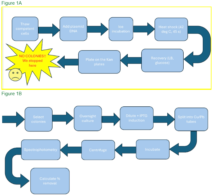 |
|---|
Bacterial strain and plasmids
| Figure 2: “Plasmid Construction Workflow” (Visual representation of plasmid construct and gene insert) | 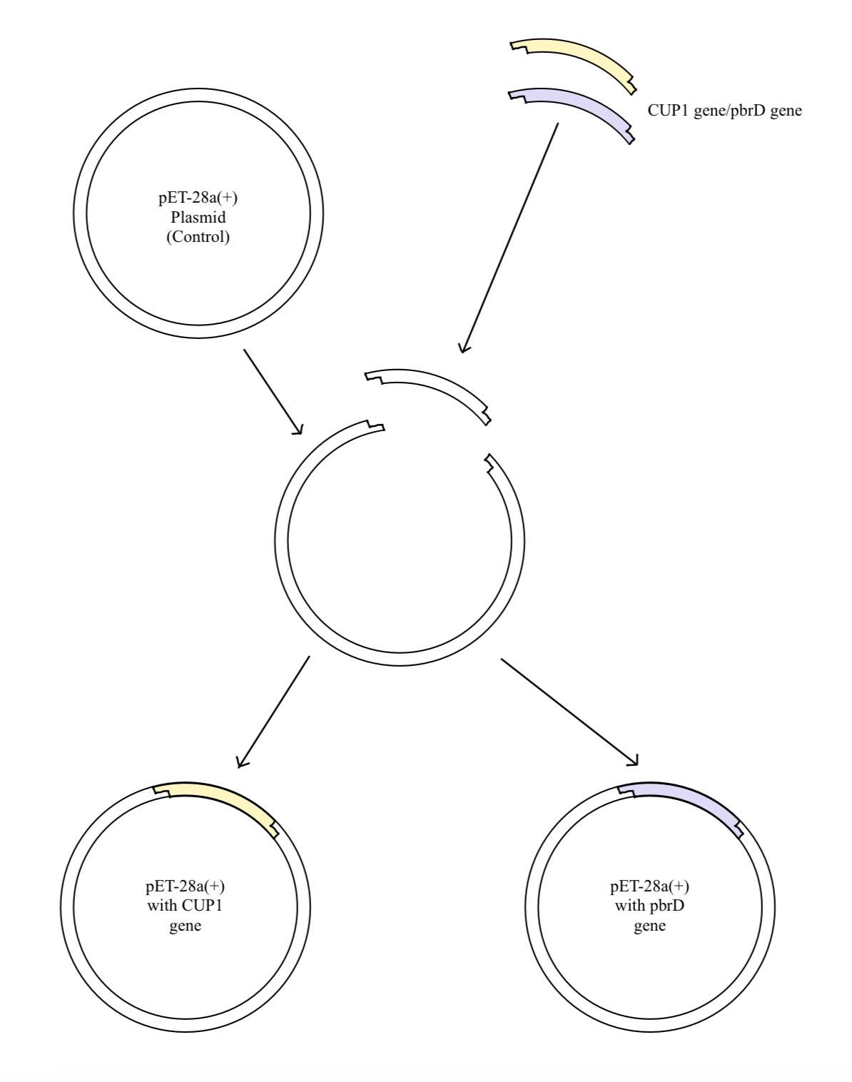 |
|---|
We used E. coli BL21(DE3) as the expression host due to its compatibility with the T7 promoter-reliant expression system relative to other strains (Elbing & Brent, 2019; Tuttle et al., 2021). We used three plasmid constructs based on the pET-28a(+) backbone (GenScript, n.d.; SnapGene, n.d.): (1) an empty vector control lacking an insert (designated X), (2) pET-28a(+) carrying the CUP1 gene (designated C), and (3) pET-28a(+) carrying the PbrD gene (designated P). All constructs contained a kanamycin resistance (KanR) cassette to facilitate the selection of successfully transformed colonies on antibiotic-containing plates. All nucleotide and protein sequences were sourced from GenBank and can be found in Table S1. Constructed plasmid maps can be found in Figure S1 and Figure S2 for Plasmid C and Plasmid P, respectively. The custom plasmid was synthesized and verified by GenScript. Visual mapping and sequence auto-annotation were performed using PlasMapper 3.0 (Wishart et al., 2023).
Table 1: Plasmid constructs used. X, C, and P represent the positive control, CUP1-transformed, and PbrD-transformed plasmids used for experimentation, respectively
| Plasmid | Gene Insert | Purpose | |
|---|---|---|---|
| X | pET-28a(+) | None | Positive control |
| C | pET-28a(+) | CUP1 | Cu2+ sequestration |
| P | pET-28a(+) | PbrD | Pb2+ sequestration |
| Figure 3: pET-28a(+) Plasmid Map Visual mapping and sequence auto-annotation were performed using PlasMapper 3.0 (GenScript n.d., Wishart et al., 2023) | 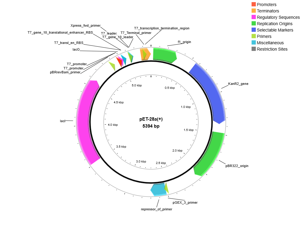 |
|---|
Transformation procedure
Competent E. coli BL21(DE3) cells (200 µL total) were removed from -80°C storage and thawed on ice for 10-15 minutes. The cells were aliquoted into 4 sterile microcentrifuge tubes (50 µL per tube) corresponding to three experimental conditions and one negative control lacking plasmid DNA. 2 µL of the appropriate plasmid DNA (targeting 1 to 10 ng) was added to each experimental tube, and tubes were gently flicked to mix. Mixing through pipetting was avoided to preserve membrane integrity and maximum cell competency (New England Biolabs, n.d.-a; New England Biolabs, n.d.-c)
All tubes were incubated on ice for 30 minutes, then heat-shocked at 42°C for 45 seconds, and immediately returned to ice for 2 to 3 minutes (New England Biolabs, n.d.-a; New England Biolabs, n.d.-c). Recovery was initiated by adding 950 µL of pre-warmed LB broth (supplemented with 50 µL of 20 mM glucose) to each tube. Note that many standard transformation protocols instead recommend SOC medium for post-heat-shock recovery (Invitrogen, n.d.; QIAGEN, n.d.). Cells were then incubated while shaking at room temperature for 120 minutes. It should be noted that standard transformation protocols typically specify incubation at 37°C for 60 minutes in a shaking incubator for E. coli BL21(DE3); Due to equipment limitations, we could only choose one among shaking or 37°C incubation, as our shaking platform was unable to fit within the incubator
As such, the standard 37°C incubation was replaced with incubation at room temperature (approximately 20-22°C), and the time was extended to approximately 120 minutes as a compensatory modification.
Following recovery, we plated cells onto LB agar made with 50 µg/mL kanamycin in two conditions. To start, 50 µL was spread directly onto one plate. Next, the remaining culture volume was subject to centrifugation at 11,000 RPM for 30 seconds, and the supernatant was discarded. The pellet was resuspended in residual volume of LB, and 50 µL was plated in a concentrated area on a second plate. This yielded eight plates in total. All plates were labeled with construct ID (X, C, or P), date, and volume, then incubated inverted at 37°C for 16 h.
Intended metal sequestration protocol (not executed)
The following describes the metal sequestration experiment as designed. It was not carried out due to the transformation failure, and is included here to fully document the intended experimental workflow.
Successfully transformed single colonies from each construct would have been used to inoculate 5 mL of LB + kanamycin (50 µg/mL), with three independent biological replicates per construct—nine cultures total (X1, X2, X3, C1, C2, C3, P1, P2, P3). After overnight growth, each culture would have been diluted in a 1:100 ratio into fresh LB + kanamycin (50 µL overnight culture + 4.95 mL fresh medium), then induced by adding 20 µL of 100 mM IPTG to achieve a 1 mM final concentration (Marbach & Bettenbrock, 2012). Each induced 5 mL culture would then have been split into two 2.5 mL aliquots—one for lead exposure, one for copper exposure—for a total of 18 tubes. Copper concentrations would also need to remain below levels that compromise E. coli culturability during metal-exposure experiments (Grey & Steck, 2001).
Metal ions would have been introduced by adding 25 µL of 1 mM copper nitrate – Cu(NO3)2 -or lead nitrate – Pb(NO3)2 – stock to each respective tube, achieving a nominal final concentration of approximately 10 µM. Cultures would then have been incubated at room temperature with shaking for 12 to 48 h, double the standard 6 to 24 h to compensate for the lower incubation temperature. Sub-lethal lead concentrations (0.01, 0.25, and 0.5 mM) would have been identified through prior toxicity testing with wild-type E. coli BL21(DE3) to prevent cell lysis, which would release sequestered ions back into solution and confound results.
Following incubation, cultures would have been centrifuged at 3,000 to 4,000 x g for 10 minutes to pellet cells, and approximately 2.3 to 2.5 mL of supernatant per tube would have been collected for metal quantification. Residual metal ion concentrations would have been measured using spectrophotometry with a metal-specific colorimetric reagent and fitted to a standard curve (Kharde et al., 2024; Lalmi et al., 2025; Mohammed et al., 2025). Percent metal removed per group would have been calculated relative to the known initial concentration, and statistical analysis would have been performed using a Student’s t-test, with significance set at p < 0.05.
| Figure 4: “E. coli growth on 0.01 mg/mL lead solution, 24 h” (The observed growth of E. coli at a lead concentration of 0.01 mg/mL establishes the baseline minimum inhibitory concentration for the wild-type strain. Successful transformation with the PbrR gene is hypothesized to enhance lead biosorption and intracellular sequestration. Consequently, the recombinant strain is expected to maintain viability while effectively immobilizing lead ions at the specified concentration of 0.01 mg/mL.) | 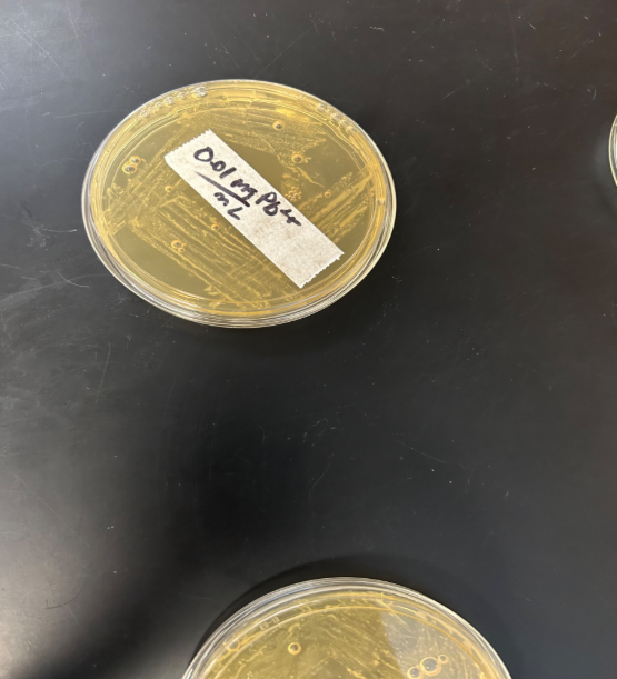 |
|---|
| Figure 5: “E. coli growth on 4 mM copper solution, 24 h” (This image confirms that the E. coli maintains metabolic activity at the experimental concentration. Following the successful integration and expression of the CUP1 gene, it is anticipated that the bacteria will exhibit an enhanced capacity for copper sequestration, providing a biological mechanism for heavy metal accumulation at the 4 mM threshold.) | 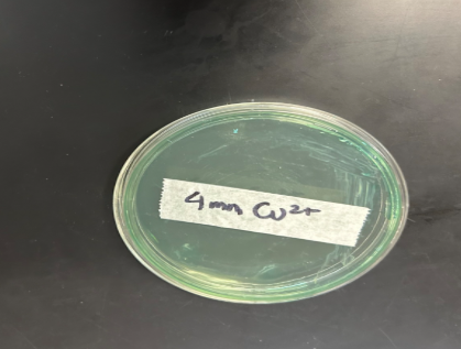 |
|---|
Results
Transformation outcome
After a full night of 37°C incubation, no colonies were observed in any of the eight kanamycin-selective plates. This includes negative control plates (contained cells with no plasmids), both of plating volumes (50 µL direct and concentrated pellet), and all three experimental constructs (X, C, P). With all eight plates missing colonies, the results indicated that transformation was unsuccessful.
Table 2: “Transformation plate summary” (Conditions of plates used for post-transformational growth and observed growth results for each plate. It is clear that an issue occurred throughout the procedure such that the kanamycin resistance was not conferred to the E. coli)
| Plate | Construct | Volume Plated (mL) | Plate Condition | Colonies Grown |
|---|---|---|---|---|
| 1 | X | 50 | Kan 50 μg/mL | 0 |
| 2 | X | 150 | Kan 50 μg/mL | 0 |
| 3 | C | 50 | Kan 50 μg/mL | 0 |
| 4 | C | 150 | Kan 50 μg/mL | 0 |
| 5 | P | 50 | Kan 50 μg/mL | 0 |
| 6 | P | 150 | Kan 50 μg/mL | 0 |
| Figure 6: “Kan Plates with no growth of Post-Transformation E. coli after 72 h” | 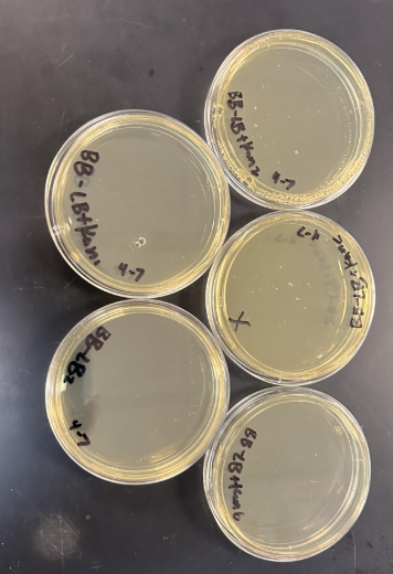 |
|---|
Survival on kanamycin-selective media requires active expression of the KanR resistance gene; the complete absence of growth across all plates therefore indicates that plasmid uptake either did not occur or occurred at frequencies too low to detect under the plating conditions used.
Metal sequestration data
Metal sequestration could not be conducted, and so no related data were collected. All following data and analysis are regarding the unsuccessful process of transformation and practical improvements that can be implemented for future trials.
Discussions
Interpreting the result
The complete absence of colonies on our kanamycin plates is a definite indicator of transformation failure. Although low colony counts or inconsistencies may suggest partial success or variability in techniques, the lack of growth on all eight plates despite variations in plasmid constructs and plating volumes signals a more fundamental issue with the transformation process itself. Below, we analyze the most probable causes of failure within our procedure and what these results suggest about the critical variables in chemical transformation.
The recovery temperature deviation
The most likely source of error was the deviation from standard protocol, specifically the use of room temperature (approximately 20 to 22°C) instead of 37°C during the post-heat-shock recovery incubation. This includes a critical period where transformed cells must repair heat-shock-induced membrane damage, resume normal metabolism, and begin expressing the plasmid-encoded kanamycin resistance gene before encountering the antibiotic on the plates. At 37°C, the optimal growth temperature for E. coli, this process occurs efficiently within 60 minutes (Elbing & Brent, n.d.-b). At room temperature, transcription and translation rates are substantially reduced. This likely resulted in insufficient KanR expression before exposure to kanamycin.
Doubling the incubation time to 120 minutes was an intuitive attempt to compensate, but metabolic rate and time are not interchangeable, especially for a process that crucially depends on reaching a threshold protein concentration before plating.
This is likely the primary reason for our transformation failure, and it is also the most instructive: it illustrates how a single, seemingly inconsequential environmental variable, temperature, can drastically influence the growth and success of bacterial transformation experiments. For any future uses, access to a 37°C shaking incubator for the recovery step should be required to induce appropriate transformation and translation before plating.
Heat shock precision
The heat shock step is among the most technically sensitive in the entire protocol. The 45-second exposure to exactly 42°C momentarily increases membrane permeability and allows plasmid DNA to enter the cell. Even small deviations in temperature or timing can greatly reduce this effect. Low temperatures may fail to adequately increase membrane permeability, while excessive temperature or duration of incubation may damage or lyse cells. In a limited high school laboratory environment lacking a calibrated heat block, achieving a precise, constant 42°C is difficult. Any deviations in the temperature or lag time between the movement of test tubes between the heat source could have further reduced our lab’s transformation efficiency.
Competent cell handling
Chemically competent cells are extremely sensitive to temperature changes before heat shock. Accidentally allowing the cells to warm up even briefly before use may weaken their membrane and reduce overall competency. Additionally, although mixing was performed by flicking rather than pipetting, any unintended mechanical agitation during setup could have further compromised cell integrity. Best practice is to work quickly, keep cells on ice at all times until the moment of heat shock, and pre-chill all tubes and tips.
Plasmid integrity
Another potential contributor to the transformation failure was the concentration and condition of the plasmid DNA. In our experiment, we directly used the plasmid stocks as received and did not independently verify the DNA concentration or integrity prior to use. If the plasmid DNA had degraded prior to use (e.g., during shipping, storage, or freeze-thaw cycling), the number of intact molecules available for uptake would have been reduced. As a result, our transformation efficiency and recovery of cells could have been compromised.
Alternatively, it is possible that our plasmid was faulty from the start, which would make possible that transformation had occurred successfully, but the plasmid did not provide adequate protection from kanamycin. If the plasmid DNA had degraded prior to use, our transformation efficiency and recovery of cells could have been compromised (OriGene Technologies, n.d.). However, given the long-standing reliability of GenScript for usage in biological research, this is an unlikely scenario. Nevertheless, verification of the plasmid DNA via direct sequencing and gel electrophoresis before use is a straightforward step that can catch these issues early and is strongly recommended for future attempts.
The compounding nature of small errors
Perhaps the most important lesson from this experience is that chemical transformation (even under ideal conditions) is an inherently low-efficiency process that depends on multiple technically demanding steps. No single procedural issue can be identified with absolute certainty as the only cause of failure. Instead, the complete lack of colonies most likely resulted from the compounding of small inefficiencies at multiple steps simultaneously. The recovery temperature deviation, potential heat-shock variability, possible cell-handling errors, and unverified plasmid quality each likely reduced our transformation efficiency only partially. Together, however, these reductions appear to have pushed efficiency below the threshold needed to observe any surviving colonies under kanamycin selection.
This compounding dynamic is important for student researchers to understand and consider in their own work. Written or pre-existing protocols make transformation appear straightforward, but in practice, experimental success is highly dependent on the maintenance of specific biological and physical conditions at every step in the process. As such, in a high school laboratory, where equipment access may be limited and procedures may be performed for the first time, small improvisations can have drastic effects. This failure underscores the necessity of specially designing synthetic biology projects around available equipment constraints and adhering strictly to each step of established protocols.
Recommendations for fellow researchers
Based on our experience, we offer the following cautionary recommendations to other student researchers undertaking bacterial transformation for the first time:
Attempt to document every deviation from protocol as it happens. In the event of failure, retrospective troubleshooting becomes far more difficult without a clear record of what was actually done.
The transformation process should be treated as its own critical experiment rather than as a simple preliminary step. For many studies, including ours, failure during the transformation phase renders further experimentation void.
Do not substitute incubation temperature without understanding the biological consequences; if the required equipment is unavailable, obtain it before proceeding as strict adherence to incubation protocols is required for optimal metabolic recovery.
If materials allow, include a positive control transformation using a known-working plasmid and the exact standard protocol. A control group run simultaneously with your experimental conditions can be used to determine whether failure was caused by the plasmid, the cells, or the transformation procedure itself.
Verify competent cell viability using the supplier-provided control plasmid before investing experimental plasmids. Competent cells lose efficiency over time and with improper handling.
Also verify plasmid integrity and concentration using nanodrop and gel electrophoresis tests before transformation. Do not assume that a stock is perfectly intact simply because it was received from a supplier.
The heat shock step should be performed using calibrated equipment (e.g. calibrated heat block) capable of maintaining 42°C accurately for the required time. A water bath, in comparison, is harder to maintain at a precise temperature, especially for brief intervals.
Cells should be kept on ice at all times barring the heat shock step. Given this short window, it is essential that all materials, including pre-chilled tubes and tips, are prepared in advance.
Next steps
The following outlines our team’s specific action plan for repeating the transformation, directly addressing the protocol deviations identified above:
Secure access to a 37°C shaking incubator for the 60-minute recovery incubation, following the standard manufacturer protocol without modification.
Verify plasmid quality and concentration via direct sequencing and gel electrophoresis prior to transformation. In future iterations, culture density should also be normalized by OD600 before induction and metal exposure comparisons (Blue-Ray Biotech, 2024).
Run a positive control transformation with the supplier-provided control plasmid first to confirm that competent cells are viable and the procedure is working before committing experimental constructs.
Use a calibrated heat block for the 42°C heat shock step to ensure precise, consistent temperature delivery across all tubes.
Identify the heat shock temperature that maximizes the growth of the E. coli strain through controlled testing aiming to compare colony growth for different temperatures of heat shock used for transformation.
If transformation is ultimately successful, proceed with the metal sequestration protocol as designed: IPTG induction, exposure to sub-lethal concentrations of lead and copper nitrate, and spectrophotometric or AAS quantification of residual metal ions, with statistical comparison across wild-type, CUP1, and PbrD groups.
For further development of E. coli based metal bioremediation, we will:
Construct a controlled holding chamber for transformed bacteria. Water will be allowed to flow past a filtering chamber, after which it flows back out clean of metal contaminants. This allows bioremediation while minimizing risks of releasing E. coli into the environment, and provides a safe and simple method to remove the system from its environment following usage.
To improve filtration and sequestration ability, sodium alginate gel coats can be formed around the free floating E. coli to compartmentalize them into manageable “packaged” units. The coat not only shields groups of E. coli from harsh environmental conditions, but is also, when properly formed, porous enough to allow metal ions like lead to diffuse into the bead and be sequestered by the contained bacterium. Sodium alginate itself also contains many negatively charged regions due to the presence of carboxylates, potentially strengthening the pull for heavy metal ions and improving sequestration ability (This forms a concentration gradient that in conjunction with the lead sequestering E. coli held within, aids in limiting metal ion release back into water) Furthermore, the bead contributes to a larger volume, which makes filtration more efficient in minimizing released E. coli.
The alginate, which has its own metal sequestration ability, thus demands further experimentation to validate and isolate the individual contribution of both alginate + modified bacterium sided systems. Our current metal sequestration protocol can be adapted to test the following experimental and control groups.
EX1: Alginate beads containing CUP1/PbrD transformed E. coli
EX2: Free CUP1/PbrD transformed E. coli (Comparative experimental group to determine the effect of alginate addition)
C1: Empty alginate beads (Material control for determining the quantity of lead sequestered by the coat alone)
C2: Alginate beads containing unmodified E. coli (Biological control for determining the innate sequestration ability of bacterial biomasses in the absence of specialized genes)
C3: Baseline lead concentration with no treatment (Negative control) Note: Appropriate sodium alginate and Ca2+ concentrations are currently TBD. Effective concentrations of E. coli across groups will be kept constant to accurately measure sequestration/biomass effects.
Author contributions
All authors contributed to performing research and generating research ideas. EW served as project lead, overseeing experimental design, coordination, and directed drafting of the manuscript. EW and SC discussed sponsorships and acquired necessary material donations for the experiment from Dr. Elizabeth Berger at Wayne State University. EW, NL, and DC detailed the specifics of the procedure. All members drafted and proofread the manuscript.
Acknowledgements
We want to express our deep gratitude to Mrs. Rebecca Brewer, AP Biology teacher at Troy High School for giving us this opportunity and allowing us to use her classroom and lab equipment. We are also greatly thankful for our mentor, Mr. Nick Nolan, MIT PhD candidate, for all the guidance and advice along the way. Finally, we would like to give our appreciation to Dr. Elizabeth Berger and the Wayne State University School of Medicine, whose generous donations made our lab vision a reality.
References
Blue-Ray Biotech. (2024, June 1). OD600 measurement: A key technique in the microbiology laboratory. Blue-Ray Biotech. https://www.blue-raybio.com/en/blog/OD600-Measurement-A-Key-Technique-in-the-Microbiology-Laboratory
Borremans, B., Hobman, J. L., Provoost, A., Brown, N. L., & van der Lelie, D. (2001). Cloning and functional analysis of the pbr lead resistance determinant of Ralstonia metallidurans CH34. Journal of Bacteriology, 183(19), 5651-5658. https://doi.org/10.1128/JB.183.19.5651-5658.2001
Butt, T. R., Sternberg, E. J., Gorman, J. A., Clark, P., Hamer, D., Rosenberg, M., & Crooke, S. T. (1984). Copper metallothionein of yeast, structure of the gene, and regulation of expression. Proceedings of the National Academy of Sciences of the United States of America, 81(11), 3332-3336. https://doi.org/10.1073/pnas.81.11.3332
Elbing, K. L., & Brent, R. (2019). Recipes and tools for culture of Escherichia coli. Current Protocols in Molecular Biology, 125(1), e83. https://doi.org/10.1002/cpmb.83
Ezell, J. M., & Chase, E. C. (2021). A population-based assessment of physical symptoms and mental health outcomes among adults following the Flint water crisis. Journal of Urban Health, 98(5), 642-653. https://doi.org/10.1007/s11524-021-00525-2
GenScript. (n.d.). pET-28a(+) 5.4 kb. GenScript. https://www.genscript.com/location.php?href=%2Fgsfiles%2Fvector-map%2Fbacteria%2FpET-28a.pdf
Grey, B., & Steck, T. R. (2001). Concentrations of copper thought to be toxic to Escherichia coli can induce the viable but nonculturable condition. Applied and Environmental Microbiology, 67(11), 5325-5327. https://doi.org/10.1128/AEM.67.11.5325-5327.2001
Invitrogen. (n.d.). S.O.C. medium. Invitrogen. https://assets.fishersci.com/TFS-Assets/LSG/manuals/soc_man.pdf
Kharde, H. S., Rutuja, T., Nikita, N., & Sonali, P. (2024). Spectrophotometric determination of lead by different methods. International Journal of Novel Trends and Innovation, 2(3). https://rjpn.org/ijnti/papers/IJNTI2403008.pdf
Lalmi, A., Amrane, C., Tamersit, S., Hamadouche, S., & Ouahab, A. (2025). Spectrophotometric determination of lead in wastewater from the battery industry based on the interaction with EDTA disodium salt. Journal of Analytical Chemistry, 80(5), 822-828. https://doi.org/10.1134/S1061934824603943
Marbach, A., & Bettenbrock, K. (2012). lac operon induction in Escherichia coli: Systematic comparison of IPTG and TMG induction and influence of the transacetylase LacA. Journal of Biotechnology, 157(1), 82-88. https://doi.org/10.1016/j.jbiotec.2011.10.009
Mohammed, G. I., Nassar, S., & Farghaly, T. A. (2025). Spectrophotometric method for detecting lead (II) ions using a chemosensor of the thiosemicarbazone-linked phenyl sulphone moiety. Results in Chemistry, 15, 102174. https://doi.org/10.1016/j.rechem.2025.102174
National Center for Biotechnology Information. (n.d.). Gene ID: 4036387, pbrD lead resistance protein [Cupriavidus metallidurans CH34]. NCBI Gene. https://www.ncbi.nlm.nih.gov/gene/4036387
New England Biolabs. (n.d.-a). Chemical transformation tips. New England Biolabs. https://www.neb.com/en-us/tools-and-resources/usage-guidelines/chemical-transformation-tips
New England Biolabs. (n.d.-b). High efficiency transformation protocol (NEB #C2987H/#C2987I). New England Biolabs. https://www.neb.com/protocols/high-efficiency-transformation-protocol-c2987?pdf=true
New England Biolabs. (n.d.-c). Transformation protocol for BL21(DE3) competent cells (C2527).
New England Biolabs. https://www.neb.com/en-us/protocols/transformation-protocol-for-bl21-de3-competent-cells-c2527
OriGene Technologies. (n.d.). Plasmid reconstitution protocol. OriGene Technologies. https://www.origene.com/support/learning-resources/protocols/plasmid-reconstitution-protocol
QIAGEN. (n.d.). What is the recipe for SOC medium? QIAGEN. https://www.qiagen.com/us/resources/faq/798
Ruckart, P. Z., Ettinger, A. S., Hanna-Attisha, M., Jones, N., Davis, S. I., & Breysse, P. N. (2019). The Flint water crisis: A coordinated public health emergency response and recovery initiative. Journal of Public Health Management and Practice, 25, S84-S90. https://doi.org/10.1097/PHH.0000000000000871
SnapGene. (n.d.). pET-28a(+) sequence and map. SnapGene. https://www.snapgene.com/plasmids/pet_and_duet_vectors_%28novagen%29/pET-28a%28%2B%29
Tuttle, A. R., Trahan, N. D., & Son, M. S. (2021). Growth and maintenance of Escherichia coli laboratory strains. Current Protocols, 1(1), e20. https://doi.org/10.1002/cpz1.20
Wishart, D. S., Ren, L., Leong-Sit, J., Saha, S., Grant, J. R., Stothard, P., Singh, U., Kropielnicki, A., Oler, E., Peters, H., & Gautam, V. (2023). PlasMapper 3.0—a web server for generating, editing, annotating and visualizing publication quality plasmid maps. Nucleic Acids Research, 51(W1), W459–W467. https://doi.org/10.1093/nar/gkad276
World Health Organization. (2024). Lead poisoning. World Health Organization. https://www.who.int/news-room/fact-sheets/detail/lead-poisoning-and-health
Zhu, N., Zhang, B., & Yu, Q. (2020). Genetic engineering-facilitated coassembly of synthetic bacterial cells and magnetic nanoparticles for efficient heavy metal removal. ACS Applied Materials & Interfaces, 12(20), 22948-22957. https://doi.org/10.1021/acsami.0c04512