
Julie Bae, Alivia Chen, Erika Chen, Ethan K. Chen, Mia Chen, Jocelyn Chiang, Conroy Chiu, William Feung, Annabelle Huang, Jacquelyn Inocencio, Woojoo Jeong, Anya Kao, Eva Lee, Laura Lee, Adriel Leung, Felisha Liang, Keera Tsai, Austin Wang, Tristin Wang, Florence Wu, Queenie Yeh, and Myron Yu, Taipei American School, Taipei, Taipei, Taiwan
Reviewed on 9 May 2026; Accepted on 22 June 2026; Published on 26 October 2026
With help from the 2026 BioTreks Production Team.
Ice crystallization causes cellular and material damage in subzero environments, presenting challenges in medicine, food preservation, and cryopreservation. Antifreeze proteins (AFPs) are specialized polypeptides produced by cold-adapted organisms that inhibit ice growth by binding specific ice planes and inducing thermal hysteresis without altering melting points. This project aims to purify and characterize AFPs from Lycodichthys dearborni, recombinantly expressed in Escherichia coli. We will express and purify the AFP proteins using immobilized metal ion affinity chromatography. We plan to evaluate the function of these AFP proteins in cryopreservation, food preservation, and anti-icing applications. We have developed preliminary assays including freeze–thaw survival testing in E. coli and injections in Eisenia fetida to assess tolerance for future cryopreservation studies. We performed initial thermal hysteresis assays with glycerol as a cryoprotectant, which demonstrated reduced freezing rates. Our next steps will involve evaluating purified AFP proteins with our various frameworks targeted to control ice formation in biological and industrial systems.
Keywords: Antifreeze proteins, AFPs, cryopreservation, thermal hysteresis
Authors are listed in alphabetical order. Jonathan Hsu mentored the group. Please direct all correspondence to mailto:hsujo@tas.edu.tw.


Background
Antifreeze proteins (AFPs) are a subset of ice-binding proteins found in a wide range of organisms. Through specific interactions with ice crystals, AFPs depress the freezing point of aqueous solutions without substantially altering the melting point, thereby inhibiting ice crystal growth and reducing freeze-related cellular damage (DeVries & Cheng, 2005). Due to this phenotype, AFPs are of interest not only for understanding how organisms survive under extreme conditions, but also for their potential applications in cryopreservation and freeze prevention (Gharib et al., 2022).
Ice crystallization is a major challenge in both biological and practical settings, as the formation of ice crystals can rupture cell membranes, disrupt tissues, and reduce survival during freezing and thawing (Kiani & Sun, 2011). In non-biological systems, ice crystallization can also contribute to frost buildup and structural damage (Gharib et al., 2022). To address these issues, researchers have investigated a variety of cryoprotective agents, including glycerol and ice-binding proteins (Murray & Gibson, 2022). In this study, we focus on a type III antifreeze protein derived from the Antarctic eelpout L. dearborni (Yu et al., 2005). The overall goal is to isolate the AFP, evaluate its activity, and assess its potential protective effects under freezing conditions.
Purification is a critical step before AFP activity assays because the protein of interest must be obtained with sufficient purity, yield, and activity for functional testing (Kuiper et al., 2003). AFPs have been isolated from a variety of cold-adapted organisms, including fish such as eelpout and flounder, which makes them useful candidates for investigating freeze tolerance and cryoprotection (Salvay et al., 2010; Gourlie et al., 1984). In this study, we performed a purification process to isolate the AFP and then subjected the purified protein to multiple functional tests.
One of the first assays used to assess AFP function was an ice crystallization assay. In this assay, a drop of water, glycerol solution, or AFP-containing sample was placed on a slide and cooled on a dry ice stage under a dissecting microscope (Ampaw et al., 2021). Ice crystal formation, morphology, and growth rate were observed and recorded. Pure water provides a clear and controlled model system, so it allows direct comparison of crystal formation between treatments without interference from other variables. In our study, the AFP-treated samples showed a significant reduction in ice formation compared to the water control, demonstrating functional antifreeze activity.
Another measure of AFP function is thermal hysteresis (TH), defined as the difference between the melting point and crystallization temperature of a sample (López Ortiz et al., 2021) AFPs bind to specific surfaces of ice crystals through their ice-binding regions, inhibiting further growth and creating a gap between the freezing and melting points. A larger TH value indicates stronger antifreeze activity (Vollmer & Bäumchen, 2012). This property makes TH a useful way to quantify AFP function and compare the activity of different preparations.
In addition to structural and physical assays, AFPs can also be evaluated for their protective effects in biological systems. Cell viability assays assess the proportion of live, metabolically active cells after exposure to freezing and thawing conditions with or without AFPs present. In the context of AFP testing, this type of assay provides a direct measure of how effectively AFPs reduce cryogenic damage and improve survival.
The potential for biological application of AFPs can also be explored in whole-organism models. Earthworms, such as E. fetida (the red wiggler earthworm), are ectothermic organisms and therefore especially vulnerable to cold conditions (Holmstrup & Zachariassen, 1996). When exposed to low temperatures, ice formation can damage cells by rupturing membranes and ultimately reduce organismal survival. Because of their simple physiology and well-characterized responses to environmental stress, red wigglers provide a useful model for evaluating the protective effects of AFPs in vivo (Greiner et al., 2011). In this study, AFPs dissolved in solution were injected into the coelom of the worms, allowing the proteins to diffuse through internal tissues before the organisms were exposed to low-temperature conditions that would normally be lethal to untreated individuals.
Overall, this study aims to investigate the antifreeze properties of a purified type III AFP from L. dearborni using a combination of ice crystallization, thermal hysteresis, cell viability, and whole-organism testing. Together, these experiments are designed to assess both the functional activity of the protein and its potential application in cryoprotection and food preservation.
Materials and methods
Transformation and expression testing
AFP-expression plasmid purchased from Genomics – labeled Eel which was named for the anti-freeze genes taken from L. dearborni – were first transformed into E. coli DH5alpha (Geneaid) cells for amplification. Following confirmation of plasmid integrity, the plasmid was subsequently transformed into E. coli BL21(DE3) cells (Geneaid) for expression purposes. Protein expression was assessed using isopropyl β-D-1-thiogalactopyranoside (IPTG) induction as well with a control of non-IPTG, where overnight cultures of E. coli BL21 containing the AFP plasmid were diluted into fresh lysogeny broth (LB) medium and incubated overnight. Afterward, the bacteria cultures were diluted in a 1 to 10 ratio and allowed to grow for 4-5 hours to maintain cells in the log phase. After doing so, the bacteria culture was induced by IPTG (Sigma Aldrich) at a concentration of 0.5 mM and allowed to grow for another 4-5 hours before it was finally harvested.
For the preparation of nickel-nitrilotriacetic acid (Ni-NTA) sepharose beads, binding buffer and elution buffer were used. The binding buffer was prepared by adding 10 mM sodium phosphate NaH2PO4, monobasic (Choneye Pure Chemicals) buffer, of 50 mM sodium chloride NaCl (Choneye Pure Chemicals), and 0.68 g imidazole (Sigma Aldrich) with H2O until the total volume was 500 mL. The pH of the solution was also adjusted to 7.4 with HCl and NaOH. The elution buffer was prepared by adding 10 mM sodium phosphate buffer, 50 M sodium chloride (NaCl), and 17.02 g imidazole to H2O until 500 mL. The pH was then adjusted to 7.4 with NaOH and HCl.
To prepare 1 mL 50% slurry, 625 µL of the fully resuspended sepharose beads was mixed with 375 µL of dH2O in a micro centrifuge tube. The bead solution was then centrifuged at 6000 RPM for 30 seconds. After centrifugation, the ethanol supernatant was removed. The remaining 500 µL of beads were resuspended in 1000 µL dH2O. Then the bead solution was gently shook for a minute and resedimented by centrifugation at 6000 RPM. The supernatant was removed. The beads were resuspended in a 500 µL binding buffer to wash the beads, gently shook for a minute, and the supernatant was removed. Washing was repeated several times. The beads were resuspended in a 500 µL binding buffer to make the final 50% slurry.
To purify AFPs, E. coli BL21 cells were transformed with the AFP-expression plasmid. Overnight cultures were diluted with a ratio of 1:10 and grown at 37°C shaking for 3 hours and induced with 0.5 mM IPTG and grown for an additional 3 hours. Cells were harvested through centrifugation at 12000 RPM, and lysed by sonication in a lysis buffer (20 mM sodium phosphate, 500 mM sodium chloride, 10 mM imidazole). Crude lysate was clarified by centrifugation at 4°C at 15000 RPM for 10 minutes. Cleared lysate was added to a column containing a 50% slurry of Ni-NTA resin. After binding, the slurry was washed two times with a wash buffer (20 mM sodium phosphate, 500 mM sodium chloride, 20 mM imidazole). Lastly, His-Tagged-AFP were eluted with an elution buffer (20 mM sodium phosphate, 500 mM sodium chloride, 500 mM imidazole).
Ice crystallization assays
Visualization of ice crystallization was conducted using a dissecting microscope equipped with bottom illumination to enhance visibility of crystal formation (Sazaki et al., 2010). Solutions were prepared in 10 µL volumes on micro cover glasses and sealed with a cover slip. Four types of solutions were tested separately: distilled water, glycerol solutions, antifreeze proteins (AFP), and elution buffer.
Distilled water was first tested as a control. This was followed by glycerol solutions prepared by diluting glycerol with distilled water to concentrations of 10%, 5%, 2.5%, and 1.25%. For each trial, 1 µL of food coloring was added to the solutions and mixed directly on the cover glass to enhance visual contrast between ice formation and liquid solution.
For polarized light imaging, a polarizing filter was taped to the camera lens, and an additional polarizing lens was attached to the microscope eyepiece.
To induce freezing and initiate crystallization, a cube of dry ice was placed on the microscope stage plate below a transparent plastic container, and the prepared cover glass, positioned on top of the plastic container, directly on top of the dry ice.
The camera was aligned with the eyepiece, and the crystallization process was recorded in video format at 30 frames per second.
Thermal hysteresis assays
A temperature-controlled freezing and melting experiment was conducted to measure thermal hysteresis in distilled water and glycerol solutions. Two water baths were prepared. Beaker 1 contained a mixture of ice, distilled water, and salt to create a subzero environment, while Beaker 2 contained only ice and distilled water to maintain a temperature near 0°C.
Samples were prepared by adding 15 mL of either distilled water, 5% glycerol, or 10% glycerol into separate test tubes. A Vernier thermocouple probe was inserted into each sample, ensuring that the probe tip was fully submerged without contacting the sides of the test tube.
Each test tube was first placed in the ice–salt bath to induce cooling and freezing. Temperature data were recorded at 0.5-second intervals. After approximately 245.5 seconds, the test tube was removed from the ice–salt bath and transferred to the ice–water bath to allow controlled warming.
Data collection continued until 380 seconds. The recorded temperature versus time data were plotted to determine the freezing and melting points, and to calculate the thermal hysteresis gap for each solution.
Viability assays
A 3 mL overnight culture was prepared in 3 mL LB broth with 100 µg/mL ampicillin. A pipette tip was then used to collect cells from another colony to grow in a test tube. The tube would then be placed into a shaking incubator at 37°C and 195 RPM overnight. In order to establish a standard baseline for the experiment to test cell viability, the optical density (OD600) of E. coli bacterial stock solution was measured. However, because the initial concentration of the stock solution exceeded the spectrometer’s accurate detection limit, it was first diluted to a 1:10 ratio with 300 µL of the stock overnight culture solution and 2700 µL of distilled water in a cuvette. The resulting absorbance level was then recorded and used to calculate the concentration of the original stock solution. The stock solution was then diluted to a final concentration of OD of 1.0 in a 1 mL solution using the C1V1 = C2V2 dilution equation. This step ensured that the subsequent experimental trials all had the same uniform starting concentration.
From this standardized OD of 1.0 solution, a systematic 1:10 serial dilution was done. This was achieved by adding 100 µL of the 1.0 OD bacterial solution to the 900 µL of distilled water. Across 6 subsequent serial dilutions, concentrations ranging from 10-1 to 10-6 were produced. In between each dilution, the new concentration would be vortexed to ensure that the suspension would be homogeneous, with the bacteria evenly distributed, before being transferred to the next tube. The 5th and 6th dilutions were then split into three groups: control, freeze, and glycerol. To establish the cryoprotectant condition, the bacterial suspension was combined with a 300 µL 50% glycerol stock with 300 µL of the two concentrations, resulting in a final glycerol concentration of 25%. Additionally, the control and freeze groups were once again diluted with 300 µL of bacteria and 300 µL of distilled water. The freeze group and glycerol groups were then frozen in a -80°C freezer for 48 hours, and the control group was plated on ampicillin-laced plates with 100 µL of bacterial solution. Finally, after 48 hours, the other two groups would be thawed and plated.
E. fetida assays
Specimens of E. fetida were used in all experiments. Materials included a 7% (v/v) ethanol solution, 5% (w/v) sodium chloride solution, tweezers, syringes, Petri dishes, beakers, distilled water, small plastic containers, shredded cardboard, rubber bands, plastic wrap (Saran wrap), and a laboratory freezer maintained at −22°C.
Injection Procedure:
Worms were first anesthetized by submersion in a 7% ethanol solution for a minimum of 10 minutes. Following anesthesia, each worm was rinsed thoroughly with distilled water to remove residual ethanol. The worm was then placed in a Petri dish and gently secured near the posterior region using tweezers. Using a sterile syringe, approximately 0.1–0.3 mL of the designated solution (depending on worm size) was injected into the coelomic cavity via the posterior region near the base of the tail.
Freezing Exposure and Recovery Assessment:
Following injection, the worms were placed in the containers and then placed in a freezer set to −22°C for 16 min. After cold exposure, containers were removed from the freezer, and damp, shredded cardboard was added to the containers to provide moisture and a substrate. Containers were sealed with plastic wrap and secured using rubber bands. Small օդholes were created in the plastic wrap to allow for ventilation, and it was kept at room temperature overnight.
Viability Assessment:
Worm survival was assessed the following day. Each specimen was tested for responsiveness to physical stimuli (e.g., gentle prodding with tweezers). Worms exhibiting movement or reflexive responses were classified as alive, while those showing no response were considered non-viable.
Results
Protein purification
Whole cell lysates from IPTG-induced cultures were transformed with the AFP eel plasmid and were analyzed on a sodium dodecyl sulfate–polyacrylamide gel electrophoresis (SDS-PAGE) gel to test if the protein was successfully induced and expressed. We express the protein by transforming the AFP plasmid into E. coli BL21 (DE3) cells, followed by IPTG induction. The SDS-PAGE analysis of the induced sample revealed a band consistent with the expected molecular weight of the EelL AFP in the elution column at 7.86 kDa, indicating successful protein expression. We used the Sonicator Microtip Probe to lyse the cells. His-tagged AFP was captured using Ni-NTA resin and eluted with the elution buffer. Eluted samples were run on a 5-15% gradient SDS-PAGE gel. The eluted sample was visualized with Coomassie blue and matched the expected size of 7.86 kDa (Figure 1).
| Figure 1. Purification of Eel antifreeze protein (AFP) by nickel affinity purification. Fractions from each purification step were resolved by SDS-PAGE and visualized by Coomassie staining. Lane labels: Ladder, molecular weight marker; L, lysate (total cell lysate prior to purification); P, pellet (insoluble fraction following centrifugation); FT, flow-through (unbound protein that did not interact with the HIC resin); W1, wash 1 (first wash to remove weakly bound contaminants); W2, wash 2 (second wash); E1, elution 1 (first elution fraction); E2, elution 2; E3, elution 3; E4, elution 4. A band consistent with the expected molecular weight of Eel AFP (7.86 kDa) is observed in the E1 fraction, indicating successful enrichment of the target protein. The absence of visible protein in elution fractions E2–E4 suggests AFP eluted primarily in E1. | 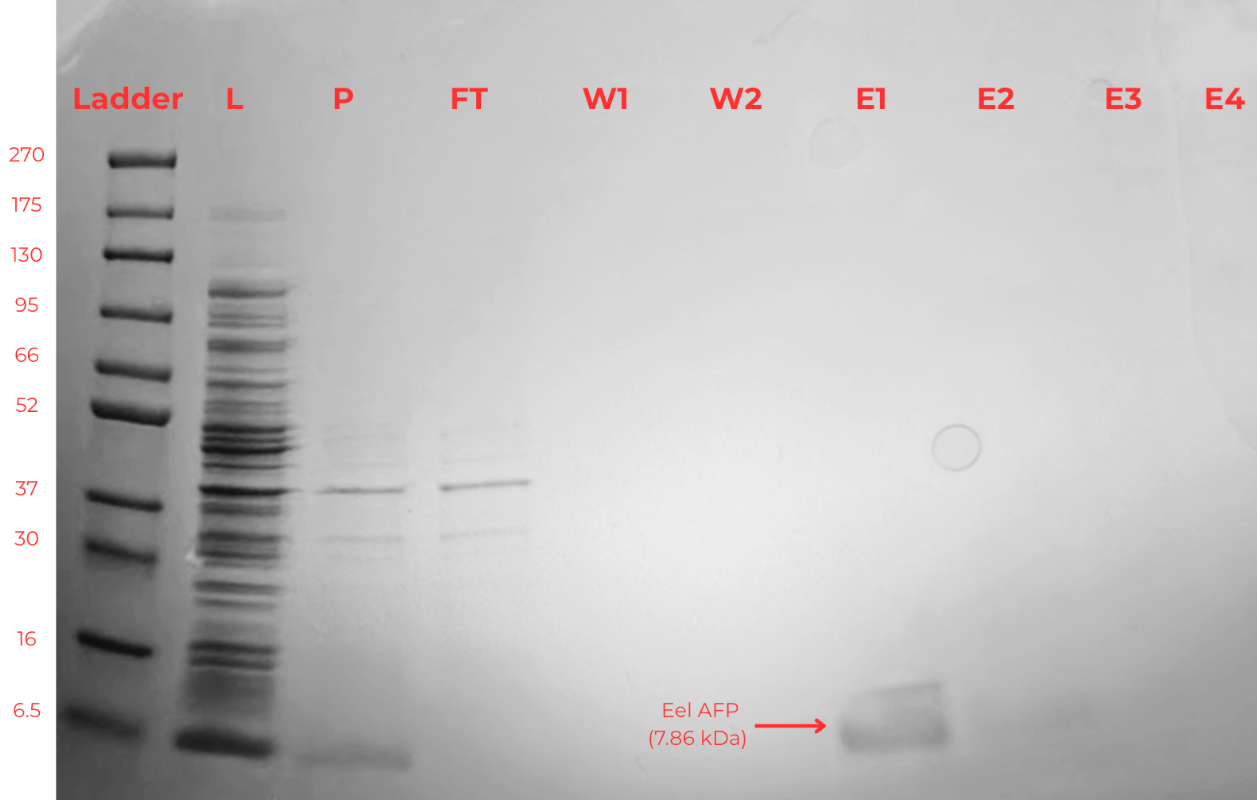 |
|---|
Ice Crystallization Microscopy
To determine whether our purified AFPs were functional, we visualized ice crystal formation using a microscope and a cold stage. In Figure 2A, when no AFP was added, the ice crystals formed elongated, feathery dendritic structures. In Figure 2B, the 10% glycerol solution produced larger, more compact, and blunt spike-like ice crystals with reduced branching. In contrast, the buffered solution in Figure 2C crystallized in broader, planar structures with flat, streak-like morphology. In Figure 2D, the AFP solution with buffer displayed highly uniform crystallization, characterized by fine, densely packed, and predominantly thin, linear ice structures, indicating a more controlled crystal growth pattern, suggesting that our purified AFPs were functional. However, more tests are required to confirm.
| Figure 2. Different shapes of ice crystallization based on the substances. (A. water freezing with food coloring, B. 10% Glycerol, C. Elution buffer only, D. AFP with elution buffer) | 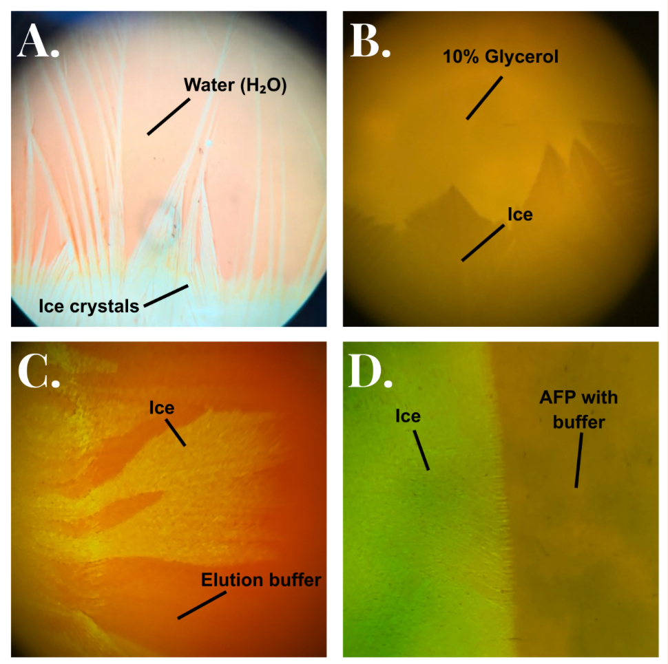 |
|---|
Thermal Hysteresis
To further confirm whether our purified AFPs are functional, we developed a preliminary thermal hysteresis assay to verify that there is a significant gap between the melting point and freezing point of water. Red line: 4.77°C. Yellow line: 2.70°C.
| Figure 3. Mean cooling curves of distilled water (blue), 5% glycerol (yellow), and 10% glycerol (red). Temperature was recorded over 380 s at 0.5 s intervals; data are shown to 300 s for clarity. At approximately 245.5 s, samples were removed from the ice–salt bath and transferred to an ice–water bath. | 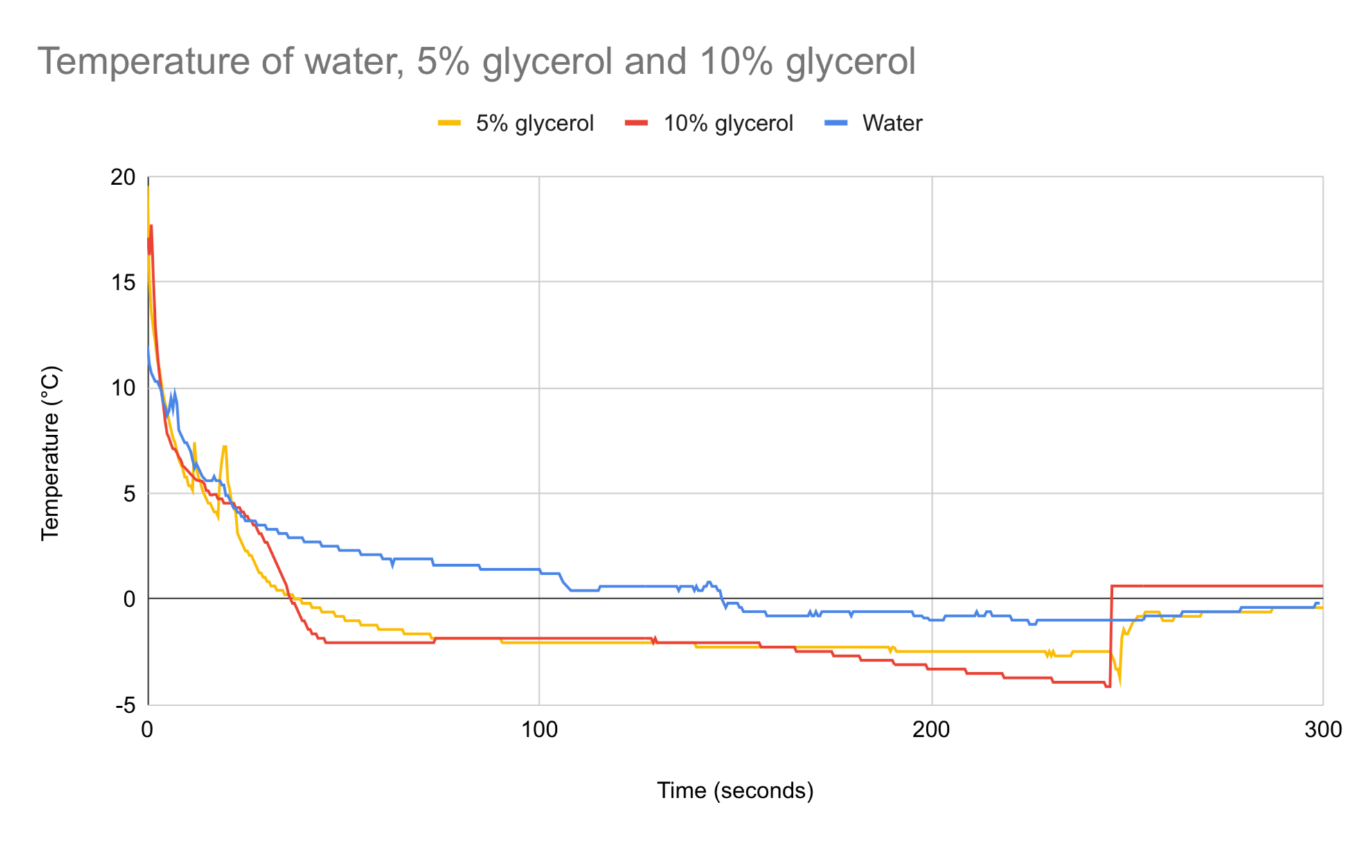 |
|---|
To determine whether our purified AFPs could act as a functional cryoprotectant in living cells, we developed a cell viability assay using E. coli cells. As a proof of concept, we first used glycerol treatment, an unknown cryoprotectant, to test whether E. coli cells were still viable after freeze-thaw cycle. Cells treated with 25% glycerol had a 73% survival rate compared to cells that were not treated with glycerol which had a survival rate of 58% (Figure 4). shown in Figure 4, the positive control had the most cell growth, while the negative control reflected the least amount of cell growth. Lastly, the group with treatment indicated an increase in cell growth when compared to the negative control, but had less growth than the positive control.
| Figure 4. Percentage of cell viability by treatment. E. coli treated with 25% glycerol had 73% viability after freeze-thaw, whereas the cells that were not treated with glycerol (negative control) had a 58% viability. (n=21 for each treatment) | 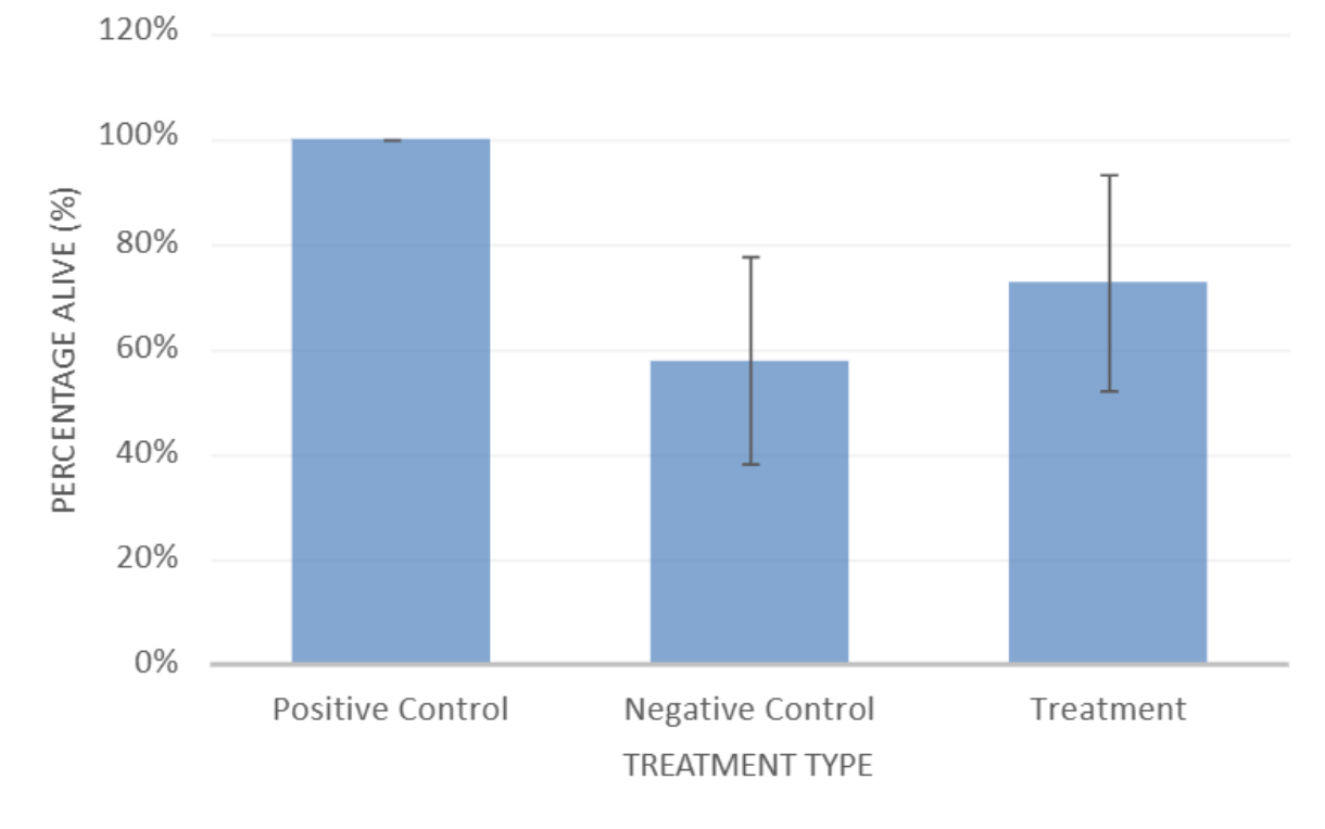 |
|---|
In order to test the effects of AFPs and cryoprotectants in the cryopreservation of multicellular organisms, earthworms were exposed to varying freezing temperatures and durations, followed by injections of glycerol and saline solutions.
Worms exposed to room temperature, 4°C, 0°C, and -20°C for 10 minutes showed 100% survival, while worms exposed to -80°C for 10 minutes showed 0% survival (Figure 5).
| Figure 5. Survival of earthworms following 10 minute exposure periods to different temperatures | 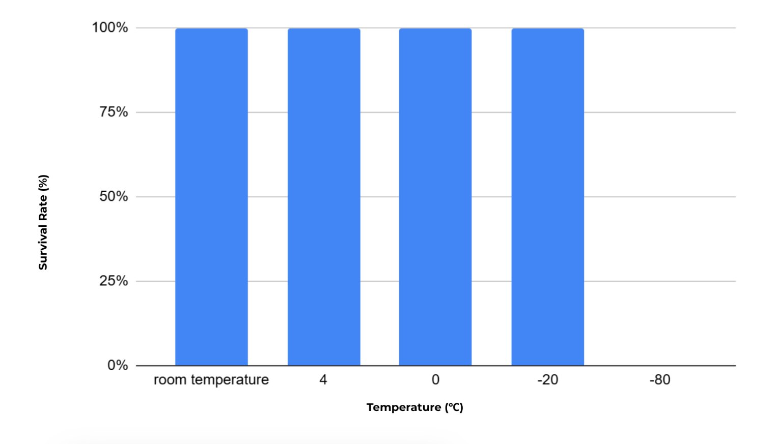 |
|---|
To further determine the freezing threshold at -20°C, worms were frozen for increasing lengths of time. Worms frozen for 10 minutes showed 100% survival. Worms frozen for 14 minutes showed 0% survival, while worms frozen for 15 and 16 minutes showed approximately 33% survival. At 20 minutes, none of the worms survived (Figure 6).
| Figure 6. Effect of freezing duration at -20°C on earthworm survival | 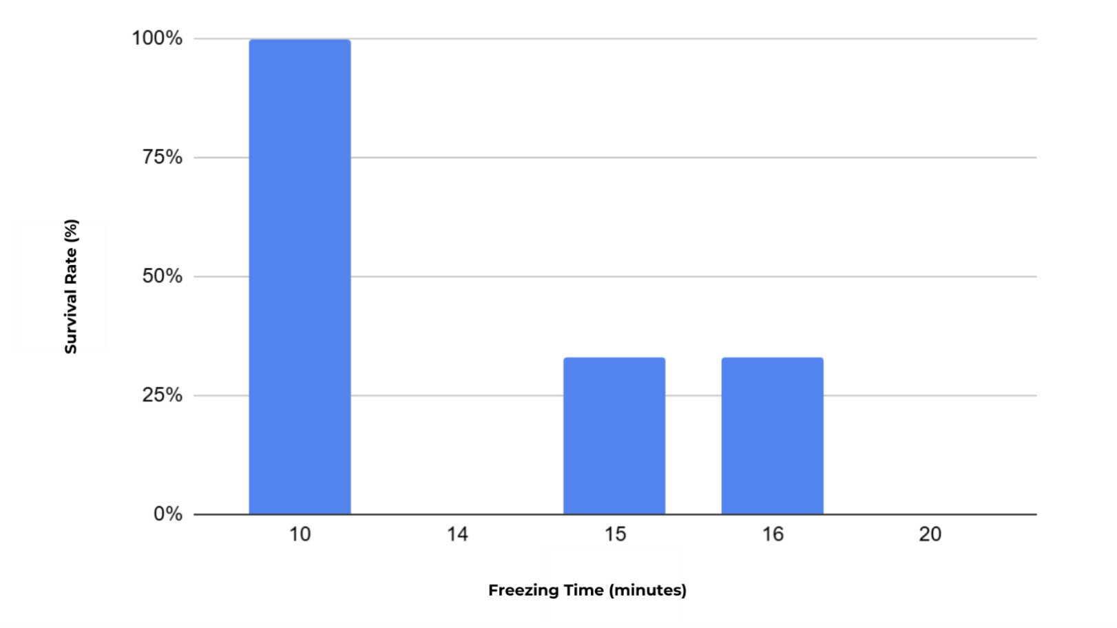 |
|---|
Since purified AFPs will be used to inject earthworms, we determined the amount of time required to anesthetize earthworms with 10% ethanol so that they could be injected with cryoprotectants. Worms exposed to 10% ethanol for 10 minutes showed high survival, while longer exposure times reduced survival. Worms exposed for 20 minutes resulted in only partial survival, and prolonged exposure caused complete mortality (Figure 7).
| Figure 7. Effect of ethanol concentration and exposure time for earthworm survival | 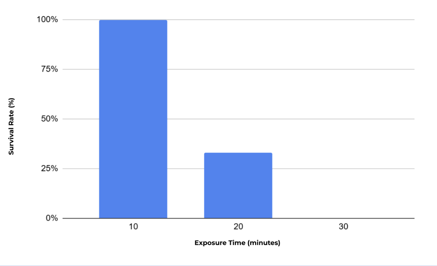 |
|---|
Following the identification of lethal freezing conditions and anesthetization times, we injected and anesthetized worms with glycerol solutions and froze them at -22°C for 14-16 minutes and compared them with ones injected with salt (control) The salt room temperature control group showed 100% survival, while the frozen salt group showed 0% survival. All glycerol treated earthworms exposed to freezing conditions were non viable regardless of freezing duration (Figure 8).
| Figure 8. Effect of different conditions of glycerol and salts on earthworm survival | 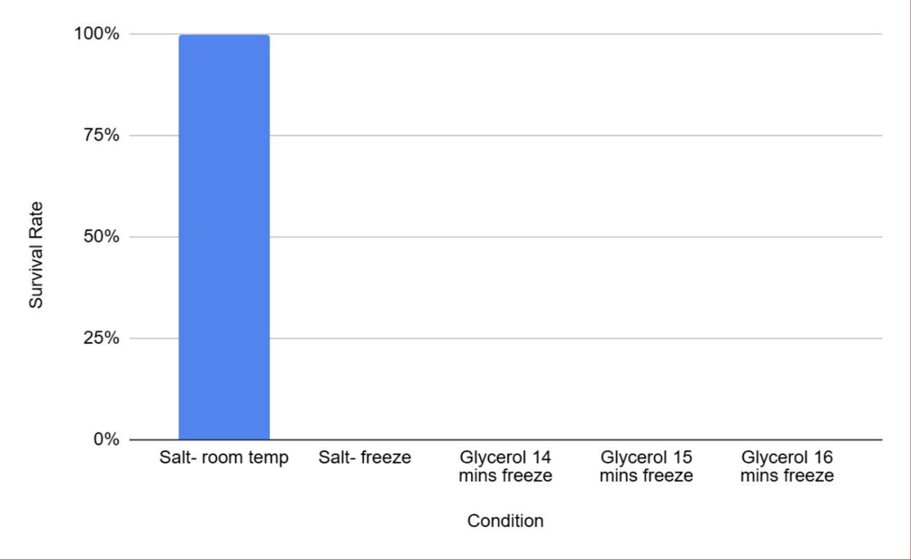 |
|---|
Discussions
The fundamental aim of our project is to utilize antifreeze proteins (AFPs) for present-day applications. Their primary function is to prevent the growth and crystallization of ice crystals, protecting cells and tissues from freeze-induced damage (Wu et al., 2021; Kim et al., 2017). Successful purification of eel-derived AFPs is crucial to providing the foundation to evaluate their practical applications and biological effectiveness (Kim et al., 2017).
By reducing ice crystal formation, AFPs protect living cells from membrane rupture and intracellular damage during freezing and thawing, making them highly valued for cryopreservation applications in medical, agricultural, and reproductive biology (Wu et al., 2021). Building on this, our viability tests provide initial evidence supporting this advantage. Based on the average percentage of cell survival, our results are consistent with our hypothesis for the 10-5 dilution, which claims that our treatment group would have higher cell viability compared to our negative control. However, even though Figure 4 illustrates a higher percentage of cell survival in our treatment group compared to the negative control, this difference is not statistically significant. As seen from the slight overlap in error bars, this gap could be due to biological variation in viability or growth state across the culture (Figure 4). This inconsistency was slightly improved by standardizing cell concentrations to an optical density of 1 at the beginning of each trial (21 replications), ensuring less variation in resulting cell viability counts. Moreover, we utilized glycerol to simulate the addition of AFPs, which is known to successfully inhibit cell death from low temperatures (Figure 4) (Murray & Gibson 2022; Wu et al., 2021). For next steps, we will replace glycerol with actual AFPs to demonstrate their ability to prevent cell death under freezing temperatures.
In addition to viability testing, we evaluated thermal hysteresis (TH) to establish a baseline for AFP activity, further highlighting their potential benefits over simple cryoprotectants like glycerol. Our results demonstrate that glycerol solutions exhibit thermal hysteresis, while distilled water does not, and that the magnitude of this effect increases with glycerol concentration. In this experiment, the TH gap becomes apparent at approximately 245.5 s, when the samples were transferred from the subzero ice-salt bath to the ~0°C ice-water bath, revealing a separation between freezing and melting temperatures (Figure 3). The freezing temperature was taken as the lowest point reached before the temperature rapidly increased, indicating crystallization. The melting temperature was identified as the first stable plateau during the warming phase after transfer. Distilled water showed little to no thermal hysteresis, with freezing and melting temperatures occurring at approximately −0.8°C, close to 0°C but slightly depressed due to experimental conditions such as probe calibration and solution impurities. This is expected, as pure water undergoes a reversible phase change without mechanisms that inhibit ice crystal growth (Kiani & Sun, 2011). Both 5% and 10% glycerol solutions exhibit thermal hysteresis. For the 5% glycerol solution, the freezing temperature is approximately −3.32°C, while the melting temperature is approximately −0.62°C; the thermal hysteresis gap was calculated using Equation 1 and was determined to be approximately 2.70°C.
For the 10% glycerol solution, the freezing temperature is approximately −4.15°C, and the melting temperature is approximately 0.62°C; the thermal hysteresis gap was calculated using Equation 2 and was determined to be approximately 3.53°C.
This behavior can be explained by colligative properties and molecular interactions, where glycerol lowers the freezing point by disrupting the ice lattice (Rall et al., 1978). However, several sources of experimental error may have influenced the results, including initial data fluctuations due to probe equilibration and kinetic energy from manual transfers. If AFPs were introduced, a significantly larger TH gap would be expected, as AFPs bind directly to ice crystal surfaces and inhibit further growth, leading to greater separation between freezing and melting temperatures.
To directly evaluate AFP function at a molecular level and confirm successful purification, we also performed ice recrystallization inhibition assays through visualization and characterization of ice crystals (Graham et al., 2018). The reason we think AFP was successfully purified is because of the uniform and flat ice expansion direction observed (Ye et al., 2020). Compared to water and 10% glycerol, which had thick spikes and lines that can easily penetrate and damage cells, Figure 2A shows that AFP does not have strong spikes nor dendritic lines, exhibiting the characteristic flat and uniform crystals of AFPs. AFPs bind to ice crystals and inhibit their expansion, making them less threatening; thus, Figure 1 supports that AFPs exist in the buffered solution (Ampaw et al., 2021). In addition, Figure 2C (only buffer) shows randomly expanding jagged lines, suggesting that buffer solution is significantly different from how AFP crystallizes (Figure 2D). Several challenges explain why the visualization was not fully successful, including unknown AFP concentration, use of food coloring, lack of a standardized scale bar, and differences in solvent. Since the AFP concentration was not standardized, it may have been too low to produce a strong crystallization pattern, and food coloring could have diluted the solution further. Inconsistency in dissecting microscope magnification also hindered comparisons; for instance, Figures 2A–2C had similar magnification for direct comparison, but Figure 2D (AFP) was at lower magnification. Moreover, the AFP sample was dissolved in buffered solution rather than pure water, so low AFP concentration allowed buffer effects to dominate, making it somewhat similar to buffer-only while differing from glycerol. The lack of replicate trials compounded this. In the future, we will utilize polarizing lenses to eliminate food coloring, standardize concentrations and imaging, and conduct multiple trials for robust visualization.
To extend findings beyond the molecular level, we conducted earthworm experiments assessing AFP effectiveness in improving survival under freezing conditions. The primary finding is that earthworms exhibit extreme sensitivity to sub-zero temperatures; exposure to −22°C for 16 minutes resulted in 100% death rate across initial trials, likely due to ice crystal formation disrupting cellular membranes. Glycerol trials (5%, 7.5%, 10%) failed to provide protection at 14–16 minute marks, suggesting insufficient concentration or poor systemic distribution via injection. Control groups support this: worms injected with 5% salt solution at room temperature all survived, confirming the injection and salt were not harmful, while frozen salt-injected worms died primarily from freezing. Similarly, AFP-injected but unfrozen worms survived, indicating AFP is not toxic under normal conditions (Tran-Guzman et al., 2022).
Overall, these results underscore the benefits of eel-derived AFPs in cryopreservation such as superior ice crystal inhibition and potential cell protection (Wang, 2000; Kim et al., 2017), while revealing challenges like purification variability, assay standardization, and organismal sensitivity (Shan et al., 2023).
Next steps
For visualizing the ice crystals, there are various methods to improve the results. Rather than using food coloring, using polarizing lights will minimize the dilution caused by the food coloring.
Using a dialysis bag could be a potential way to transfer the AFP in water so the solvent stays consistent throughout the experiment. In addition, freeze-drying the AFP solution to obtain the solid protein and concentrating it with desired concentration is another alternative way to maximize the concentration of the AFP when freezing the crystals.
Moreover, future improvements include AFP viability tests on earthworms (exposing them to AFP solution under controlled mild freezing, then comparing survival rates, tissue damage, and movement recovery to controls), replacing glycerol with purified AFPs in cell assays, optimizing transformation for other AFP types, standardizing ice crystal imaging with polarizing lenses and replicates, and quantifying AFP concentrations for precise TH and recrystallization assays.
We will also be doing a circular dichroism spectroscopy and specialized ice-shuttling assays to analyze the secondary structure of the protein, evaluate whether it maintains proper folding under different conditions, and assess its ability to interact with and bind specific ice planes (Kun & Mastai, 2007).
Author contributions
MY, WJ, WF, FL, and MC worked on transformation, expression testing, and purifying of the AFP proteins. AC, JB, AH, AK, EL, and TW studied the visualization of ice crystallization. JC, JI, and EC studied thermal hysteresis. CC, AL, KT, and FW worked on cell viability. EKC, LL, AW, and QY studied cryopreservation via earthworms.
Acknowledgements
References
Amornwittawat, N., Lee, J., Lee, H. Y., & Park, H. J. (2022). Antifreeze proteins: Novel applications and navigation towards their clinical application in cryobanking. International Journal of Molecular Sciences, 23(5), 2664. https://pubmed.ncbi.nlm.nih.gov/35269780/
Ampaw, A. A., Sibthorpe, A., & Ben, R. N. (2021). Use of Ice Recrystallization Inhibition Assays to Screen for Compounds That Inhibit Ice Recrystallization. Methods in molecular biology (Clifton, N.J.), 2180, 271–283. https://doi.org/10.1007/978-1-0716-0783-1_9
Bar Dolev, M., Braslavsky, I., & Davies, P. L. (2016). Ice-Binding Proteins and Their Function. Annual review of biochemistry, 85, 515–542. https://doi.org/10.1146/annurev-biochem-060815-014546
Chakraborty, A., & Jana, B. (2020). The properties, biotechnologies, and applications of antifreeze proteins. Biotechnology Advances, 43, 107578. https://pubmed.ncbi.nlm.nih.gov/32156540/
Correia, L. F. L., Alves, B. R. C., Batista, R. I. T. P., Mermillod, P., & Souza-Fabjan, J. M. G. (2021). Antifreeze proteins for low-temperature preservation in reproductive medicine: A systematic review over the last three decades. Theriogenology, 176, 94–103. https://doi.org/10.1016/j.theriogenology.2021.09.025
DeVries, A. L., & Cheng, C.-H. C. (2005). Antifreeze proteins and organismal freezing avoidance in polar fishes. In J. F. Steffensen (Ed.), Fish physiology: Physiology of polar fishes (Vol. 22, pp. 155–201). Academic Press. https://doi.org/10.1016/S1546-5098(04)22004-0
Drori, R., & Stevens, C. A. (2024). Divergent Mechanisms of Ice Growth Inhibition by Antifreeze Proteins. Methods in molecular biology (Clifton, N.J.), 2730, 169–181. https://doi.org/10.1007/978-1-0716-3503-2_12
Ekpo, M. D., Xie, J., Hu, Y., Liu, X., Liu, F., Xiang, J., Zhao, R., Wang, B., & Tan, S. (2022). Antifreeze Proteins: Novel Applications and Navigation towards Their Clinical Application in Cryobanking. International journal of molecular sciences, 23(5), 2639. https://doi.org/10.3390/ijms23052639
Gourlie, B., Lin, Y., Price, J., DeVries, A. L., Powers, D., & Huang, R. C. (1984). Winter flounder antifreeze proteins: a multigene family. The Journal of Biological Chemistry, 259(23), 14960–14965. https://doi.org/10.1016/S0021-9258(17)43582-6
Graham, L. A., & Davies, P. L. (2005). Glycosylated fish antifreeze proteins and thermal hysteresis. Annual Review of Physiology, 67, 359–383. https://doi.org/10.1146/annurev.physiol.67.040403.103317
Graham, L. A., Agrawal, P., Oleschuk, R. D., & Davies, P. L. (2018). High-capacity ice-recrystallization endpoint assay employing superhydrophobic coatings that is equivalent to the ‘splat’ assay. Cryobiology, 81, 138–144. https://doi.org/10.1016/j.cryobiol.2018.01.011
Greiner, H. G., Stonehouse, A. L., & Tiegs, S. D. (2011). Cold tolerance among composting earthworm species to assess their potential for invasion of northern temperate forests. The American Midland Naturalist, 166(2), 349–362. https://doi.org/10.1674/0003-0031-166.2.349
Holmstrup, M., & Zachariassen, K. E. (1996). Physiology of cold hardiness in earthworms. Comparative Biochemistry and Physiology Part A: Physiology, 115(1), 91–101. https://doi.org/10.1016/0300-9629(96)00010-2
Kiani, H., & Sun, D.-W. (2011). Water crystallization and its importance to freezing of foods: A review. Trends in Food Science & Technology, 22(8), 407–426. https://doi.org/10.1016/j.tifs.2011.04.005
Kim, H. J., Lee, J. H., Hur, Y. B., Lee, C. W., Park, S. H., & Koo, B. W. (2017). Marine Antifreeze Proteins: Structure, Function, and Application to Cryopreservation as a Potential Cryoprotectant. Marine drugs, 15(2), 27. https://doi.org/10.3390/md15020027
Kuiper, M. J., Lankin, C., Gauthier, S. Y., Walker, V. K., & Davies, P. L. (2003). Purification of antifreeze proteins by adsorption to ice. Biochemical and biophysical research communications, 300(3), 645–648. https://doi.org/10.1016/s0006-291x(02)02900-5
Kun, H., & Mastai, Y. (2007). Activity of short segments of Type I antifreeze protein. Biopolymers, 88(8), 807–814. https://doi.org/10.1002/bip.20844
Liu, Y., & others. (2017). Marine antifreeze proteins: Structure, function, and application to cryopreservation. Marine Drugs, 15(2), 30. https://doi.org/10.3390/md15020030
López Ortiz, J. I., Quiroga, E., Narambuena, C. F., Riccardo, J. L., & Ramirez-Pastor, A. J. (2021). Thermal hysteresis activity of antifreeze proteins: A model based on fractional statistics theory of adsorption. Physica A: Statistical Mechanics and its Applications, 574, 125962.
Murray, K. A., & Gibson, M. I. (2022). Chemical approaches to cryopreservation. Nature Reviews Chemistry, 6, 579–593. https://doi.org/10.1038/s41570-022-00407-4
Rall, W. F., Mazur, P., & Souzu, H. (1978). Physical-chemical basis of the protection of slowly frozen human erythrocytes by glycerol. Biophysical Journal, 23(1), 101–120. https://doi.org/10.1016/S0006-3495(78)85436-8
Salvay, A. G., Gabel, F., Pucci, B., Santos, J., Howard, E. I., & Ebel, C. (2010). Structure and interactions of fish type III antifreeze protein in solution. Biophysical journal, 99(2), 609–618. https://doi.org/10.1016/j.bpj.2010.04.030
Sazaki, G., Zepeda, S., Nakatsubo, S., Yokoyama, E., & Furukawa, Y. (2010). Elementary steps at the surface of ice crystals visualized by advanced optical microscopy. Proceedings of the National Academy of Sciences, 107(46), 19702–19707. https://doi.org/10.1073/pnas.1008866107
Shan, Y., Liao, L., Xu, Y., & Chen, B. (2023). Recent progress in the characterization of multidimensional activities of antifreeze proteins. Chinese Journal of Polar Research, 35(3), 490–497. https://doi.org/10.13679/j.jdyj.20220424
Tran-Guzman, A., Moradian, R., Walker, C., Cui, H., Corpuz, M., Gonzalez, I., Nguyen, C., Meza, P., Wen, X., & Culty, M. (2022). Toxicity profiles and protective effects of antifreeze proteins from insect in mammalian models. Toxicology Letters, 368, 9–23. https://doi.org/10.1016/j.toxlet.2022.07.009
Vollmer, J., & Bäumchen, O. (2012). Thermodynamic analysis of thermal hysteresis: Mechanistic insights into biological antifreezes. Journal of Chemical Thermodynamics, 52, 46–53. https://doi.org/10.1016/j.jct.2012.04.028
Wang, J. H. (2000). A comprehensive evaluation of the effects and mechanisms of antifreeze proteins during low-temperature preservation. Cryobiology, 41(1), 1–9. https://doi.org/10.1006/cryo.2000.2265
Wu, X., Yao, F., Zhang, H., & Li, J. (2021). Antifreeze proteins and their biomimetics for cell cryopreservation: Mechanism, function and application-A review. International journal of biological macromolecules, 192, 1276–1291. https://doi.org/10.1016/j.ijbiomac.2021.09.211
Ye, Q., Eves, R., Campbell, R. L., & Davies, P. L. (2020). Crystal structure of an insect antifreeze protein reveals ordered waters on the ice-binding surface. Biochemical Journal, 477(17), 3271–3286. https://doi.org/10.1042/BCJ20200626
Yu, J., Cheng, C.-H., DeVries, A. L., & Chen, L.-B. (2005). Characterization of a multimer type III antifreeze protein gene from the Antarctic eel pout (Lycodichthys dearborni). Glycobiology, 15(10), 1143–1152. https://doi.org/10.1093/glycob/cwj045