
Sajani Brown, Kirsten Choi, and Tanishka Singh, Weston High School, Weston, Massachusetts, United States
Reviewed on 9 May 2026; Accepted on 22 June 2026; Published on 26 October 2026
With help from the 2026 BioTreks Production Team.
Hematopoietic stem cells (HSCs) comprise less than 0.01 percent of bone marrow, yet they are essential for bone marrow transplantation and lifelong blood production. Currently, HSCs cannot be significantly expanded in vitro, meaning clinicians can only transplant what a donor provides. As a result, many patients, especially those from underrepresented ethnic backgrounds with reduced access to compatible donor matches, die waiting for a compatible match. The central barrier is our inability to recreate the functional bone marrow stem cell niche outside the body to keep HSCs in an undifferentiated state. In vivo, endothelial cells regulate HSC quiescence through localized paracrine signaling. However, existing culture systems rely on externally added growth factors that degrade rapidly and fail to replicate sustained niche signaling. We propose a programmable microfluidic niche model in which a basal layer composed of endothelial cells is transfected with lipid nanoparticle-encapsulated mRNAs (mRNA LNP) encoding necessary proteins and factors in order to mimic paracrine signaling. We hypothesize this approach will preserve CD34-positive stemness in the HSC cells and enable controlled, sustained expansion of HSCs while maintaining their undifferentiated state. By enabling scalable HSC expansion in an in vitro system, this platform could help improve the accessibility of bone marrow transplantation for patients with limited donor availability.
Keywords: Stem cell niches, microfluidic device, hematopoietic stem cells, mRNA-LNPs, induced paracrine signaling
Authors are listed in alphabetical order. *Neurath mentored the group. Please direct all correspondence to mailto:neurathr@weston.org.
Background
HSCs are the foundation of bone marrow transplantation, a life-saving treatment for leukemia and other blood disorders. These cells reside in a specialized stem cell niche in the bone marrow, where endothelial cells regulate their self-renewal through localized paracrine signaling. While transplantation is highly effective, it is severely limited by a shortage of human leukocyte antigen (HLA) matched donors. HLAs are specialized proteins found on the surface of nearly all cells in the human body, allowing the immune system to identify whether a cell is foreign or not. If there are any unknown proteins displayed, the immune system reacts to remove any potential threats. However, HLAs are encoded by the most polymorphic genes in the human body, meaning there is a great number of variations from person to person. In the case of transplantations, when the HLA does not match, the immune system can get triggered and cause rejection within the body, leading to illnesses such as graft-versus-host disease (GVHD) (Park, 2012). Another problem that limits bone marrow HSC transplantations is that it depends greatly on the quantity of HSCs being transplanted, which is inherently insufficient, as HSCs make up only 0.01 percent of bone marrow (Mayle et al., 2012). Traditional approaches attempt to bypass these hurdles by using non-identical donor sources or intensive post-transplant immunosuppression, yet they cannot replicate the natural bone marrow niche signals required for safe, large-scale cell expansion (Khaddour et al., 2023). These strategies ultimately fail to overcome the dual barriers of graft rejection and insufficient cell dosing.
Furthermore, existing in vitro culture systems rely on externally added growth factors that degrade rapidly and fail to replicate sustained niche signaling (Sharma et al., 2012). As a result, HSCs often lose stemness and differentiate prematurely, preventing their therapeutic use. Patients with blood disorders like leukemia must rely on donor bone marrow, which carries risks of painful procedures, immune complications, and limited HSC counts.
To address this, we propose a synthetic biology-based microfluidic system that models the stem cell niche in vitro by mimicking paracrine signaling, enabling HSC culture and expansion for transplantation. By utilizing mRNA-LNPs, we would instruct a set of feeder cells to constantly produce the essential proteins rather than adding proteins through perfusion. This would provide the sustained signaling required to keep the stem cells from losing their potency. Developing a reliable method to transform somatic cells into functional, transplant-ready HSCs could enable autologous transplantation, which utilizes the patient’s own cells to eliminate the risk of immune rejections. Engineering a niche that maintains HSC multipotency is a critical step toward safer, scalable transplantation therapies.
Systems level
Our proposed solution relies on repurposing Organ-on-a-Chip (OoC) models, microfluidics devices typically used for disease modeling, to recreate the Hematopoietic Stem Cell niche in vitro. The Organ-on-a-Chip (OoC) platform investigates the use of mRNA lipid nanoparticles to create a more accurate representation of an in vivo bone marrow (BM) HSC niche. OoCs are a form of microtechnology that ultimately mimic the natural environment of cells in vitro through the use of microfluidics and cell culturing (Emulate, 2023). By utilizing the microtechnology of the Emulate BM Chip, which is shown in Figure 1, we aim to transform mature somatic cells into healthy, stable HSCs that would be viable for BM transplants in leukemia patients (Emulate, 2018). The main goal of the experiment is to compare two distinct methods for driving the differentiation of iPSCs into HSCs. Specifically, we are evaluating the impact of directly delivering exogenous differentiation factors via perfusion against a simulated paracrine approach, which uses mRNA LNPs to prompt endothelial cells to produce those same factors
| Figure 1. Illustrates the general structure of the Emulate BM OoC which we will be using. The green area represents the marrow channel while the yellow area represents the vascular channel. | 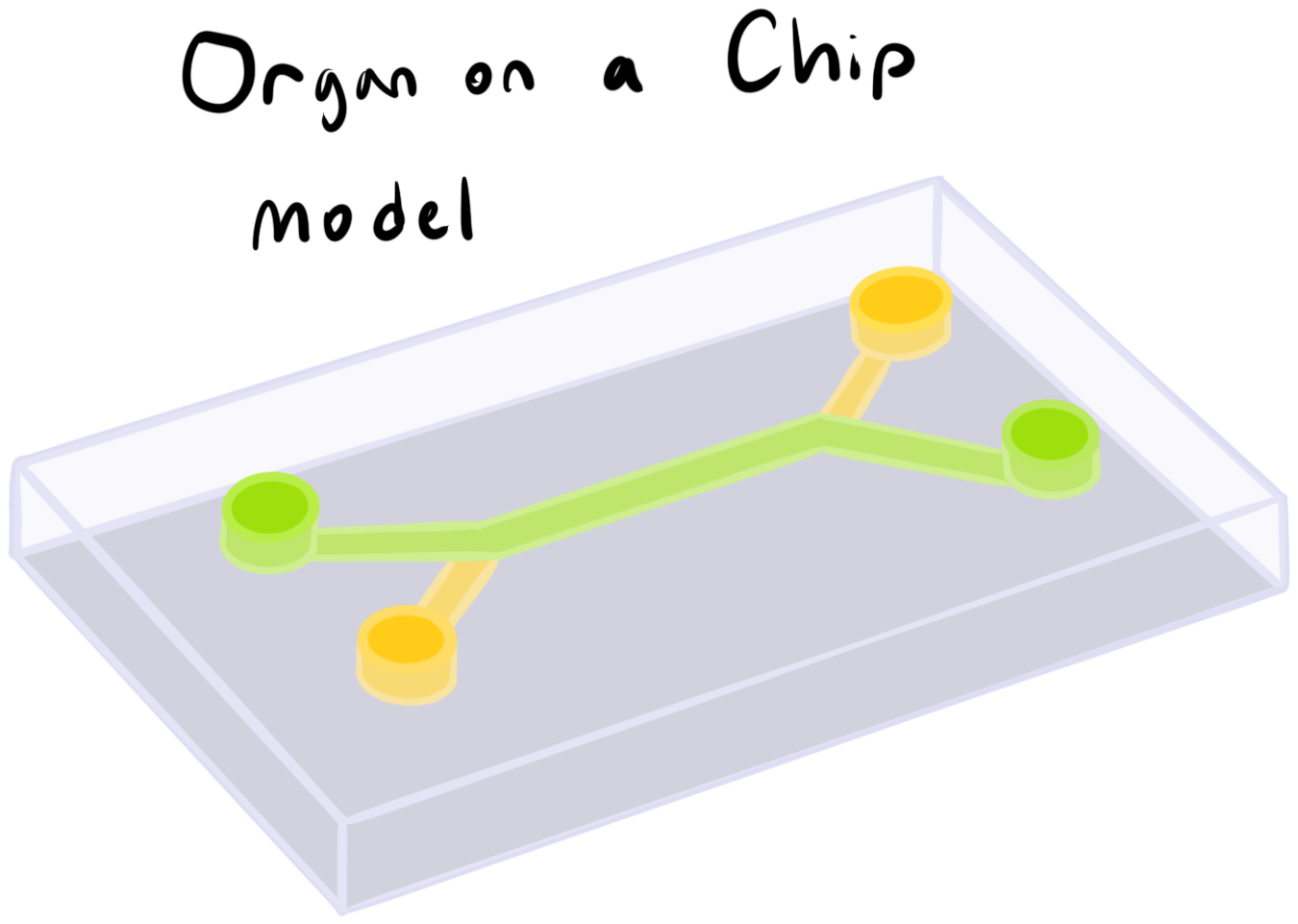 |
|---|
Cellular Source and Microtechnology Assembling
Primary Umbilical Vein Endothelial Cells (HUVEC; ATCC PCS-100-013 ™) will be utilized as the foundational cell source. HUVECs are selected for their strong response to inflammatory signals and their established role as a substitute for the bone marrow vascular niche. Following the standardized protocols for the Emulate BM-OoC, cells will be recovered from cryopreservation and seeded into a dual-channel microfluidic device (Chou et al., 2020). There are two channels within the Emulate BM-OoC: the vascular channel (bottom) and the marrow channel (top) (use Figure 1. as a reference). The bottom channel will be lined with HUVECs to mimic the bone marrow sinusoids, while the top channel will be constructed with a secondary layer of HUVECs adjacent to a 3D fibrin gel scaffold, mimicking the extravascular hematopoietic space (see Figure 2 for a better understanding of the structure). Using the perfusion strategy from Chou et al., the system is fed via the bottom vascular channel to establish nutrient gradients and physiological shear stress (Chou et al., 2020).
| Figure 2. Breaks down the overall structure of the OoC model, specifically for the experimental group. The created HSCs are on the top channel with a green colored perfusion while the endothelial feeder cells are on the bottom channel with a yellow perfusion containing mRNA LNPs. | 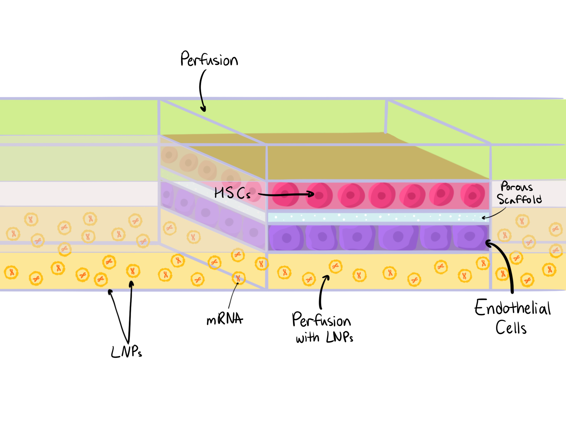 |
|---|
Turning the HUVEC into iPSC
To conduct the undifferentiation from HUVECs into iPSCs, Yamanaka factors (Oct4, Sox2, Klf4, and c-Myc) are introduced to the system via the top perfusion layer. While iPSC reprogramming generally resets a cell's epigenetic and phenotypic state, it doesn’t correct somatic genetic mutations. Therefore, to derive healthy HSCs, the backbone of BM transplants, the starting somatic cells must be sourced from a non-malignant lineage. iPSCs derived directly from acute myeloid leukemia cells retain any leukemia-based mutations and express the leukemic phenotype upon redifferentiation (Kotini et al., 2023). Thus, the success of this method relies on the utilization of unaffected endothelial cells. Because testing this is not the main idea of the experiment, the use of HUVECs is acceptable.
Hematopoietic differentiation
Once the HUVECs are undifferentiated, the iPSCs in the top layer are re-differentiated into HSCs by delivering the specific differentiation factors mentioned in the Parts level section. This mimics the transition from hemogenic endothelial cells to HSCs within a controlled environment. Use live-cell imaging to track structural changes as the plate iPSC-derived cells group into round, non-adherent HSCs.
Maintaining HSC multipotency
The experiment will have one experimental group and one control group. The experimental group (OoC 1) will introduce the necessary proteins and factors via mRNA LNPs into the bottom vascular channel (more details about which proteins/factors in the Parts level). Ideally, the bottom HUVECs will uptake the LNPs and will translate & secrete the stabilizing proteins through the gel layer. The control group will be exposed to the same factors; however, the delivery method will differ, with the proteins being delivered directly via perfusion into the top channel.
Evaluation and data collection
To evaluate whether the LNP-mediated paracrine approach sustains stemness better than exogenous protein perfusion, the chips will be analyzed using a combination of confocal imaging and flow cytometry.
First, the total hematopoietic output must be quantified without interference from the underlying HUVEC layer. Because both HUVECs and HSCs are CD34+, standard CD34 screening cannot accurately distinguish between the two cell types in this co-culture system. Instead, fluorescently labeled anti-CD45 antibodies will be perfused into the marrow channel, as CD45 is exclusively expressed on cells of the hematopoietic lineage. Confocal Z-stacking microscopy will then be used to capture 3D images of the fibrin gel scaffold, allowing us to count the distinct CD45+ fluorescent rings and calculate total cell yields (Rix et al., 2022).
Following imaging, cells will be harvested from the marrow channel to confirm true long-term HSC identity and rule out premature differentiation. To maintain cost-effectiveness while ensuring high accuracy, the harvested cells will be screened using a streamlined 6-marker human panel. Successful expansion of undifferentiated HSCs will be verified via positive selection for CD34+ (the clinical baseline), EPCR+ (a marker for in vitro expansion), and GPRC5C+ (a marker confirming a dormant, quiescent state) (Rix et al., 2022). Concurrently, downstream multipotent progenitors and mature blood lineages will be excluded via negative selection for Lin-, CD38-, and CD45RA-.
Ultimately, we will compare the total yield of this precise Lin-, CD38-, CD45RA-, CD34+, EPCR+, GPRC5C+ population between the experimental LNP group (OoC 1) and the protein-perfused control group. A higher yield of these quiescent cells in the LNP group will demonstrate that continuous paracrine signaling successfully replicates the native bone marrow niche mechanics required to preserve stem cell potency.
Device level
To model the hematopoietic stem cell (HSC) niche in vitro, we propose a microfluidic organ-on-chip system based on the Emulate Bone Marrow (BM) Chip. This platform will be adapted to recreate key structural and biochemical features of the native bone marrow microenvironment to support HSC maintenance, expansion, and controlled differentiation.
The proposed device consists of two parallel microchannels separated by a semi-permeable porous membrane. The upper channel will be seeded with HSCs derived from patient-specific iPSCs, while the lower channel will contain feeder cells. This compartmentalized architecture is intended to mimic the spatial organization of the bone marrow niche, allowing distinct cell populations to interact through soluble signaling while remaining physically separated.
Continuous microfluidic perfusion will establish physiologically relevant flow conditions within both channels. This dynamic flow is expected to support nutrient delivery, waste removal, and the formation of stable signaling gradients. The porous membrane between channels will enable paracrine exchange of factors, allowing feeder cells to regulate HSC behavior through the secretion of cytokines, growth factors, and other niche-specific signals.
To engineer sustained and tunable paracrine signaling within the device, we propose the use of mRNA-loaded lipid nanoparticles (mRNA-LNPs) to feeder cells. These nanoparticles will deliver synthetic mRNA encoding key niche factors, enabling feeder cells to act as in situ producers of essential proteins. mRNA will be generated using plasmid construct #222990 as a template for in vitro transcription (Chen et al., 2023).
By integrating mRNA-LNP-mediated protein expression with a microfluidic co-culture system, the proposed device aims to more accurately simulate the dynamic signaling environment of the bone marrow. The device is designed to guide iPSC-derived cells toward functional HSC states, while preserving their multipotency. This proposed microfluidic device represents a modular and scalable approach for engineering synthetic stem cell niches, with potential applications in regenerative medicine, disease modeling, and cell therapy development.
| Figure 3. This diagram shows the overall chemical processes to give a full visual of the main biological checkpoints of the experiment. | 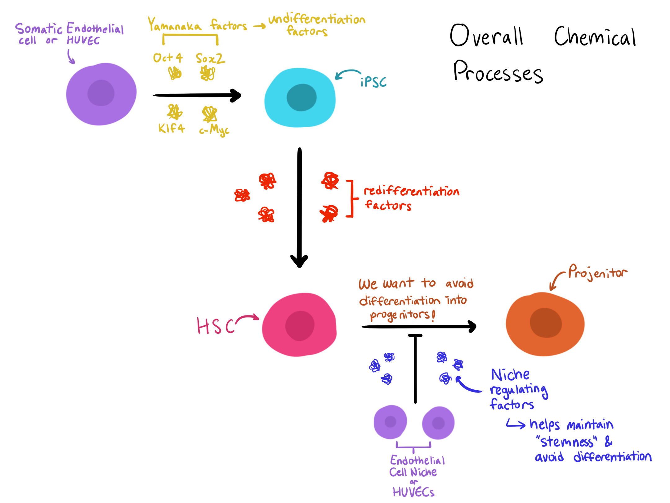 |
|---|
Parts level
In our method, there is first the re-programming of patient-derived somatic cells (HUVECs) into iPSCs, the differentiation of those iPSCs into HSCs, and finally, signaling to maintain those HSCs in a quiescence state ( refer to Figure 3 diagram). Our proposed idea utilizes mRNA LNPs and feeder cells to mimic in vivo paracrine signaling. All of these biological processes and the creation of mentioned factors are addressed in this section in detail.
| Figure 4. This diagram magnifies membrane fusion or the process in which the mRNA LNP is taken up by the endothelial cell. | 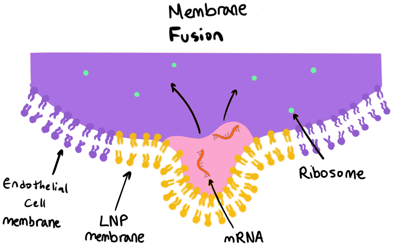 |
|---|
mRNA LNP production
We utilized Addgene-sourced plasmids shown in Table 1 as templates for independent in vitro transcription of each mRNA needed. Producing each mRNA species in isolation minimized the risk of cross-contamination and unwanted molecular interference prior to LNP formulation.
The “gold standard” for constructing an LNP that is taken in by endothelial cells involves blending four primary ingredients: an ionizable lipid, a helper lipid, cholesterol, and PEG-functionalized lipids at carefully optimized ratios. The LNPs would be made out of SM-102, phosphatidylcholine, cholesterol, and polyethylene glycol lipid which would be assembled with a NanoAssemblr Ignite (McKenzie et al., 2023). To ensure the LNP’s transfection into the HUVECs is sufficient, the mRNA LNP would be conjoined to anti-CD31 using maleimide-thiol chemistry and subsequently purified (Leo et al., 2026). A visual of proper transfection is depicted by Figure 4. All methods of creating the mRNA LNPs (except for the mRNA contents and the mentioned plasmid) will follow the process used by Leo et al. (2026).
Table 1. This is a list of the proteins delivered into the OoC and their respective plasmid and/or protein source.
| Protein or Signal | Addgene Plasmid | Protein Order |
|---|---|---|
| Oct4 | R&D Systems (Recombinant Human OCT4/POU5F1, Cat# 9445-OD, Minneapolis, MN) | |
| Sox2 | Thermo Fisher Scientific (Gibco Human Sox2 Recombinant Protein, PeproTech, Cat# 100-03-25UG, Waltham, MA) | |
| Klf4 | R&D Systems (Recombinant Human KLF4, Cat# 10243-K4, Minneapolis, MN) | |
| c-Myc | R&D Systems (Recombinant Human c-Myc, Cat# 10563-MC, Minneapolis, MN) | |
| bFGF (Basic Fibroblast Growth Factor) | Thermo Fisher Scientific (Gibco Human FGF-basic 154 aa Protein, PeproTech, Cat# 100-18B-50UG, Waltham, MA) | |
| Laminin | Sigma-Aldrich (Laminin from human fibroblasts, Cat# L4544, St. Louis, MO) | |
| Collagen I | Thermo Fisher Scientific (Gibco Collagen I, Rat Tail, Cat# A1048301, Waltham, MA) | |
| Entactin | R&D Systems (Recombinant Human Nidogen-1/Entactin, Cat# 2570-ND, Minneapolis, MN) | |
| Heparan sulfate proteoglycan | Sigma-Aldrich (Heparan Sulfate Proteoglycan from basement membrane, Cat# H4777, St. Louis, MO) | |
| BMP4 (Bone Morphogenetic Protein 4) | R&D Systems (Recombinant Human BMP-4, Cat# 314-BP, Minneapolis, MN) | |
| Collagen IV | Sigma-Aldrich (Collagen from human placenta, Bornstein and Sage Type IV, Cat# C7521, St. Louis, MO). | |
| Fibronectin | Sigma-Aldrich (Fibronectin from human plasma, Cat# F2006, St. Louis, MO) | |
| SCF (Stem Cell Factor) | pCS2-zfKitlgb was a gift from Petr Bartunek (Addgene plasmid # 168200 ; http://n2t.net/addgene:168200 ; RRID:Addgene_168200) | R&D Systems (Recombinant Human SCF, Cat# 255-SC, Minneapolis, MN) |
| Flt-3 Ligand (Fms-like tyrosine kinase 3 receptor ligand) | R&D Systems (Recombinant Human Flt-3 Ligand, Cat# 308-FK, Minneapolis, MN) | |
| TPO (Thrombopoietin) | pAc-His-zfTpo was a gift from Petr Bartunek (Addgene plasmid # 64309 ; http://n2t.net/addgene:64309 ; RRID:Addgene_64309) | R&D Systems (Recombinant Human Thrombopoietin/THPO, Cat# 288-TP, Minneapolis, MN) |
| Interleukins: IL-3 & IL-6 | R&D Systems (Recombinant Human IL-3, Cat# 203-IL, Minneapolis, MN) R&D Systems (Recombinant Human IL-6, Cat# 206-IL, Minneapolis, MN) | |
| HoxB4 | HOXB4-WG-SP65 was a gift from Corey Largman (Addgene plasmid # 20986 ; http://n2t.net/addgene:20986 ; RRID:Addgene_20986) | |
| AGM (Aorta-gonad-mesonephros) | pscALPS-SIV-AGM-VERAGM3 vpr was a gift from Jeremy Luban (Addgene plasmid # 115826 ; http://n2t.net/addgene:115826 ; RRID:Addgene_115826) | |
| CXCL12 | pcDNA3-CXCL12-sfGFP was a gift from Erik Procko (Addgene plasmid # 98961 ; http://n2t.net/addgene:98961 ; RRID:Addgene_98961) | |
| CXCL4 | PF4-bio-His was a gift from Gavin Wright (Addgene plasmid # 53411 ; http://n2t.net/addgene:53411 ; RRID:Addgene_53411) | |
| TGF-β | pMT-TAK1-V5-His was a gift from Cyril Gueydan (Addgene plasmid # 63753 ; http://n2t.net/addgene:63753 ; RRID:Addgene_63753) |
Stage 1 (reprogramming – iPSCs)
Reprogramming Somatic Cells into iPSCs through the use of Yamanaka factors
The reprogramming of somatic cells is achieved by the introduction of four transcription factors, known as the Yamanaka factors, into the patient-derived somatic cells. These factors, Oct4, Sox2, Klf4, and c-Myc, override the cell’s existing purpose and induce a pluripotent state (Aguirre et al., 2023). Oct4 is the most critical factor, as it activates the genes responsible for pluripotency and suppresses genes that define a specialized cell type (Radzisheuskaya & Silva, 2014). Sox2 works along with Oct4, helping to open regions of DNA that are typically in mature cells, which allows pluripotency to be activated. Klf4 stabilizes the process by preventing the cell from undergoing apoptosis during reprogramming, while also reinforcing the activity of Oct4 and Sox2 (Narayan et al., 2017). c-Myc acts as an accelerant, increasing overall gene expression and making chromatin more accessible, which speeds up reprogramming (Li et al., 2013). Each Yamanaka factor is encoded on a separate plasmid. mRNA is synthesized via in vitro transcription, then purified, and finally encapsulated in LNPs for delivery. As mRNA degrades naturally after use, the reprogramming signal is temporary and self-limiting.
| Figure 5. This diagram demonstrates the three different methods of introducing each protein or factor to the cells. | 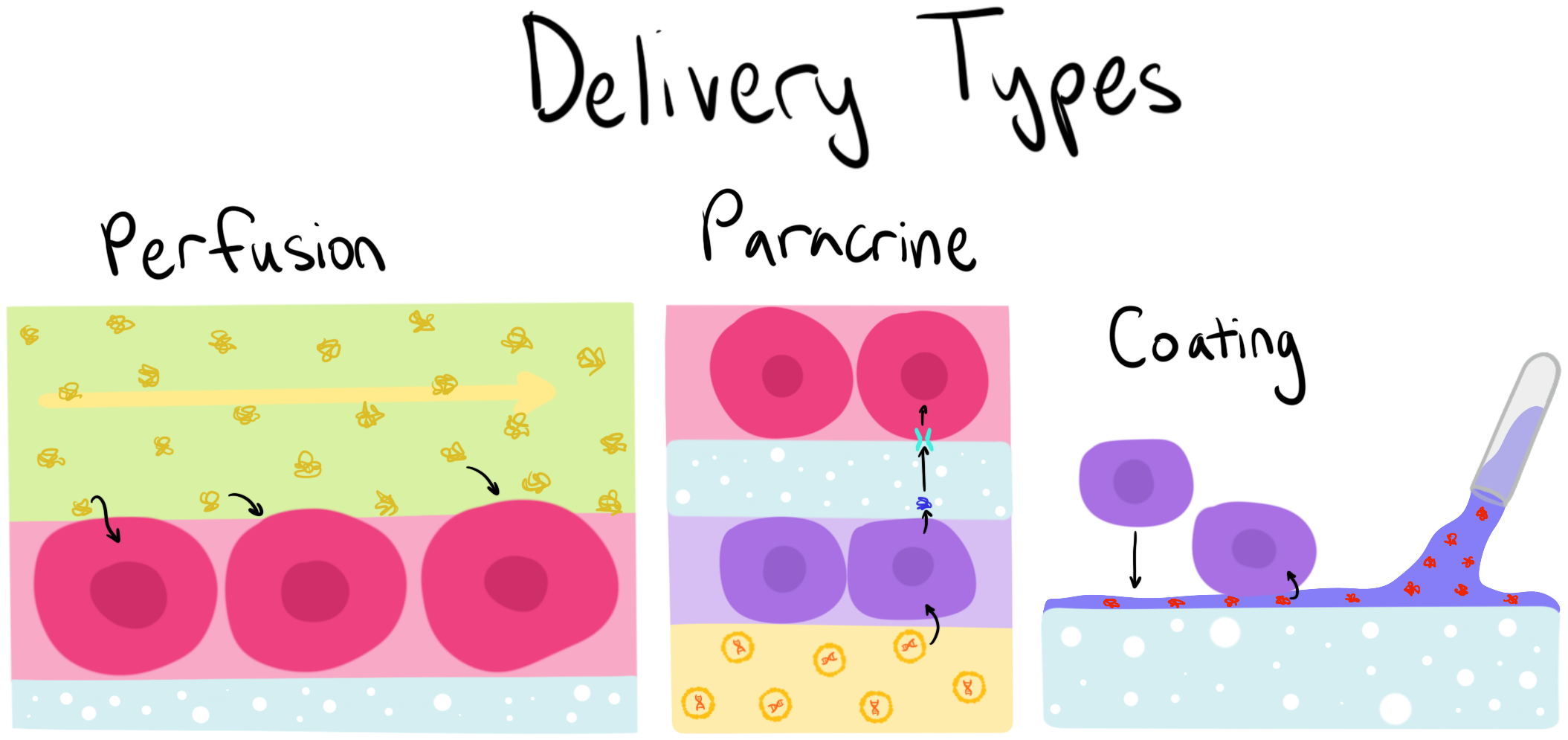 |
|---|
Stage 2 (differentiation – HSCs)
Once the iPSCs are established, they must go through a series of stages to become HSCs. As shown in Figure 5, there would be three forms of delivery: perfusion, paracrine, and coating. Perfusion delivery is when the wanted factors are put through the solution that goes through a channel of the OoC. Paracrine delivery refers to the usage of mRNA LNPs to create the necessary factors in feeder cells and deliver them to the targeting cells. Coating occurs before the cells are adhered to the scaffold and often carries factors that would help in current or later processes. It’s important to note that while some deliveries must occur simultaneously, others must be stopped before others start.
The first step is to maintain the pluripotency of the iPSCs after establishment. It’s important to add bFGF through the top perfusion but once ready to start the initiation of differentiation, the perfusion must be replaced by a solution without it.
The next step is mesoderm induction and early specification where BMP4 should be delivered through the perfusion. This is an important step because it is the first major “commitment” where the cells it can now differentiate into are narrowed down into tissue like blood, bone, muscle, or heart rather than brain or gut tissue. Mesodermal cells can be characterized by the gene Brachyury (Lim et al., 2013).
Afterward, the prior perfusion should be replaced by a “hematopoietic cocktail” medium which would include the soluble factors SCF, Flt-3 Ligand, TPO, IL-3, and IL-6. This specifies the mesodermal cells into the hematopoietic precursors hemangioblast then into hematopoietic progenitor cells, forcing the cells to “commit” to a blood-forming pathway. At this stage, the cells begin expressing surface markers like CD34 (Lim et al., 2013).
Lastly, the factors HoxB4 and AGM would be delivered through paracrine signaling to ensure the progenitors turn into true HSCs. This is the most critical step for bone marrow transplants because only HSCs have the ability to live in an adult host and continuously create all types of blood cells for the rest of the patient’s life (Lim et al., 2013). Look over Table 2 for more detailed information about the chosen factors.
Table 2. Separates the necessary proteins and factors into each step/procedure and delivery type and gives a description of what each does for the transformation of cell types. All of the information is from Lim et al. (2013).
| Step/Procedure | Protein or Signal | Purpose | Delivery: Perfusion (Soluble) | Delivery: Paracrine (Cell-Secreted) | Delivery: Coating (Substrate) |
|---|---|---|---|---|---|
| Maintenance of Pluripotency | bFGF (Basic Fibroblast Growth Factor) | Maintains iPSCs in an undifferentiated, self-renewing state before induction | Yes | ||
| Mesoderm Induction | Laminin, Collagen I, Entactin, & Heparan sulfate proteoglycan | Major matrix components required for initial differentiation of iPSCs into mesodermal cells | Yes | ||
| BMP4 (Bone Morphogenetic Protein 4) | Recapitulates early embryonic signaling to help drive mesoderm emergence and early erythropoiesis | Yes | |||
| Specification to Precursors (HPCs) | Collagen IV | Directs the generation of Flk1+ mesoderm and accelerates transition into hemangioblasts and progenitors | Yes | ||
| Fibronectin | Provides physical support for the generation of CD34+ hematopoietic progenitors | Yes | |||
| SCF (Stem Cell Factor) | Critical factor for hematopoietic development and the proliferation of immature hematopoietic cells | Yes | |||
| Flt-3 Ligand (Fms-like tyrosine kinase 3 receptor ligand) & TPO (Thrombopoietin) | Promotes the expansion and proliferation of immature hematopoietic progenitors | Yes | |||
| Interleukins: IL-3 & IL-6 | Supports early hematopoietic development and the induction of myeloid/erythroid progenitors | Yes | |||
| Definitive HSC Maturation | HoxB4 | Transcription factors that provide definitive engraftment potential and regulate the maturation of HSCs (upregulating CD34) | Yes | ||
| AGM (Aorta-gonad-mesonephros) | Provides the favorable niche microenvironment and secreted factors required to acquire long-term HSC activity | Yes |
Maintaining HSCs
To maintain the HSCs, both paracrine signaling and perfusion must be used for the most accurate representation of in vivo settings. The proteins and signals needed are listed and described in Table 3. For the experimental group, all proteins and factors that are delivered through paracrine signaling in vivo would be sent into the bottom channel of the OoC via mRNA LNPs and under paracrine signaling according to Figure 5 and Figure 6 while TPO would be delivered through the top perfusion channel. The control group would not use paracrine delivery and only use perfusion through the top channel using a solution of all of the listed proteins and signals in Table 3.
| Figure 6. Shows the process of protein synthesis and delivery from the endothelial cell to the HSC. It helps to illustrate how the use of mRNA LNP would help model paracrine signaling. | 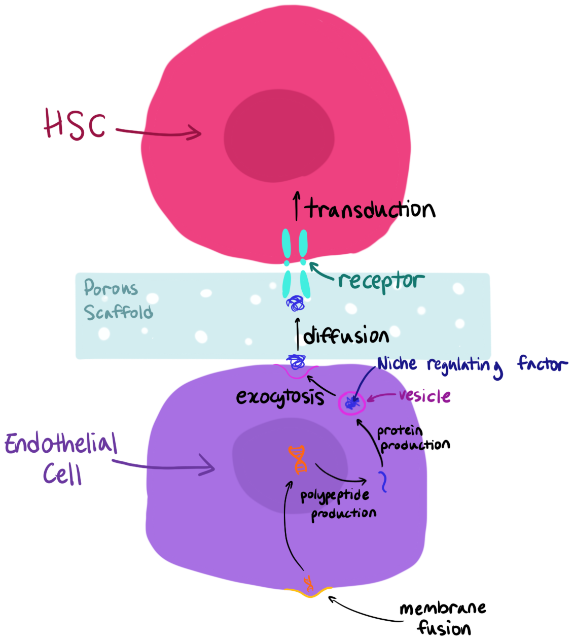 |
|---|
Table 3. Lists all of the proteins and signals, its purpose, and delivery type necessary for maintaining the HSCs in a quiescence state.
| Protein or Signal | Purpose | Delivery Type |
|---|---|---|
| SCF (Stem Cell Factor) | Essential cytokine for the maintenance of HSCs | Paracrine (Produced locally by CAR/LepR+ cells and sinusoidal endothelial cells) |
| CXCL12 | Essential for HSC maintenance and critical for maintaining stem cell quiescence | Paracrine (Produced locally by CAR/LepR+ cells and sinusoidal endothelial cells) |
| TPO (Thrombopoietin) | Essential for maintaining HSC numbers and their potential | Perfusion (Synthesized by hepatocytes in the liver and acts as a systemic hormone) |
| CXCL4 | Specifically regulates homeostatic HSC quiescence | Paracrine (Secreted by megakaryocytes within the bone marrow) |
| TGF-β | Required for maintaining HSC quiescence | Paracrine (Produced by non-myelinating Schwann cells in the niche) |
Safety
Main idea
Safety is an important consideration at every stage of this proposed design and encompasses ethical standards, laboratory practice, and the responsible disposal of materials.
Ethical considerations
The reprogramming iPSC approach utilized in this design raises two ethical considerations. First, collecting somatic cells from a patient requires consent. All donors must be fully informed of the intended use of their cells, and samples must be handled in accordance with the IRB guidelines. Second, iPSC research does avoid the ethical concerns usually associated with embryonic stem cells, but the use of c-Myc as a reprogramming factor does introduce theoretical oncogenic risk, as c-Myc is a proto-oncogene. To mitigate this, it may be possible to use l-Myc as a substitute, and this should be evaluated. Additionally, the reach of this research is limited to therapeutic applications and does not extend to germline modification.
Ethical clearance
To ensure that the experiment remains ethical with deriving and using iPSCs, researchers must undergo an NIH investigation and provide documents along with an additional IRB protocol number to then have IRB Approval. Not only that, but because this experiment utilizes iPSCs in vitro, all iPSCs must be reviewed and approved by the Institutional Biosafety Committee. There is also a form that must be completed along with restrictions on the use of iPSCs. One restriction states that human iPSC should not be introduced to non-human primate blastocyst (National Institutes of Health: Office of Intramural Research, n.d.). Overall, it is very important that researchers get the ethical clearance necessary to treat and use derived iPSCs with respect.
Laboratory safety
All mRNA synthesis and LNP formulation procedures must be conducted within a certified biosafety restriction to prevent the contamination of RNase and to protect researchers from exposure. Full PPE is required at all times during LNP preparation, as ionizable lipids can be cytotoxic at elevated concentrations. Because this design uses mRNA, a non-viral vector, there is no risk of generation of replicated particles, which reduces the biosafety burden overall (Hardee et al., 2017). All cell cultures must be performed under BSL-2 conditions, given the use of human-derived cells.
Disposal
All biological waste must be autoclaved pre-disposal. Any materials that have come in contact with human-derived cells must be treated as biohazardous and disposed of accordingly. Chemical waste generated during the LNP formulation must be collected and removed by a licensed hazardous materials contractor. (Environmental Protection Agency, n.d.) Finally, under no circumstances will biological or chemical waste enter the standard waste stream.
Discussions
Using OoC models to culture HSCs in order to proliferate them in vitro has many important applications in biology. It has the potential to change how we approach transplantation procedures for diseases such as leukemia, potentially allowing patients a less painful and far more successful recovery, as the use of their own somatic cells eliminates, or at least greatly diminishes, the chances of an acute immune response to the newly transplanted cells.
One matter that must be addressed is the use of HUVECs in the proposed experiment. HUVECs are cells found in the umbilical vein of an umbilical cord, which can only be derived from newborns, not adults. Therefore, they may not fully reflect the behavior of cells derived from patients as they are a differing cell type with differing properties. They were the chosen cell type due to their accessibility and because most research on endothelial cells utilized HUVECs as their main cell type. This means that we will be able to compare our results with other research to better understand the results. Such variations, however, can cause some fundamental changes to how the cells react in the experiment. HUVECs generally adhere to scaffolding due to their characteristic of responding well to lab coatings (Sgarioto et al., 2012). Other types of endothelial cells tend to have trouble adhering to surfaces unless such environments are encoded specifically to mimic in vivo settings due to the diversity between each type (Chi et al., 2003). Due to these differences, the procedure to adhere to the cells may change depending on the cell type. Additionally, because HUVECs are macrovascular while other endothelial cells (such as those from bone marrows) are microvascular, they experience high-shear stress and thus require a significantly higher laminar flow (Garcia-Polite et al., 2016). While these differences may be significant, the main goal for this experiment is to see if mRNA LNPs can help HSCs maintain “stemness”, therefore, these differences may be temporarily undermined and looked into later into development.
Some other potential issues to address include unsupervised cell signaling from the feeder cells to the HSCs and the complexity of the system which may make it hard to identify the root of problems. In the event that these issues arise, they may be addressed by changing certain aspects of the experiment, such as the cell types, cell environment, protein factors, etc. in order to work towards achieving the desired results.
Next steps
This experiment will ideally allow researchers to culture HSCs in vitro, enabling controlled proliferation and retention of a stem-like state. The next steps will include further refining the model to more accurately replicate the niche, increase HSC proliferation, and ensure cost-effectiveness.
Refining the model will include making small changes to specific components of the device in order to test whether the changes resulted in the creation of a more accurate niche environment. One of the factors that will be changed is the laminar flow conditions. The microfluidic niche flow conditions will be modified to ensure the device can replicate the shear stress these cells are exposed to in their natural niche environment. The flow rate, direction, and pressure can be modified as needed to ensure maximum HSC proliferation. The composition of the solutions being used in the channels can also be experimented with in order to improve the fidelity of the niche and ensure cost-effectiveness.
Another avenue that might be explored to improve cost-efficacy is experimenting with the combination and concentration of the factors used to mimic paracrine signaling. The proposed model uses the “gold standard” which increases the true positive rate when determining the presence of HSCs. This is made possible by testing for multiple HSC markers (CD34+, EPCR+, CD49f+) and transfecting the feeder layer of cells with multiple proteins (SCFs, CXCL-12) encoded for by LNP encapsulated mRNA. This increases the chances of culturing and identifying true HSCs from other cell types that may be present, however, it also increases the overall cost, which can lead to challenges with accessibility of this technology.
Lastly, the most pressing issue to address following the experiment is determining how to obtain the cells from the OoC model and identifying the optimal transplantation method. It is imperative that the method for removal and use is precise and intentional since the goal is to have as many HSCs as possible to use for transplantation. Experimenting with varying protein factors, delivered through mRNA LNPs, to achieve higher success rates in iPSC proliferation and differentiation represents another promising avenue for future research.
Author contributions
TS worked on the Abstract, Next steps, and Device level sections. TS contributed to the Systems level and Parts level. KC worked on the Background, Parts, and Systems level. SB worked on the safety level and contributed to the Parts level. All images were drawn by KC, however, TS contributed to Figure 3.
Acknowledgements
We would like to sincerely thank Peter Horanyi for taking the time to meet with us, for offering his insights, and for sharing his knowledge on this topic. We are also deeply grateful to Rachel Neurath for encouraging us to pursue this opportunity and for providing us with the knowledge, resources, and support to help bring this project to life.
References
A human platelet receptor protein microarray identifies FcepsilonR1alpha as an activating PEAR1 ligand. Sun Y, Vandenbriele C, Kauskot A, Verhamme P, Hoylaerts MF, Wright GJ. Mol Cell Proteomics. 2015 Feb 23. pii: mcp.M114.046946. 10.1074/mcp.M114.046946 PubMed 25713122
Aguirre, M., Escobar, M., Forero Amézquita, S., Cubillos, D., Rincón, C., Vanegas, P., Tarazona,
M. P., Atuesta Escobar, S., Blanco, J. C., & Celis, L. G. (2023). Application of the Yamanaka Transcription Factors Oct4, Sox2, Klf4, and c-Myc from the Laboratory to the Clinic. Genes, 14(9), 1697. https://doi.org/10.3390/genes14091697
American Type Culture Collection. (n.d.). Primary Umbilical Vein Endothelial Cells; Normal, Human, Pooled (HUVEC). ATCC. https://www.atcc.org/products/pcs-100-013
Asbrock, N., Chen, C., & Chu, V. (n.d.). HumanKine® Thermostable bFGF. sigmaaldrich. https://www.sigmaaldrich.com/US/en/technical-documents/technical-article/cell-culture-and-cell-culture-analysis/stem-cell-culture/thermostable-bfgf
Chi, J.-T., Chang, H. Y., Haraldsen, G., Jahnsen, F. L., Troyanskaya, O. G., Chang, D. S., Wang, Z., Rockson, S. G., Rijn, M. v. d., Botstein, D., & Brown, P. O. (2003, September 08). Endothelial cell diversity revealed by global expression profiling. pnas. https://www.pnas.org/doi/10.1073/pnas.1434429100
Chou, D. B., Frismantas, V., Milton, Y., David, R., Pop-Damkov, P., Ferguson, D., MacDonald, A., Bölükbaşı, Ö. V., Joyce, C. E., Moreira Teixeira, L. S., Rech, A., Jiang, A., Calamari, E., Jalili-Firoozinezhad, S., Furlong, B. A., O'Sullivan, L. R., Ng, C. F., Choe, Y., Marquez, S., … Ingber, D. E. (2020, February 12). On-chip recapitulation of clinical bone marrow toxicities and patient-specific pathophysiology. DOI. https://doi.org/10.1038/s41551-019-0495-z
Dissection of vertebrate hematopoiesis using zebrafish thrombopoietin. Svoboda O, Stachura DL, Machonova O, Pajer P, Brynda J, Zon LI, Traver D, Bartunek P. Blood. 2014 Jul 10;124(2):220-8. doi: 10.1182/blood-2014-03-564682. Epub 2014 May 28. 10.1182/blood-2014-03-564682 PubMed 24869937
Emulate. (2018, November). Bone Marrow-Chip. Emulatebio. https://emulatebio.com/bone-marrow-chip/
Environmental Protection Agency. (n.d.). Incident Waste Decision Support Tool (I-WASTE DST). epa. https://iwaste.epa.gov/guidance/natural-disaster/fact-sheets/types-of-waste?id=biohazard-waste
Garcia-Polite, F., Martorell, J., Del Rey-Puech, P., Melgar-Lesmes, P., O'Brien, C. C., Roquer, J., Ois, A., Principe, A., Edelman, E. R., & Balcells, M. (2016, October 04). Pulsatility and high shear stress deteriorate barrier phenotype in brain microvascular endothelium. journals.sagepub. https://doi.org/10.1177/0271678X16672482
Hardee, C. L., Arévalo-Soliz, L. M., Hornstein, B. D., & Zechiedrich, L. (2017). Advances in
Non-Viral DNA Vectors for Gene Therapy. Genes, 8(2), 65.
https://doi.org/10.3390/genes8020065
Incident Waste Decision Support Tool (I-WASTE DST). (n.d.). Iwaste.epa.gov.
https://iwaste.epa.gov/guidance/natural-disaster/fact-sheets/types-of-waste?id=biohazard-waste
Khaddour, K., Hana, C. K., & Mewawalla, P. (2023, May 6). Hematopoietic Stem Cell
Transplantation (Bone Marrow Transplant). PubMed; StatPearls Publishing.
https://www.ncbi.nlm.nih.gov/books/NBK536951/
Kotini, A. G., Carcamo, S., Cruz-Rodriguez, N., Olszewska, M., Wang, T., Demircioglu, D., Chang, C.-J., Bernard, E., Chao, M. P., Majeti, R., Luo, H., Kharas, M. G., Hasson, D., & Blood Cancer Discovery. (2023, July 05). Patient-Derived iPSCs Faithfully Represent the Genetic Diversity and Cellular Architecture of Human Acute Myeloid Leukemia Open Access. aacrjournals. https://doi.org/10.1158/2643-3230.BCD-22-0167
Leo, L., Papp, T. E., Chen, C.-C., Pham, J., Jeong, S., Halilovic, F., Thompson, J., Akyianu, A., Zeng, J., Rompolas, P., Marcos-Contreras, O. A., & Parhiz, H. (2026, June 09). Targeted LNP-mediated delivery of VE-cadherin mRNA reduces vascular hyperpermeability in vivo. ashpublications. https://ashpublications.org/bloodadvances/article/10/11/3820/567184/Targeted-LNP-mediated-delivery-of-VE-cadherin-mRNA
Li, Y., Shen, Z., Shelat, H., & Geng, Y.-J. (2013). Reprogramming somatic cells to pluripotency:
A fresh look at Yamanaka’s model. Cell Cycle, 12(23), 3594–3598. https://doi.org/10.4161/cc.26952
Lim, W. F., Inoue-Yokoo, T., Tan, K. S., Lai, M. I., & Sugiyama, D. (2013, June 18). Hematopoietic cell differentiation from embryonic and induced pluripotent stem cells. stemcellres. http://stemcellres.com/content/4/3/71
Liu, G. W., Guzman, E. B., Menon, N., & Langer, R. S. (2023, February 03). Lipid Nanoparticles for Nucleic Acid Delivery to Endothelial Cells. nih. https://pmc.ncbi.nlm.nih.gov/articles/PMC9897626/
Liu, Y., Huang, Y., He, G., Guo, C., Dong, J., & Wu, L. (2024). Development of mRNA Lipid
Nanoparticles: Targeting and Therapeutic Aspects. International Journal of Molecular
Sciences, 25(18), 10166. https://doi.org/10.3390/ijms251810166
Mapping Interaction Sites on Human Chemokine Receptors by Deep Mutational Scanning.
Heredia JD, Park J, Brubaker RJ, Szymanski SK, Gill KS, Procko E. J Immunol. 2018
Apr 20. pii: jimmunol.1800343. doi: 10.4049/jimmunol.1800343. 10.4049/jimmunol.1800343 PubMed 29678950
Mayle, A., Luo, M., Jeong, M., & Goodell, M. A. (2012, June 16). Flow cytometry analysis of murine hematopoietic stem cells. wiley. https://doi.org/10.1002/cyto.a.22093
McKenzie, R. E., Minnell, J. J., Ganley, M., Painter, G. F., & Draper, S. L. (2023, September). mRNA Synthesis and Encapsulation in Ionizable Lipid Nanoparticles. nih. https://pubmed.ncbi.nlm.nih.gov/37747354/
Narayan, S., Bryant, G., Shah, S., Berrozpe, G., & Ptashne, M. (2017, October 19). OCT4 and SOX2 work as transcriptional activators in reprogramming human fibroblasts. nih. https://pmc.ncbi.nlm.nih.gov/articles/PMC5648000/
National Institutes of Health: Office of Intramural Research. (n.d.). Induced Pluripotent Stem Cells (iPSCs) Use in the Intramural Research Program. nih. https://oir.nih.gov/sourcebook/ethical-conduct/special-research-considerations/human-stem-cell-use/induced-pluripotent-stem-cells-ipscs-use-intramural-research-program
Omatsu, Y. (2023, February 21). nih. Cellular niches for hematopoietic stem cells in bone marrow under normal and malignant conditions. https://pmc.ncbi.nlm.nih.gov/articles/PMC9942337/
Park, M., & Seo, J. J. (2012, October 02). Role of HLA in Hematopoietic Stem Cell Transplantation. Wiley Online Library. https://doi.org/10.1155/2012/680841
Primate immunodeficiency virus proteins Vpx and Vpr counteract transcriptional repression of proviruses by the HUSH complex. Yurkovetskiy L, Guney MH, Kim K, Goh SL, McCauley S, Dauphin A, Diehl WE, Luban J. Nat Microbiol. 2018 Dec;3(12):1354-1361. doi: 10.1038/s41564-018-0256-x. Epub 2018 Oct 8. 10.1038/s41564-018-0256-x PubMed 30297740
R&D Systems. (2026). Recombinant Human BMP-4 Protein (Cat# 314-BP). Minneapolis, MN.
Retrieved from https://www.bio-techne.com/p/recombinant-human-bmp-4-protein_314-bp
R&D Systems. (2026). Recombinant Human c-Myc Protein (Cat# 10563-MC). Minneapolis, MN.
Retrieved from
https://www.bio-techne.com/p/recombinant-human-c-myc-protein_10563-mc
R&D Systems. (2026). Recombinant Human Flt-3 Ligand Protein (Cat# 308-FK). Minneapolis,
MN. Retrieved from
https://www.bio-techne.com/p/recombinant-human-flt-3-ligand-protein_308-fk
R&D Systems. (2026). Recombinant Human IL-3 Protein (Cat# 203-IL). Minneapolis, MN.
Retrieved from https://www.bio-techne.com/p/recombinant-human-il-3-protein_203-il
R&D Systems. (2026). Recombinant Human IL-6 Protein (Cat# 206-IL). Minneapolis, MN.
Retrieved from https://www.bio-techne.com/p/recombinant-human-il-6-protein_206-il
R&D Systems. (2026). Recombinant Human KLF4 Protein (Cat# 10243-K4). Minneapolis, MN.
Retrieved from https://www.bio-techne.com/p/recombinant-human-klf4-protein_10243-k4
R&D Systems. (2026). Recombinant Human Nidogen-1/Entactin Protein (Cat# 2570-ND).
Minneapolis, MN. Retrieved from
https://www.bio-techne.com/p/recombinant-human-nidogen-1-entactin-protein_2570-nd
R&D Systems. (2026). Recombinant Human OCT4/POU5F1 Protein (Cat# 9445-OD).
Minneapolis, MN. Retrieved from
https://www.technedirect.com/products/human-oct4-pou5f1-recombinant-protein-9445-od
R&D Systems. (2026). Recombinant Human SCF Protein (Cat# 255-SC). Minneapolis, MN.
Retrieved from
https://www.rndsystems.com/products/recombinant-human-scf-protein_255-sc
R&D Systems. (2026). Recombinant Human Thrombopoietin/THPO Protein (Cat# 288-TP).
Minneapolis, MN. Retrieved from
https://www.bio-techne.com/p/recombinant-human-thrombopoietin-thpo-protein_288-tp
Radzisheuskaya, A., & Silva, J. C. R. (2014). Do all roads lead to Oct4? The emerging concepts
of induced pluripotency. Trends in Cell Biology, 24(5), 275–284. https://doi.org/10.1016/j.tcb.2013.11.010
Rix, B., Maduro, A. H., Bridge, K. S., Grey, W., & Frontiers in Physiology. (2022, September 30). Markers for human haematopoietic stem cells: The disconnect between an identification marker and its function. PMC. https://pmc.ncbi.nlm.nih.gov/articles/PMC9564379/
Sgarioto, M., Vigneron, P., Patterson, J., Malherbe, F., Nagel, M.-D., & Egles, C. (2012, August 21). Collagen type I together with fibronectin provide a better support for endothelialization. Comptes-Rendus. https://comptes-rendus.academie-sciences.fr/biologies/articles/en/10.1016/j.crvi.2012.07.003/#:~:text=Nagel%3B%20Christophe%20Egles.-,Collagen%20type%20I%20together%20with%20fibronectin%20provide%20a%20better%20support,j.crvi.2012.07.003
Sharma, M. B., Limaye, L. S., & Kale, V. P. (2012, May). Mimicking the functional hematopoietic stem cell niche in vitro: recapitulation of marrow physiology by hydrogel-based three-dimensional cultures of mesenchymal stromal cells. nih. https://pmc.ncbi.nlm.nih.gov/articles/PMC3342965/
Sigma-Aldrich. (2026). Collagen from human placenta, Bornstein and Sage Type IV (Cat#
C7521). St. Louis, MO. Retrieved from
https://www.sigmaaldrich.com/US/en/product/sigma/c7521
Sigma-Aldrich. (2026). Fibronectin from human plasma (Cat# F2006). St. Louis, MO. Retrieved
from https://www.sigmaaldrich.com/US/en/product/sigma/f2006
Sigma-Aldrich. (2026). Heparan Sulfate Proteoglycan from basement membrane of Engelbreth-Holm-Swarm mouse sarcoma (Cat# H4777). St. Louis, MO. Retrieved from https://www.sigmaaldrich.com/US/en/product/sigma/h4777
Sigma-Aldrich. (2026). Laminin from human fibroblasts, cell culture derived (Cat# L4544). St. Louis, MO. Retrieved from https://www.sigmaaldrich.com/US/en/product/sigma/l4544
Thermo Fisher Scientific. (2026). Gibco Collagen I, Rat Tail Protein (Cat# A1048301). Waltham, MA. Retrieved from https://www.thermofisher.com/order/catalog/product/A1048301
Thermo Fisher Scientific. (2026). Gibco Human FGF-basic (FGF-2/bFGF) (154 aa) Protein, PeproTech (Cat# 100-18B-50UG). Waltham, MA. Retrieved from https://www.thermofisher.com/proteins/product/Human-FGF-basic-FGF-2-bFGF-154-aa-Recombinant-Protein/100-18B-50UG
Thermo Fisher Scientific. (2026). Gibco Human Sox2 Recombinant Protein, PeproTech (Cat# 100-03-25UG). Waltham, MA. Retrieved from https://www.thermofisher.com/proteins/product/Human-Sox2-Recombinant-Protein/110-03-25UG
What are Organ-Chips? (2023, April 27). Emulate. Retrieved May 26, 2026, from https://emulatebio.com/an-introduction-to-organ-on-a-chip-technology/
Zebrafish Kit ligands cooperate with erythropoietin to promote erythroid cell expansion. Oltova J, Svoboda O, Machonova O, Svatonova P, Traver D, Kolar M, Bartunek P. Blood Adv. 2020 Dec 8;4(23):5915-5924. doi: 10.1182/bloodadvances.2020001700. 10.1182/bloodadvances.2020001700 PubMed 33259600