
Ibrahim B. Abumohsen, Luka Ascencio, Havish Bathina, Madeline Boram-Rella, Brayden Carty, Alyssa Gay, Isaac J. Pare, and Priyansh Shah, Athens Drive Magnet High School, Raleigh, North Carolina, United States of America
Reviewed on 9 May 2026; Accepted on 22 June 2026; Published on 26 October 2026
With help from the 2026 BioTreks Production Team.
Colorectal cancer (CRC) is the second most commonly diagnosed cancer, with an estimated 154,270 individuals diagnosed in 2025 alone, and 52,900 deaths in the US. Yet of all Americans, only 60% of the at-risk population will be tested. This hesitancy is in part due to current diagnostic methods (e.g., fecal immunochemical testing, colonoscopy, computed tomography colonography), which are either expensive, invasive, or require both specialized medical equipment and operational expertise. Live biologic-based diagnostic technologies show great promise for closing the diagnostic gap in CRC. In particular, a sensor capable of detecting genetic mutants that employs the chassis organism Acinetobacter baylyi (A. baylyi). This bacterium acquires DNA from its environment, such as the gastrointestinal tract. When combined with a modified CRISPR counterselection system, this organism can detect critical mutations in the kRAS oncogene with extraordinary specificity, capable of distinguishing DNA base pair alterations. Here, we developed a design for a novel diagnostic technology, which detects three of the most prevalent CRC-associated gene mutations (APC, TP53, and kRAS) while fixed within a calcium alginate hydrogel applied on a nondigestible pill. This results in the expression of an AmilCP reporter, allowing for a color change in the presence of genetic mutants. This biologic system aims to minimize the diagnostic gap of CRC by facilitating easy at-home detection and providing patient-interpretable results.
Keywords: Biosensor pill, CRISPR-Cas3 detection, colorectal cancer screening, Acinetobacter baylyi, cell-free DNA, cfDNA, CRISPR
Authors are listed in alphabetical order. Kaitlin M. Dailey mentored the group. Please direct all correspondence to mailto:kaitlin.dailey@uri.edu.



Background
Cancer is one of the most prevalent causes of death worldwide, accounting for 613,349 recorded deaths in the US alone during 2023 (Cancer Data and Statistics | Cancer, 2026). Colorectal cancer, or CRC, is the second most common cause of cancer death – largely due to the lack of early diagnosis (Colorectal Cancer Statistics, 2023, n.d.). During the early stages of CRC, it is very easily treatable, and can have up to a 91% 5-year survival rate when the cancer is still localized, which is why it is best for the cancer to be detected as soon as possible (Colorectal Cancer Survival Rates | Colorectal Cancer Prognosis, 2026). However, when the cancer spreads to other parts of the body in later stages, the 5-year survival rate drops to approximately 8% (Table 1). Unfortunately, only 1 in 3 cases are diagnosed during the initial stages (I and II), when the cancer is still localized (About Colorectal Cancer, n.d.). This is largely attributed to lack of patient compliance with recommended CRC screening processes. In fact, the CDC reports that approximately 33% of adults aged 50-64 have an outdated CRC screening (Use of Colorectal Cancer Screening Tests | Colorectal Cancer, 2025). This lack of regular screening is at least in part due to current CRC screening methods’ complexity and invasiveness. In 2005, a multi-institution surveyed 660 patients with open ended questions regarding CRC screening barriers (Devers et al., 2010). 74% responded that the most important barrier to CRC screening was fear or anxiety surrounding the process. This design aims to encourage patients to get screened by helping them evaluate their risk factor for acquiring this disease.
Table 1: Relative five year survival rate for CRC patients by stage (adapted from (O’Connell et al., 2004)).
| Stage | 5 Year Survival Rate |
|---|---|
| 1 | >90% |
| 2 | 82% |
| 3 | 60% |
| 4 | 8% |
Here, we describe a live pill (Figure 1) able to detect biomarkers of CRC, which is meant to provide a cost effective, user-friendly, at-home test meant to help patients analyze their risk factor. Although this device is not intended to replace the current clinical standard care, it will detect common signs of CRC or precancerous lesions – thereby providing prescreening, addressing both primary care provider case load and patient financial burdens. Based on whether these biomarkers are detected, the patient can make an informed decision about the need for further medical attention. A key feature of this pill is the ability to detect CRC and precancerous lesions during their early stages when the cancer is still localized and therefore easier to treat (e.g., polypectomy). CRC is a very treatable disease if detected and treated early enough, thus this novel intervention will directly have a positive patient impact.
| Figure 1: Showcasing the structure and layers of the pill. | 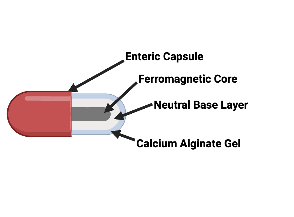 |
|---|
Briefly, to detect CRC, the pill is ingested orally and subsequently moves through the digestive tract into the large intestine. During transport, the pill repeatedly incorporates exogenous DNA from its environment (the colorectal organs) and detects sequence variants indicative of CRC. The system uses A. baylyi as a bacterial chassis, genetically engineered to serve as a living biosensor. A. baylyi has demonstrated a natural capacity to acquire DNA from its environment. When this ability is harnessed and connected to an amilCP reporter, it can thus be engineered to express amilCP upon exposure to certain genetic mutations that often serve as CRC biomarkers. When these biomarkers are detected, the pill will turn a dark blue color. After defecation, the user will be able to determine if the pill has detected indicators of CRC through this visible color change. The biomarkers chosen (APC, TP53, and kRAS) are all genes that are typically mutated in the early stages of CRC. APC and TP53 are both tumor suppressor genes that, when mutated, fail to stop the uncontrolled proliferation caused by kRAS. These genetic mutations are present in over 90% of CRC cases, making them a valuable biomarker (Su et al., 2024). This design aims to overcome the challenges faced by at-risk populations in screening for CRC and thus reduce CRC mortality rates.
Systems level
Here, a conceptual architecture for detecting colorectal cancer-associated DNA mutations and generating a visible colorimetric output is presented. This system combines the natural competency of A. baylyi to take up DNA with a CRISPR-Cas3-based counterselection system to reduce potential false positives as well as with a genetic ‘landing pad’ within the cell to detect the presence of the mutated sequence via homologous recombination (Figure 2). We selected the Type I-F Cas3 nuclease over canonical Cas9 for its dual advantages in delivery and mechanism. Because Cas3 is endogenous to the host cell, it bypasses the requirement for complex heterologous delivery systems. Furthermore, unlike the localized double-strand breaks characteristic of Cas9 or Cas12, Cas3 drives processive DNA degradation. Although less suited for high-precision editing, this robust degradation is ideal for our application, as it eliminates wild-type genomic DNA and prevents unintended homologous recombination, effectively lowering false-positive rates. A second, transcriptionally controlled reporter sequence then colorimetrically indicates the presence of a mutation. Together, these components enable a non-invasive, macroscopic indication of disease-associated mutations within the gastrointestinal tract.
| Figure 2: Schematic overview of mechanical processes behind the proposed detection method. | 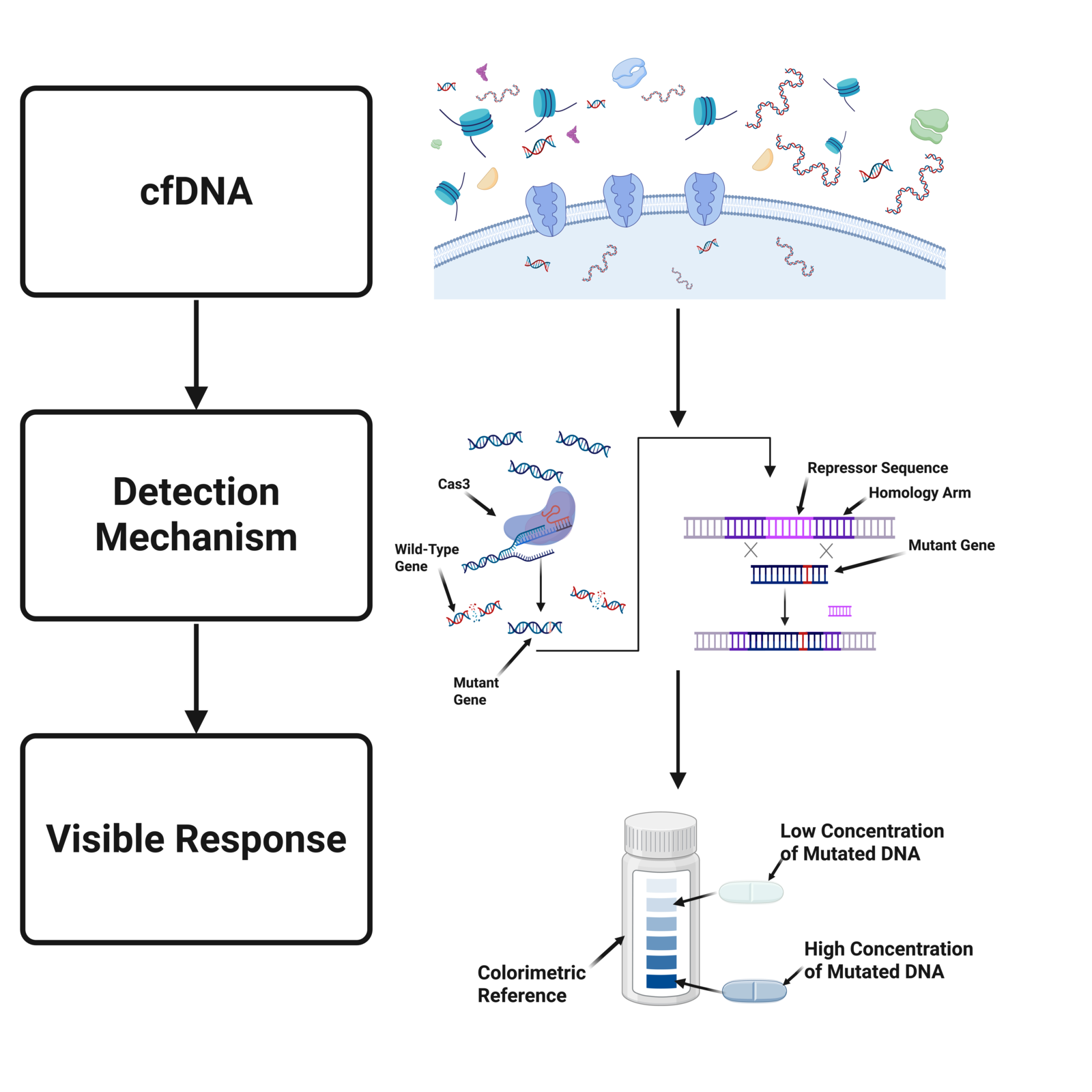 |
|---|
Following oral ingestion, the capsule passes through the stomach and into the intestinal tract, where environmental conditions enable the enteric coating to dissolve. CfDNA fragments of colorectal cancer including those associated mutations are shed from intestinal adenomas and can diffuse into the pill to interact with the engineered A. baylyi sensors. Wild-type target gene sequences are eliminated via CRISPR-Cas3 counterselection mechanism, and only mutated genes are allowed to interact with the detection system. Upon target sequence recognition, the detection module activates gene expression pathways that drive pigment production. This ultimately results in a visible color change within the pill, serving as the system’s output signal.
DNA uptake
The intake of nucleic acid sequences represents the first step of the design. A. baylyi is naturally competent, meaning it can take up DNA from its environment. When this capacity is paired with the diffusion of cell-free DNA (cfDNA) shed from the intestinal wall, DNA is taken up as short, digested 100–200 bp double-stranded sequences (Matov, 2024). Once the DNA is inside the cell, it can proceed to the next step. However, this poses a challenge: the availability of high-quality DNA impacts sensor effectiveness. Given the detection system’s reliance on homologous recombination, insufficient long homologous regions (e.g., from poor fragment length or DNA integrity) hinder the ability to verify gene identity and thus directly contribute to avoidance of both false negatives and false positives.
Selectivity via CRISPR counterselection
Following A. baylyi DNA uptake, a degradation step is utilized. Here, a CRISPR counter selection system digests wild-type genes to prevent false detection of wild-type sequences. Guide RNAs (gRNAs) are designed to specifically target and initiate digestion of the wild-type gene of interest and to place the targeted mutant gene site of interest in direct proximity to the Protospacer Adjacent Motif (PAM) site – enabling highly specific Cas3 digestion. When A. baylyi takes up a mutated version of the gene, Cas3 is unable to degrade it due to an inherent lack of sequence homology, thereby facilitating detection. By leveraging a CRISPR/Cas3 system to degrade wild-type DNA collected from the gastrointestinal tract, these sequences are prevented from undergoing homologous recombination, thereby enhancing sequence specificity and limiting potential false positives. By selecting Cas3 as our degradation pathway over the more common Cas9 or Cas12, we are able to take advantage of existing CRISPR systems within the cell. Also, Cas3 digests DNA not through a double-strand break, but by shredding DNA, whereby the Cas complex reels up one of the DNA strands, and Cas3 digests the other strand into short 10-nucleotide segments (Künne et al., 2016). The size of these fragments entirely inhibits the opportunity for homologous recombination, unlike the double strand breaks of Cas9 which leave long sections of homologous DNA.
Recombination based detection
Once the biomarker DNA sequence ( kRAS, APC, TP53) has passed through the counterselection system, it interacts with prepared genetic landing pads at a neutral cellular loci. These sites consist of homology arms specific to the target biomarker gene, allowing for recombination-sensitive control of downstream expression via a TetR repressor. The TetR repressor suppresses the bacterial reporter site. When the mutated gene is present, it can undergo homologous recombination with the corresponding homology arms subsequently resulting in the repressor sequence removal.
Reporter activation
The reporter sequence is activated upon removal of the repressor sequence. The reporter is located at a second neutral locus in the genome consisting of a PLtetO-1 promoter – notably repressed by TetR in baseline conditions. Upon repressor sequence removal from a successful recombination event, the promoter becomes transcriptionally active leading to the production of a visible pigment.
Delivery
The engineered A. baylyi are encapsulated within an alginate matrix, which serves as immobilization while still facilitating exposure to cfDNA fragments via diffusion. When the detection system is activated and the reporter is expressed, a visible color change can be detected on the surface of the capsule. The alginate gel is covered in an enteric coating selected to withstand the gastric environment and dissolve in the higher pH environment of the small intestine, where the target nucleic acids are more likely to be encountered.
Device level
The physical architecture of the pill is engineered to facilitate a ‘lab-on-a-chip’ – effectively integrating complex diagnostic workflows into a miniaturized, autonomous platform capable of operating within the complex environment of the human gastrointestinal tract. The primary engineering challenge for the device is therefore maintaining high permeability for environmental DNA, while ensuring robust biocontainment of the genetically modified A. baylyi.
Chassis bacteria
In order for the user to identify a positive result and thus an indication of predisposition for CRC disease, we selected the chassis bacteria A. baylyi engineered to emit an indigo color after exposure to a biomarker (e.g., genetic mutation of choice). A. baylyi was selected because of its natural competency, meaning it is able to take in DNA from its environment without any artificial catalysis or intervention. Here, we have genetically engineered A. baylyi to act as a living biosensor connected to an amilCP reporter and a detection mechanism. This schematic facilitates straightforward expression of amilCP when biomarkers are present.
The immobilization matrix
A calcium alginate hydrogel is utilized as the primary immobilization scaffold. Alginate, a polysaccharide derived from brown algae, undergoes rapid gelation when exposed to divalent cations like Ca2+. This matrix creates a fibrous network with pores ranging from 5 to 100 nm. Because the DNA fragments shed by colorectal tumors are roughly 50-60 nm in length (typically 150-200 base pairs), the hydrogel remains permeable to the target biomarkers while physically trapping A. baylyi, which are 1 to 1.5 µm in length. This size-exclusion principle ensures that bacteria cannot enter the gut microbiome, minimizing the risk of horizontal gene transfer.
Protective coatings and pH stability
To address the extreme stomach acidity (pH=1.5-3.5), which can lead to alginate shrinkage and degradation, the device incorporates a cellulose-acetate-based coating. Cellulose-acetate is a biocompatible, semipermeable plastic that is able to survive the acidity of the stomach and pass through the neutral pH of the intestines (Vidal-Romero et al., 2021). Due to its semi-permeability, this coating allows nutrient diffusion, thus keeping the chassis bacteria alive while simultaneously facilitating the diffusion of cell-free DNA. This layer is crucial for maintaining the viability of A. baylyi while the pill travels towards the more neutral pH of the large intestine, where tumor DNA largely accumulates.
Surface area optimization
To maximize the probability of a ‘capture event’ between a bacterium and a rare mutant DNA fragment, this device is designed with a textured surface. As an alternative to a smooth coating, we propose using alginate beads packed into a semi-permeable capsule. This significantly increases the effective surface area for diffusion, ensuring that the limited transit time through the colon is sufficient for the bacteria to take up the DNA and trigger the CRISPR-Cas3 counterselection mechanism.
Parts level
Early and accurate detection of tumor-associated mutations at the base-pair level is critical for improving patient outcomes. However, distinguishing between wild-type and mutated sequences remains a significant challenge because of the inherent high level of similarity – most mutations consist of single letter polymorphisms (SLP) in critical genes (Giglia & Chu, 2016). To address this issue, the color-change mechanism of the pill employs a modular design consisting of distinct elimination, detection, and reporting components. By coordinating these elements, the system converts molecular recognition events into a detectable signal. To better understand the rationale behind these design choices and evaluate system performance, an in-depth analysis of each genetic component is provided below. A schematic representation of the proposed system is included in Figure 3.
| Figure 3: Schematic representation of genetic constructs for proposed sensor bacteria. | 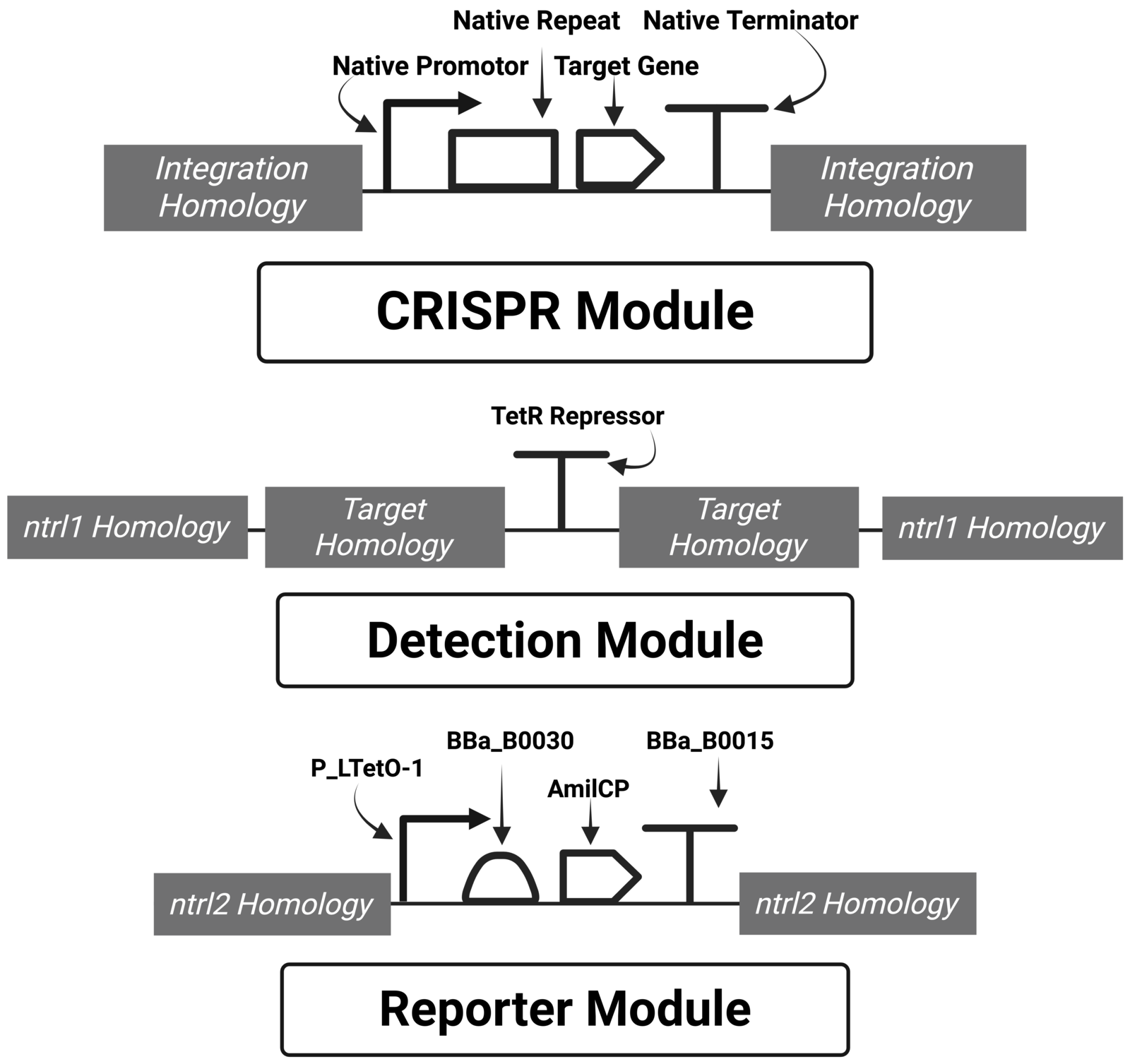 |
|---|
CRISPR counterselection
CRISPR-based elimination is a critical step in the system, as it reduces false positives by selectively degrading wild-type sequences via Cas3 activity. Once degraded, these wild-type sequences are unable to interact with downstream components. To implement this system, genetic constructs containing the CRISPR elements were assembled and transformed into A. baylyi. Successful integration would be verified using colony PCR (Cooper et al., 2023), ensuring that no selection sequences were incorporated that could interfere with pigment production.
Integration homology arms
To integrate the genetic construct into the A.baylyi genome, specialized homology arms were designed to ensure that genomic insertion did not disrupt cellular viability or essential functions. These arms (~1000 bp) were selected to target non-essential genomic regions. The CRISPR construct replaced segments of genes ACIAD_2185, ACIAD_2186, and ACIAD_2187 (Cooper et al., 2023).
Native promoter
A. baylyi already encodes the necessary machinery for a Type I-F CRISPR system, a native promoter is used to drive expression of the engineered CRISPR RNA (crRNA). This approach minimizes discrepancies between endogenous and introduced CRISPR components, avoiding issues associated with under- or overtranscription. Specifically, the 684 bp region upstream of the native CRISPR locus is then utilized and has been shown to function effectively (Cooper et al., 2023).
Native repeat
A native repeat sequence is incorporated to ensure proper crRNA processing by host machinery. Upon transcription, these repeats form characteristic hairpin loop structures required for recognition and cleavage by processing enzymes such as Cas6f to prepare the crRNA for use by Cas3.
crRNA spacer target
The CRISPR spacer sequence is derived directly from the target genes that are known biomarkers for CRC (Cooper et al., 2023). These 33 bp sequences were designed to place mutation hotspots (e.g., KRAS G12D) in close proximity to the protospacer-adjacent motif (PAM), thereby maximizing targeting specificity and degradation efficiency.
Detection module
Following elimination of wild-type sequences, mutated sequences (which are biomarkers of CRC) proceed to the detection stage. Using sequence-specific homology arms, the system enables recombination-mediated removal of an embedded repressor sequence. Loss of this repressor allows activation of a downstream promoter, which in turn drives expression of a reporter gene encoding a chromoprotein.
Integration arms into neutral site 1 (ntrl1)
The detection module was integrated into a known neutral genomic locus (ntrl1) in A. baylyi, where insertions do not compromise cellular viability. Homology arms (~1000 bp each) flanking the construct facilitated recombination into this site. The detection construct would replace gene ACIAD_2826 (Cooper et al., 2023).
Target gene homology arms
To ensure specificity, 1000 bp homology arms corresponding to regions flanking the target gene (e.g., KRAS) were incorporated into the detection construct. These sequences guide homologous recombination events, reducing the likelihood of false positives and negatives by restricting recombination to the intended genomic target.
TetR repressor
A TetR repressor cassette will be positioned between the homology arms to regulate reporter expression. TetR binds its operator with high affinity (Kd ≈ 10⁻¹²) in the absence of anhydrotetracycline (aTc), effectively repressing transcription (Ramos et al., 2005). However, experimental observations (Cooper et al., 2023) indicated that wild-type TetR was overly repressive, leading to false negatives. A modified TetR variant with reduced binding affinity, featuring an 8-amino-acid C-terminal extension and a W75R mutation, was isolated and experimentally shown to function (Cooper et al., 2023).
Reporting module
Following successful recombination and removal of the TetR repressor, intracellular TetR levels rapidly decrease, permitting activation of the reporter construct. The system is optimized to maximize chromoprotein production by combining a strong inducible promoter, an efficient ribosome binding site (RBS), and a robust transcriptional terminator.
Integration into neutral site 2 (ntrl2)
A second set of homology arms was chosen to integrate the reporter construct into another neutral genomic locus (ntrl2), ensuring no detrimental effects on cell viability. This integration replaced regions of genes ACIAD_1076 and ACIAD_1077. (Cooper et al., 2023).
P_LTetO-1 promoter
The P_LTetO-1 promoter, a well-characterized TetR-repressible promoter, was selected for its strong transcriptional activity and compatibility with the modified TetR variant. Its performance in prokaryotic systems has been extensively characterized (Lutz & Bujard, 1997)
Ribosome binding site (RBS) (BBa_B0030)
While a strong promoter enhances transcription, efficient translation requires a high-performance Ribosomal Binding Site (RBS). The BBa_B0030 RBS was selected due to its well-documented strength and reliability in promoting high levels of protein expression in E. coli and related systems.
AmilCP reporter
AmilCP, a blue-purple chromoprotein derived from Acropora millepora, was chosen as the reporter due to its distinct and easily distinguishable coloration. The gene was codon-optimized for expression in A. baylyi as A. millepora is a corals. Its blue color provides contrast against typical background pigmentation (e.g., brown, yellow, or red tones) found in the intestinal tract, thus facilitating visual detection. Furthermore, AmilCP serves as an excellent candidate for spectrophotometric quantification. Its distinct absorbance maximum (λMax) at 588 nm enables the straightforward assessment of translational efficiency.
Rho-independent terminator (BBa_B0015)
To ensure efficient termination of transcription and prevent read-through into downstream regions, the BBa_B0015 double terminator was chosen. This well-characterized rho-independent terminator enhances transcriptional efficiency and helps concentrate cellular resources on reporter production (Do, et al., 2025).
Safety
When constructing biological designs, certain design risks are required. These risks must be carefully considered and, through careful design, mitigated and, whenever possible, eliminated. The capsule in this case comes with some inherent risks as all transgenic organisms pose a potential environmental contamination risk. This particular organism and system carries additional risk associated with the ferromagnetic core. However the benefits of the ferromagnetic core, being that one will not have to dissect their feces, heavily outweighs any concern (Figure 4). This is because the ferromagnetic core will attract magnetic material meaning a magnet can be passed over any waste and the pill can easily be found for a quick color analysis. Here, through intentional and careful design, we detail plans to minimize these inherent risks. Although these risks do warrant acknowledgment the potential benefit far outweighs them by offering an opportunity to significantly increase the proportion of high risk groups which undergo regular screening, in particular in resource-constrained environments.
| Figure 4: Schematic of the pill retrieval system, showcasing the ease and cleanliness of using a magnet to find the pill. | 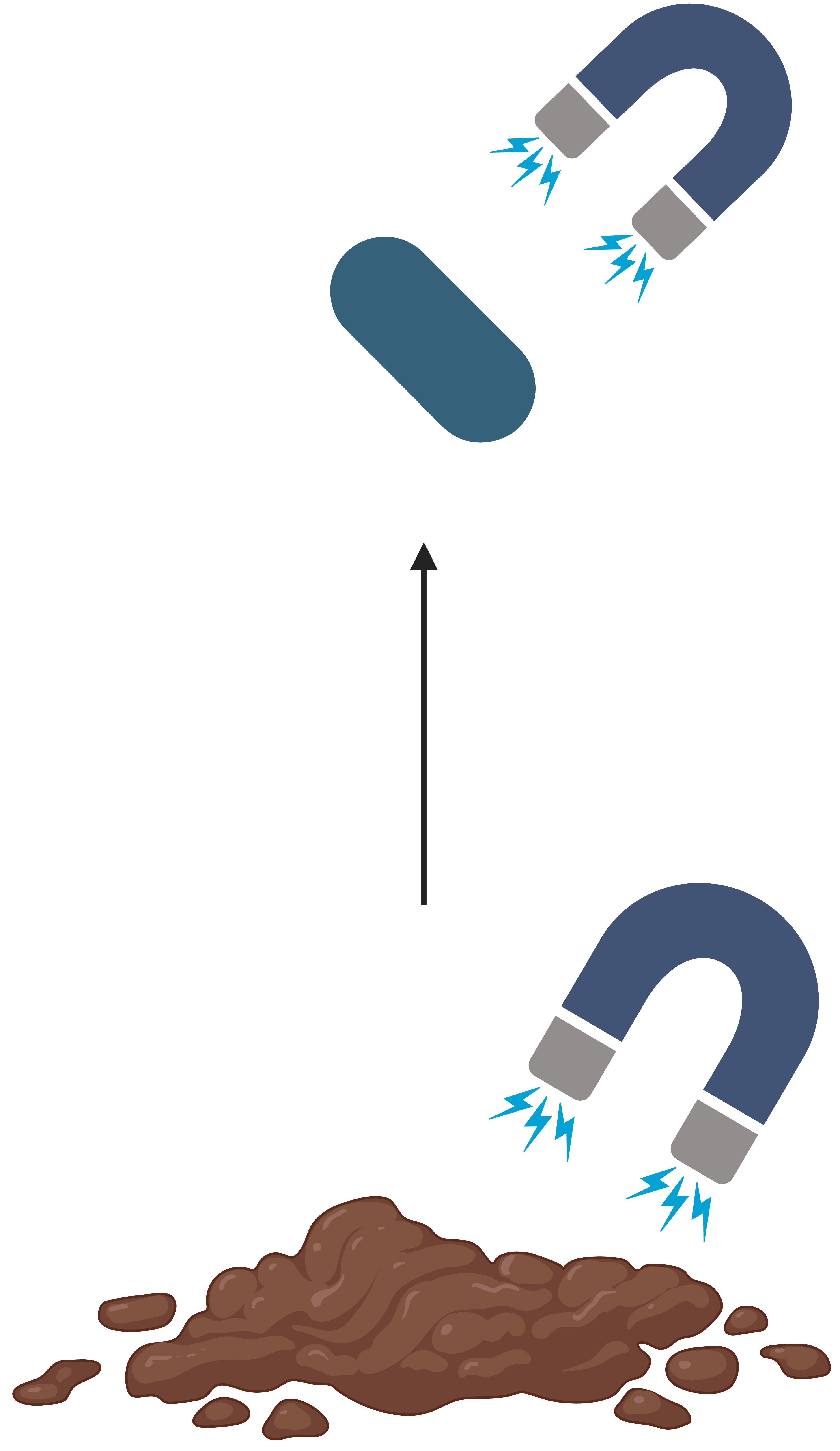 |
|---|
Potential risks associated with A. baylyi
As an Acinetobacter, A. baylyi are largely considered harmless environmental organisms found in soil and water (Santala & Santala, 2021). The genus does however contain a few opportunistic pathogens, most notably Acinetobacter baumannii, which is known for involvement in antibiotic-resistant infections. However, A. baylyi is considered a soil pathogen and has been proven to be largely safe in most circumstances (Santala & Santala, 2021). It is worth noting, in a 2008 study six highly immunocompromised patients were reported to have A. baylyi in their system (Chen et al., 2008). This was attributed to Actinobacteria’s general tendency to form biofilms within medical equipment and subsequently becoming an opportunistic pathogen. This intervention was designed with this context in mind to avoid infection by entrapping A. baylyi within an immobilizing alginate matrix. However, as a safety precaution this pill would not be clinically recommended for immunocompromised or high-risk patients.
Ferromagnetic concern: magnetic resonance imaging (MRI)
The minimal quality of the ferromagnetic material at the core of the pill, while small, could be sufficient to disrupt medical devices that depend on interactions with magnets or very high electromagnetic fields (e.g., MRI). In these situations, the amount of material inside the pill may pose an elevated risk of physical harm to the recipient as a result of rapidly accelerating (Eshed et al., 2010). As a result, it would be heavily contraindicated to have an MRI scan while the pill was still present in the GI tract. For additional safety, proximity to high electromagnetic fields would be against the suggested protocol with patients receiving thorough compliance counseling.
Allergen response
Ferromagnetic materials can elicit an immune response in individuals with metal-induced hypersensitivity (McKee & Fontenot, 2016). A condition similar to autoimmunity, metal-induced hypersensitivity occurs more frequently in immunocompromised people due to T-cell sensitization. In an attempt to avoid this, the core is encased in a variety of barrier materials such as polypropylene, chitosan, and finally a high density alginate, minimizing the majority of the risk of immune response (Küçük et al., 2025) while also ensuring the viability of the sensor bacteria.
Genetic environmental contamination
Genetic Contamination is commonly a high concern for any synthetic biological design. The risk associated with the transfer of antibiotic resistance genes, unnatural expression, virulence material, and biosynthesis pathways, giving rise to atypical phenotypic traits pose an inherent risk. A solution to these problems would be to implement biocontainment with a ‘kill’ switch via auxotrophy or timed expression construct (Chan et al., 2016). By neutralizing the bacteria naturally following activation of a specific genetic circuit for example triggered by an included activator subsequently limits the bacterial lifespan. Again, the use of an immobilization matrix aids in safety by ensuring the containment of any modified bacteria within the matrix where genetic information is less likely to migrate out from the cell. We further forgo the option of antibiotic resistance genes entirely, instead employing methods such as colony PCR we can screen for successful transformants without introducing antibiotic resistance genes which could also pose a threat to the production of the chromoprotein.
Discussions
The transition of synthetic biology from laboratory-bound systems to real-world medical diagnostics requires a fundamental shift in how we approach patient interaction and biomarker stability. By engineering a living genosensor capable of autonomous detection within the gut lumen, we address the critical ‘translational gap’ that currently hinders colorectal cancer screening efforts. This section evaluates the clinical rationale behind choosing a DNA-based detection model and examines how this technology serves as a low-barrier triage tool compared to traditional diagnostic standards.
Addressing the screening fear
The current diagnostic standard, colonoscopy, is highly accurate but suffers from low patient compliance due to its invasive nature and the requirement for bowel preparation. While fecal tests, such as Fecal Immunochemical Testing (FIT) or Cologuard, offer non-invasive alternatives, they often rely on detecting hemoglobin or broad metabolic changes that lead to false positives from diet or inflammation. Our device addresses this by targeting somatic mutations like (kRAS, TP53, and APC) which are definitive signatures of neoplastic growth and thus considered biomarkers for CRC. By providing an at-home, low-cost solution, we aim to identify high-risk individuals who would otherwise forgo screening-funneling them toward necessary clinical interventions. Mitigating operational and financial burdens on primary care infrastructure, while providing more accessible screening pathways that are lower cost and circumvents systemic barriers many uninsured and underinsured demographics when seeking screening.
Rationale for DNA-based sensing
We opted for a genosensor over protein-based biosensors for three main reasons. 1) Stability: Proteins are rapidly denatured and digested by the harsh enzymes of the GI tract. However, DNA is remarkably resilient and can be recovered from fecal matter in sufficient quantities for detection. 2) Specificity: Protein markers for CRC are often elevated in non-cancerous conditions. Particularly in chronic inflammation disorders that are poorly understood. In contrast, the CRISPR-Cas3 system we employ can be programmed to distinguish between a healthy gene and a single point mutation, offering a level of specificity that proteomic sensors have not yet matched. 3) Amplification: While proteins are static, meaning they can’t replicate, the uptake of a single mutated DNA fragment by A. baylyi triggers a biological cascade, homologous recombination followed by protein expression, essentially using the bacteria’s own machinery to amplify the signal into a visible color change.
Next steps
Several components of this design warrant further investigation during assembly. First it is necessary to determine the minimal amount of mutated DNA required for color change of the pill to occur, i.e., to establish this intervention’s sensitivity. Moreover, there is a need to consistently and reproducibly correlate pigment production with cfDNA concentration. Another important component will be the stability of the latter; hence, experiments establishing effectiveness of various methods for preserving the sensing mechanism integrity must occur to ensure stability during distribution and storage. In addition, future iterations could expand the capabilities of a singular capsule to detect multiple genes as an effective detection for several mutations simultaneously with varying reporter types and productions.
Quantification of signal thresholds
The most critical next step is determining the Minimum Detection Limit (MDL). We must establish how many copies of a mutated gene are required to trigger a color change perceptible to the human eye. We propose laboratory trials using synthetic DNA fragments simulating concentrations typically found in the gut lumen. If the AmilCP reporter expression is undetectable, we will explore the use of high-copy-number plasmids or stronger synthetic promoters to overexpress the chromoprotein once the mutation is detected. Based on prior studies utilizing analogous systems, the minimum detection limit for mutated DNA is anticipated to be approximately 3–5 pg/µL (Cooper et al., 2023). This concentration is expected to be sufficient to initiate successful recombination events and generate a discernible reporter signal.
Secretion integration
To improve the readability of the result, we plan to test the addition of a secretion peptide, like the HlyA secretion system, to the AmilCP sequence (Gentschev et al., 2002). By secreting the blue chromoprotein into the surrounding hydrogel matrix rather than keeping it inside the bacterial cytoplasm, we can create a more uniform and intense color change on the pill’s surface, reducing user error during interpretation.
Environmental resilience and lyophilization
For the device to be commercially viable, it must survive the ‘cold chain’ or be shelf-stable at room temperature. Future research will focus on the lyophilization (freeze-drying) of the bacteria-laden hydrogel beads. We will test various cryoprotectants to ensure that the bacteria can be rehydrated and become metabolically active immediately upon ingestion.
Multi-channel logic gates
Finally, we aim to evolve the system from a single-gene detector to a Boolean logic-based sensor. By engineering separate bacterial populations for APC, TP53, and kRAS, we could design a pill that only changes color if two or more mutations are present, further increasing the diagnostic accuracy and reducing the likelihood of false alarms.
Author contributions
IA wrote and researched about the system and parts of the paper and was the key leader in preparing this paper. HB and PS wrote the device level, Discussion, and Next step sections of the paper. IP finished the background portion, LA and AG wrote safety, and all individuals had a role in the ideation of the project.
Acknowledgements
This paper examines the significance of the efforts of a team that focused on a goal and worked hard to achieve it. Ibrahim Abumohsen played a key role in leading the project, with contributions from Luka Ascencio, Havish Bathina, Madeline Boram-Rella, Alyssa Gay, Isaac Pare, and Priyansh Shah.
In addition to our student team, we would like to express our gratitude towards the efforts of Dr. Kaitlin M. Dailey, Mr. Brayden Carty, and Ms. Caitlin Deleo. This research wouldn't have been possible without their constant guidance and mentorship, which has helped us immeasurably. Dr. Dailey constantly contributed by providing us with guidance, information, and editing, which has helped solidify our understanding of the topic and put our ideas on paper. The mentorship of these individuals contributed heavily to our ideation phase and the preparation of our manuscript.
References
About Colorectal Cancer. (n.d.). American Cancer Society National Colorectal Cancer Roundtable. Retrieved April 23, 2026, from https://nccrt.org/our-impact/about-colorectal-cancer/
Cancer Data and Statistics | Cancer. (2026, January 13). CDC. Retrieved March 29, 2026, from https://www.cdc.gov/cancer/data/index.html
Chan, C. T., Lee, J. W., Cameron, D. E., Bashor, C. J., & Collins, J. J. (2016). 'Deadman' and 'Passcode' microbial kill switches for bacterial containment. Nature chemical biology, 12(2), 82–86. https://doi.org/10.1038/nchembio.1979
Chen, T. L., Siu, L. K., Lee, Y. T., Chen, C. P., Huang, L. Y., Wu, R. C., Cho, W. L., & Fung, C. P. (2008). Acinetobacter baylyi as a pathogen for opportunistic infection. Journal of clinical microbiology, 46(9), 2938–2944. https://doi.org/10.1128/JCM.00232-08
Colorectal cancer statistics, 2023. (n.d.). PubMed. Retrieved April 23, 2026, from https://pubmed.ncbi.nlm.nih.gov/36856579/
Colorectal Cancer Survival Rates | Colorectal Cancer Prognosis. (2026, January 13). American Cancer Society. Retrieved April 23, 2026, from https://www.cancer.org/cancer/types/colon-rectal-cancer/detection-diagnosis-staging/survival-rates.html
Cooper, R. M., Wright, J. A., Ng, J. Q., Goyne, J. M., Suzuki, N., Lee, Y. K., Ichinose, M., Radford, G., Ryan, F. J., Kumar, S., Thomas, E. M., Vrbanac, L., Knight, R., Woods, S. L., Worthley, D. L., & Hasty, J. (2023). Engineered bacteria detect tumor DNA. Science, 381(6658), 682–686. https://doi.org/10.1126/science.adf3974
Devers, K. J., Jones, R. M., Kuzel, A. J., & Woolf, S. H. (2010, May). Patient-Reported Barriers to Colorectal Cancer Screening. Retrieved April 15, 2026, from https://www.ajpmonline.org/article/S0749-3797(10)00098-X/abstract
Do, T. D., Figueroa-Bossi, N., Black, J. C., Eveno, E., Delaleau, M., Bossi, L., & Boudvillain, M. (2025). Rho-dependent transcription termination: mechanisms and roles in bacterial fitness and adaptation to environmental changes. RNA (New York, N.Y.), 31(9), 1207–1234. https://doi.org/10.1261/rna.080486.125
Eshed, I., Kushnir, T., Shabshin, N., & Konen, E. (2010). Is magnetic resonance imaging safe for patients with retained metal fragments from combat and terrorist attacks?. Acta radiologica (Stockholm, Sweden : 1987), 51(2), 170–174. https://doi.org/10.3109/02841850903376298
Gentschev, I., Dietrich, G., & Goebel, W. (2002b). The E. coli α-hemolysin secretion system and its use in vaccine development. Trends in Microbiology, 10(1), 39–45. https://doi.org/10.1016/s0966-842x(01)02259-4
Giglia, M., & Chu, D. (2016). Familial Colorectal Cancer: Understanding the Alphabet Soup. Clinics in Colon and Rectal Surgery, 29(03), 185–195. https://doi.org/10.1055/s-0036-1584290
Küçük, K., Van Gysel, J., Del Marmol, V., & White, J. M. L. (2025). Allergic Contact Dermatitis Induced by Modern Wound Dressings: A Comprehensive Analysis of Risks and Allergenic Components. International wound journal, 22(3), e70153. https://doi.org/10.1111/iwj.70153
Künne, T., Kieper, S. N., Bannenberg, J. W., Vogel, A. I. M., Miellet, W. R., Klein, M., Depken, M., Suarez-Diez, M., & Brouns, S. J. J. (2016). Cas3-Derived Target DNA Degradation Fragments Fuel Primed CRISPR Adaptation. Molecular Cell, 63(5), 852–864. https://doi.org/10.1016/j.molcel.2016.07.011
Lutz, R., & Bujard, H. (1997). Independent and tight regulation of transcriptional units in Escherichia coli via the LacR/O, the TetR/O and AraC/I1-I2 regulatory elements. Nucleic acids research, 25(6), 1203–1210. https://doi.org/10.1093/nar/25.6.1203
Matov, A. (2024). Analysis of Genome-Wide Cell-Free DNA Fragment Length Distributions in Colorectal Cancer. openRxiv. https://doi.org/10.1101/2024.07.17.24310568
McKee, A. S., & Fontenot, A. P. (2016). Interplay of innate and adaptive immunity in metal-induced hypersensitivity. Current opinion in immunology, 42, 25–30. https://doi.org/10.1016/j.coi.2016.05.001
O’Connell, J. B., Maggard, M. A., & Ko, C. Y. (2004). Colon cancer survival rates with the new American Joint Committee on Cancer sixth edition staging. Journal of the National Cancer Institute, 96(19), 1420–1425. https://doi.org/10.1093/jnci/djh275
Ramos, J. L., Martínez-Bueno, M., Molina-Henares, A. J., Terán, W., Watanabe, K., Zhang, X., Gallegos, M. T., Brennan, R., & Tobes, R. (2005). The TetR family of transcriptional repressors. Microbiology and molecular biology reviews : MMBR, 69(2), 326–356. https://doi.org/10.1128/MMBR.69.2.326-356.2005
Santala, S., & Santala, V. (2021). Acinetobacter baylyi ADP1-naturally competent for synthetic biology. Essays in biochemistry, 65(2), 309–318. https://doi.org/10.1042/EBC20200136
Su, Z., Hage, M. E., & Linnebacher, M. (2024, September 15). Mutation patterns in colorectal cancer and their relationship with prognosis. Science Direct. Retrieved May 1, 2026, from https://www.sciencedirect.com/science/article/pii/S2405844024125819
Use of Colorectal Cancer Screening Tests | Colorectal Cancer. (2025, June 11). CDC. Retrieved April 23, 2026, from https://www.cdc.gov/colorectal-cancer/use-screening-tests/index.html
Vidal-Romero, G., Rocha-Pérez, V., Zambrano-Zaragoza, M. L., Alicia Del Real, Martínez-Acevedo, L., Galindo-Pérez, M. J., & Quintanar-Guerrero, D. (2021). Development and Characterization of pH-Dependent Cellulose Acetate Phthalate Nanofibers by Electrospinning Technique. Nanomaterials, 11(12), 3202–3202. https://doi.org/10.3390/nano11123202