
Adelaide Baran, Sanam Mody, Arya Shirsat, Lydia Stropki, and Colton Waters, Western Reserve Academy, Hudson, Ohio, United States
Reviewed on 9 May 2026; Accepted on 22 June 2026; Published on 26 October 2026
With help from the 2026 BioTreks Production Team.
Duchenne Muscular Dystrophy (DMD) is the most common form of muscular dystrophy, affecting 20,000 children worldwide yearly. Muscle degenerative conditions, including the genetic disorder DMD and age-related sarcopenia, share pathological mechanisms such as progressive muscle wasting and elevated myostatin signaling. Existing therapies rely on systemic drug administration or gene-based approaches, which can take years to develop. They also raise safety concerns, costs, and a lack of a long-term solution. We propose using the chassis Limosilactobacillus reuteri (formerly Lactobacillus reuteri) as a gut-safe probiotic. Elevated interleukin-6 (IL-6), a cytokine released during inflammation and muscle stress, signals disease activity. Our system uses an IL-6-responsive promoter to detect this state and drive expression of FST-344 (a specific follistatin protein). The resulting follistatin protein suppresses myostatin signaling by preventing receptor binding, thereby promoting muscle preservation. To ensure homeostatic control and prevent overproduction, the circuit incorporates a TetR-regulated repression loop. For additional biosafety and regulatory control, the design incorporates a mazF-based kill switch and a feedback mechanism to limit expression and trigger cell termination. This oral, sensor-regulated delivery system offers a potentially scalable, noninvasive, and adaptable alternative to traditional gene therapies and systemic drug regimens traditionally used to treat diseases like DMD and sarcopenia.
Keywords: Limosilactobacillus reuteri, Duchenne Muscular Dystrophy, myostatin, follistatin, interleukin-6
Authors are listed in alphabetical order. Vivien Marmerstein mentored the group. Please direct all correspondence to mailto:marmersteinv@wra.net.
Background
Duchenne Muscular Dystrophy (DMD) is one of the most severe forms of inherited muscular dystrophies, affecting 1 in every 3600 males worldwide (Venugopal & Pavlakis, 2023). Through advances in medical research and ongoing research, the average life expectancy for individuals with DMD has increased from the late teens to approximately 25–30 years (Venugopal & Pavlakis, 2023). Similarly, sarcopenia, an age-related condition characterized by loss of muscle mass and strength, shares mechanisms of muscle degeneration with DMD (Malatesta, 2012). Individuals with DMD and sarcopenia experience progressive muscle weakness that leads to loss of mobility, respiratory failure, and cardiac complications, significantly reducing quality of life. These conditions deteriorate the musculoskeletal system, which enables the body to move and maintain stability through the interaction of muscles and bones (Cleveland Clinic, 2022). By examining the underlying biological mechanisms of sarcopenia and DMD, our team is developing a better understanding of their shared pathways of muscle degeneration. Our team investigated these pathways in Sarcopenia and DMD to identify potential strategies to slow or prevent their progression.
At the cellular level, sarcopenia is characterized by a decline in the number and size of muscle fibers. Particularly, this decline in type II fast-twitch fibers is driven by diminished protein synthesis, reduced anabolic hormones such as insulin-like growth factor-1 (IGF-1) and testosterone, and compromised satellite cell (muscle stem cell) function, which impairs regeneration (Cruz-Jentoft et al., 2019). Exacerbating factors include physical inactivity, inadequate protein intake, insulin resistance, chronic low-grade inflammation, and denervation due to loss of motor neurons at the neuromuscular junctions (Cleveland Clinic, 2022). Clinically, patients experience reduced endurance, difficulty with walking and stair climbing, impaired balance, increased falls, and overt muscle atrophy, which heighten frailty and fracture risk and contribute to sarcopenic obesity, in which fat accumulation accompanies muscle decline (Cruz-Jentoft et al., 2019).
DMD arises from mutations in the dystrophin gene on chromosome Xp21, leading to absent or defective dystrophin protein, which normally anchors the muscle cytoskeleton to the sarcolemma during contraction (Venugopal & Pavlakis, 2023; Birnkrant et al., 2018). In the absence of dystrophin, muscle fibers exhibit membrane fragility, excessive calcium influx, and recurrent necrosis, with regenerative efforts ultimately exhausted and replaced by fibroadipose tissue (Venugopal & Pavlakis, 2023). Symptoms emerge in early childhood, around ages 3 to 5, with proximal weakness, delayed motor milestones, waddling gait, and pseudohypertrophic calves from fat infiltration; walking is typically lost by age 12, progressing to respiratory insufficiency, cardiomyopathy, and mortality in the 20 s or 30 s absent intervention (Venugopal & Pavlakis, 2023; Birnkrant et al., 2018).
While DMD is a genetic muscular dystrophy and sarcopenia is an age-related syndrome, both conditions share key pathological features such as progressive muscle degeneration, impaired regeneration, and–critically for therapeutic intervention–elevated myostatin signaling that inhibits muscle growth. (Venugopal & Pavlakis, 2023; Malatesta, 2012). DMD stems from a primary genetic mutation, whereas sarcopenia reflects multifactorial aging, yet both display nuclear anomalies such as altered pre-mRNA splicing and sequestration of RNA-binding proteins that hinder repair (Malatesta, 2012; Cruz-Jentoft et al., 2019). These convergent mechanisms enable cross-disease insights, though divergent origins demand distinct therapeutic strategies. The goal of our project is to develop a treatment for muscular dystrophy applicable to both DMD and sarcopenia.
Although scientific understanding of sarcopenia and DMD has advanced substantially, current treatments largely focus on managing symptoms rather than correcting the underlying disease processes, with limited effectiveness and safety concerns that point to the need for new approaches such as synthetic biology.
While multiple therapeutic strategies exist for muscle diseases such as DMD and sarcopenia, most are constrained by fundamental limitations that undermine their safety, accessibility, and durability. In DMD, the standard pharmacological intervention remains corticosteroid therapy, which relies on synthetic glucocorticoids such as prednisone or deflazacort to suppress inflammation and slow muscle degeneration. While these drugs can preserve muscle strength and delay loss of ambulation (Birnkrant et al., 2018), their nonspecific immune suppression and metabolic disruption impose a substantial long-term cost. Chronic use is associated with osteoporosis, weight gain, growth suppression in children, and metabolic disorders, including diabetes, making sustained treatment clinically problematic (Birnkrant et al., 2018). Gene-replacement therapy offers a more targeted strategy by attempting to restore dystrophin expression, yet it faces structural and immunological barriers that limit its effectiveness. The dystrophin gene is too large to be packaged into standard viral vectors, forcing reliance on truncated microdystrophin constructs delivered via adeno-associated viruses (AAVs). These vectors can provoke immune responses against both the viral capsid and the introduced transgene, leading to inflammation, loss of expression, or incomplete functional rescue. Compounding these biological challenges, the high cost–upwards of $3.2 million–and manufacturing complexity of AAV-based therapies have slowed clinical adoption, and recent late-stage trials have produced inconsistent gains in muscle strength (Elangkovan & Dickson, 2021; Reuters, 2024).
By contrast, sarcopenia is managed primarily through behavioral interventions such as resistance training and nutritional supplementation, including increased protein and vitamin D intake. Although these approaches can attenuate muscle loss in otherwise healthy adults, their benefits are modest in frail or chronically ill populations, and long-term adherence remains a persistent barrier (Cruz-Jentoft et al., 2019). Taken together, these limitations expose a gap between symptom management and true disease modification, underscoring the need for therapeutic systems that are precise rather than systemic, durable rather than transient, and capable of responding dynamically to physiological demand while minimizing off-target effects.
Synthetic biology provides a novel framework for improvement, enabling scientists to program living organisms to perform therapeutic tasks. Unlike traditional drugs, which act continuously or require repeated injections, engineered probiotics can colonize the gut and respond dynamically to biological signals. These living therapeutics can sense molecules associated with disease, process that information, and release a drug-like compound only when necessary, achieving spatial and temporal control – precise regulation of where and when therapy occurs (Cubillos-Ruiz et al., 2021; Mimee, Citorik, & Lu, 2016). One particularly relevant target is the myostatin-follistatin axis, a key regulatory system that controls muscle growth. Myostatin is a natural protein that limits how large muscles can grow, while follistatin is a binding protein that neutralizes
myostatin’s activity (See Figure 1). Increasing follistatin levels, or mimicking it with a follistatin fragment, a smaller peptide with similar function, can enhance muscle mass and strength (Lee & McPherron, 2001). Engineering a safe gut bacterium, such as Limosilactobacillus reuteri (formerly Lactobacillus reuteri), to secrete this peptide could provide continuous, localized inhibition of myostatin through the gut without injections. Modern synthetic biology also incorporates safety controls, such as kill switches (genetic programs that cause the engineered bacteria to self-destruct under unsafe conditions) and inducible lysis systems, which enable controlled therapeutic release in vivo (Din et al., 2016). Taken together, an oral, sensor-regulated probiotic therapeutic could be less invasive and more scalable than viral gene therapy, avoid the chronic systemic toxicity of steroids, and deliver targeted, biologically relevant myostatin inhibition that adjusts to a patient’s real-time physiology. This adaptability makes synthetic biology one of the most promising and practical approaches to addressing both genetic muscle diseases, such as DMD, and age-related conditions, such as sarcopenia.
| Figure 1. This shows how myostatin affects muscle cells. When myostatin binds to its receptor on the surface of a muscle cell, it sends a signal that slows down muscle fiber development and reduces the cell’s ability to grow and repair itself. The diagram compares healthy muscle cells, which appear full and tightly packed, to dystrophic muscle cells, which are smaller, weaker, and more irregularly shaped. | 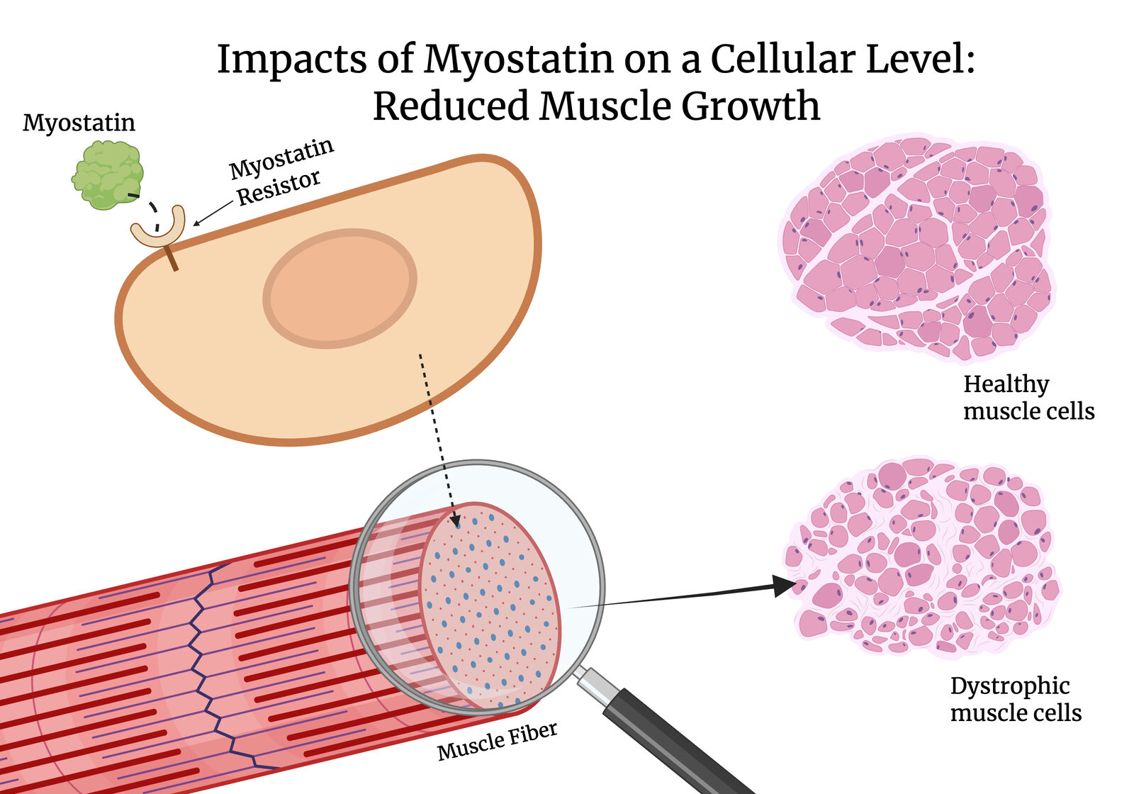 |
|---|
A naturally occurring glycoprotein, follistatin, plays a considerable role in regulating muscle growth and tissue repair. It is a single-chain glycoprotein composed of a 63-amino-acid N-terminal region and multiple cysteine-rich follistatin domains (ScienceDirect, n.d.). The function of follistatin is to primarily bind to and inhibit myostatin, a protein that suppresses muscle development. Typically, myostatin prevents excessive muscle growth by limiting the size and number of muscle fibers produced and by degrading existing muscle cells. However, when muscle-degenerative, age-related conditions, such as DMD or sarcopenia, are present, elevated myostatin activity is observed (Dao & Green, 2020). The excess myostatin causes all of these conditions. However, if follistatin is used to promote increased muscle mass and regeneration, it can block myostatin, making it a promising therapeutic candidate for muscle-related diseases.
Mysotatin inhibitors, including follistatin fragments and synthetic peptides, can enhance muscle growth without the severe side effects associated with long-term drug exposure, such as liver toxicity, infertility, cardiovascular complications, mood disorders, and psychiatric effects. There are three main types of signaling pathways: endocrine, paracrine, and autocrine. Follistatin primarily functions in the autocrine pathway (intracellular signaling). Intracellular signaling pathways occur within the cell and involve enzymes and transcription factors that respond to changes at the molecular level. By doing this, follistatin modulates receptor-mediated signaling in target cells (Zhang et al., 2023). This method is safer than other drug therapies because inhibitors don’t alter molecular structures in the body; rather, they block specific components of pathways, as shown in Figure 2, where follistatin prevents myostatin from binding its receptor.
| Figure 2. Follistatin binds to Myostatin, preventing Myostatin from interacting with the Activin receptor-like kinase (ALK) receptor on the cell surface. This inhibition blocks the subsequent intracellular signaling cascade, including Smad activation, nuclear translocation, and gene regulation that typically leads to the degradation of muscle growth factors. | 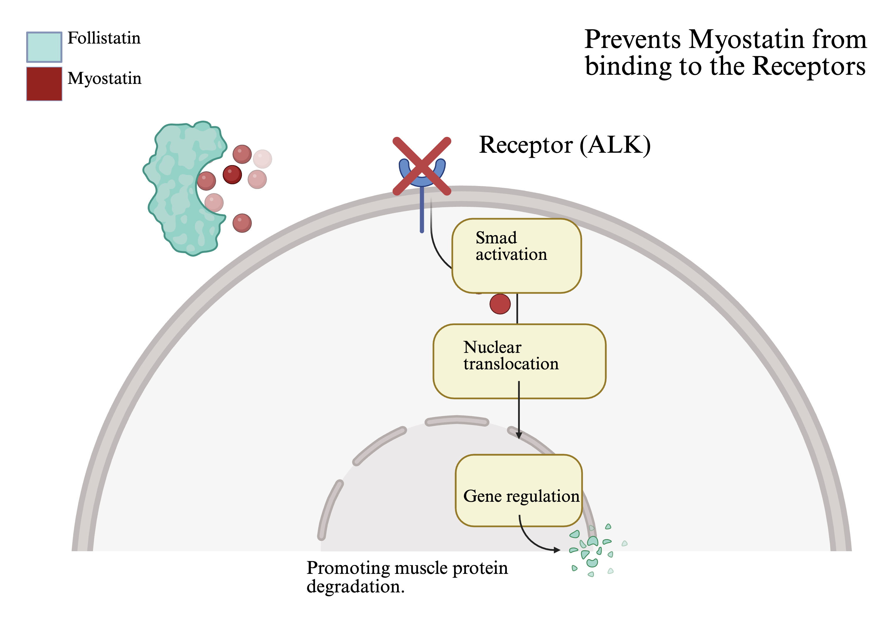 |
|---|
To ensure our engineered Limosilactobacillus reuteri safely produces the follistatin peptide, multiple biological control systems will be incorporated to regulate expression and maintain biosafety in the event of accidental release from the host environment. The core of this system is the expression of the human FST gene, producing the functional 63-amino-acid N-terminal fragment of follistatin.. The first control mechanism utilizes IL-6-based activation. IL-6 is a cytokine naturally released by the body when muscles are damaged, such as during episodes of degeneration associated with DMD or muscle deterioration in sarcopenia (Rose-John, 2020). Elevated IL-6 levels serve as a biological signal to activate the genetic switch responsible for peptide production. By incorporating the specific transcription factors, STAT3 or NF-κB, into the genetic switch, the probiotic will release the follistatin fragment only under conditions of muscle stress or injury. To prevent continuous peptide expression, a TetR regulatory loop will be implemented. TetR is a protein that functions as a transcriptional repressor, or “off switch,” turning off gene expression once peptide levels are sufficient. This feedback mechanism maintains homeostatic control and prevents overproduction, which could otherwise stress the arterial cells or disrupt normal muscle regulation (Ramos et al., 2005). As an additional biosafety measure, a mazF kill switch will be incorporated to prevent engineered bacteria from proliferating outside the gut environment. Activation of the mazF gene induces synthesis of a toxin that causes the bacteria to self-destruct (Nariya & Inouye, 2008). This biosafety feature minimizes ecological risks and strengthens containment, ensuring our design remains environmentally and clinically secure.
This synthetic biology approach offers a promising alternative for addressing muscle degradation disorders. By engineering a probiotic chassis to deliver therapeutic peptides directly through the gut, the system provides a targeted, noninvasive, and affordable solution. This design not only strengthens muscular function but also empowers patients with a treatment that adapts to their body's needs.
Systems level
Interleukin-6 (IL-6) is a signaling protein called a cytokine that cells release to communicate stress, inflammation, and immune activity. This inflammatory signal is a response to muscle stress shown in individuals with DMD. Normal IL-6 levels in healthy adults are very low, typically below 5 pg/mL (Pelosi et al., 2015). When these levels are significantly above this range, indicating an inflammatory response, our system triggers the production of our genetic components to engineer Limosilactobacillus reuteri to produce a follistatin fragment (FST-344) in response to muscle damage. A Limosilactobacillus reuteri probiotic would be administered in a clinical setting to colonize the human gut and, therefore, respond dynamically to muscle damage for individuals with Duchenne Muscular Dystrophy, as demonstrated in Figure 3 Upon sensing elevated IL-6 levels, the engineered probiotic activates its circuit to produce and secrete the follistatin fragment, which suppresses myostatin and enables muscle repair.
| Figure 3. This therapeutic workflow shows engineered L. Reuteri colonizing the gut to detect IL-6 signals released by damaged muscle tissue. Once triggered, the probiotic secretes Follistatin (FST-344) to inhibit myostatin, promoting muscle repair and regeneration. | 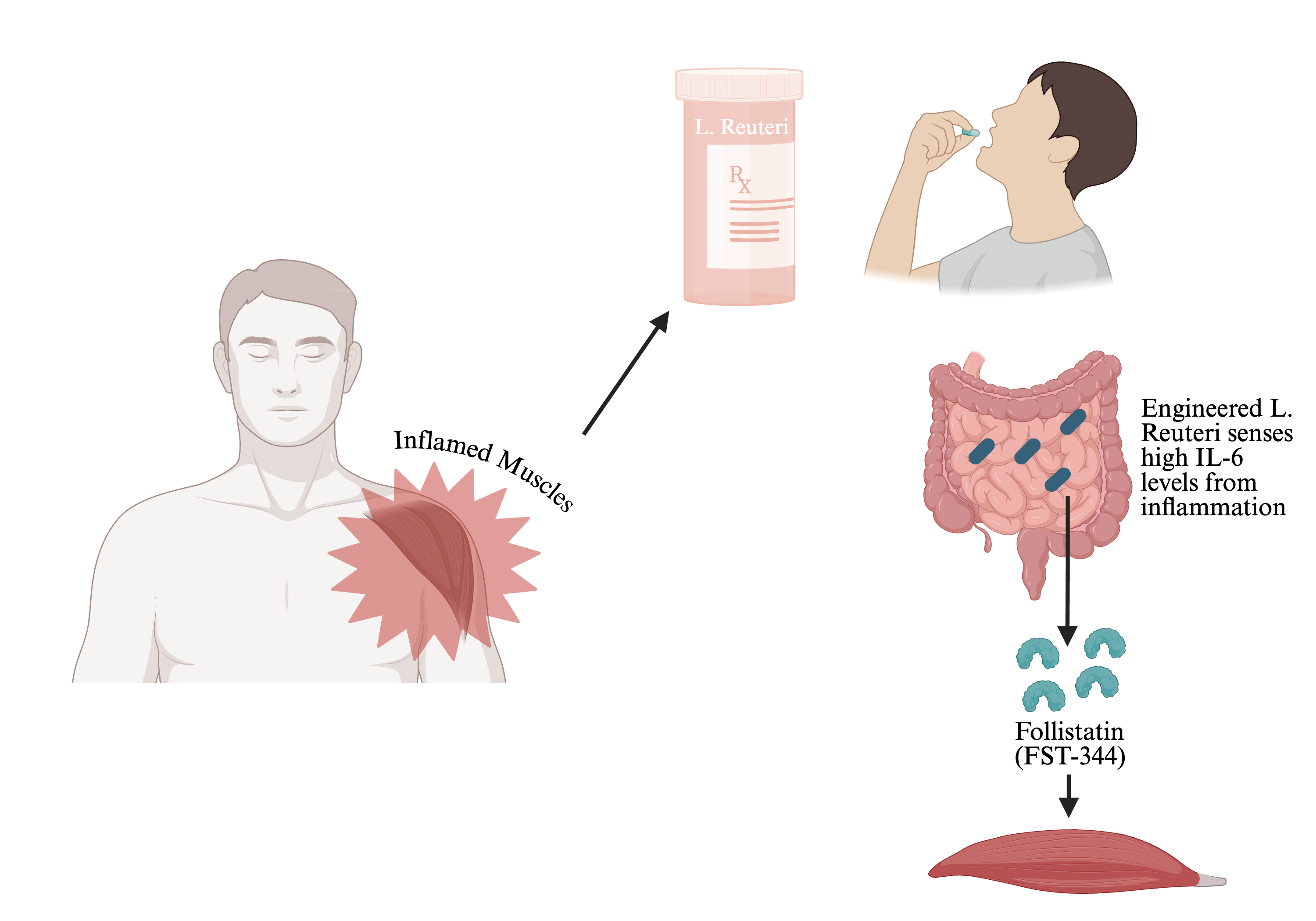 |
|---|
This system is designed to shut down when IL-6 levels return to normal (below the 5 pg/mL benchmark), the responsive promoter turns off, triggering the system's built-in kill switch, which relies on a TetR-mazF regulatory circuit. Under inflammatory conditions, TetR remains expressed and represses the mazF toxin, allowing the bacteria to survive while producing follistatin (Chan et al., 2015, p. 82). As the inflammatory response wanes, TetR expression declines, relieving repression of mazF. The system targets cell death, eliminating the engineered probiotic once FST-344 is secreted. When the probiotic is taken, it remains localized in the gut and only activates in response to elevated IL-6 levels, thereby avoiding unintended effects, providing on-demand treatment, and preventing long-term persistence in the body (Bober et al., 2018). The system promotes muscle regeneration and repair by inhibiting the natural negative regulator of muscle growth and by using a TetR-mazF regulatory circuit to remain localized and function only when necessary.
Device level
Our design uses a three-step procedure: producing follistatin only when necessary and promptly killing the probiotic when it is no longer needed. This results in a lower-risk treatment option for those suffering from DMD.
The first device is the IL-6 sensing module, which serves as the input to the genetic circuit. It utilizes a SOCS3 promoter, a transcriptional element derived from STAT-3-responsive elements in IL-6 signaling pathways that becomes active when IL-6 binds its receptor and triggers the JAK-STAT3 cascade (Rose-John, 2020). Because Limosilactobacillus reuteri does not naturally possess mammalian signaling components, these functions must be added for functionality with a bacterial synthetic circuit, commonly used in the engineering of biomarker-responsive probiotics (Bober et al., 2018). To enable IL-6 detection in bacterial chassis, this design requires the incorporation of a synthetic signaling interface, such as a chimeric receptor or engineered two-component system, capable of converting extracellular IL-6 binding into intracellular activation of STAT3-responsive transcription (Riglar & Silver; Riglar et al., 2017). Transcription is activated strictly when IL-6 exceeds the 5 pg/mL threshold established in DMD patients, meaning downstream devices remain silent under healthy baseline conditions (Pelosi et al., 2015).
The second device is the follistatin secretion module, which constitutes the system's therapeutic output. Once the sensing device activates transcription, it drives expression of the FST-344 isoform of follistatin, the active strain responsible for myostatin inhibition (Lee & McPherron, 2001). The FST gene is placed under the direct control of the SOCS3 promoter, thereby tightly coupling therapeutic protein production to the inflammatory signal. The coding sequence is fused to a secretion signal peptide compatible with Limosilactobacillus Sec or Tat translocon pathways, directing the follistatin fragment through the bacterial membrane for extracellular release and systemic uptake (Bober et al., 2018).
The third device is the safety and control module, which manages feedback regulation and biocontainment through two interlocking mechanisms. The first is a TetR repression loop that is activated under inflammatory conditions and acts as a transcriptional repressor, preventing runaway follistatin overproduction (Ramos et al., 2005). The second is a mazF-based kill switch. MazF is an mRNA interferase, meaning it cuts mRNA at a specific sequence to stop protein synthesis, in this case leading to cell death (Nariya & Inouye, 2008). While IL-6 stays elevated, TetR represses mazF, allowing the bacteria to survive and produce FST-344. This establishes a conditional survival circuit in which bacterial viability is directly linked to the presence of inflammatory signals. As inflammation subsides and TetR expression declines, mazF is released, triggering bacterial death and preventing their persistence in the gut (Cubillos-Ruiz et al., 2021).
This design enables rapid induction of therapeutic protein production in response to elevated IL-6 levels, followed by automatic downregulation and bacterial clearance as inflammation subsides, ensuring a transient and tightly controlled therapeutic response.
Parts level
The genetic components of the system are assembled into a plasmid vector to ensure compatibility with L. reuteri. To ensure controlled production of the protein (follistatin), the genetic circuit uses the TetR transcriptional repression system. TetR is a well-known bacterial transcriptional repressor that binds to specific DNA operator sequences. Figure 4 shows the plasmid backbone derived from Limosilactobacillus reuteri, starting with the IL-6-responsive promoter. This promoter is located within the synthetic cassette between positions 1000 and 7400 bp. The system triggers transcription of FST-344 in response to inflammatory biomarkers. The map also highlights the restriction enzyme sites for modular cloning. This plasmid will be inserted into the bacteria that will be in the probiotic form, as shown in Figure 4.
| Figure 4: Map of the Probiotic expression vector in Limosilactobacillus reuteri. This figure shows the 11,621 bp of the recombinant plasmid design for the localized delivery of Follistatin (FST-344). The plasmid uses the L. reuteri backbone to modulate muscle wasting. | 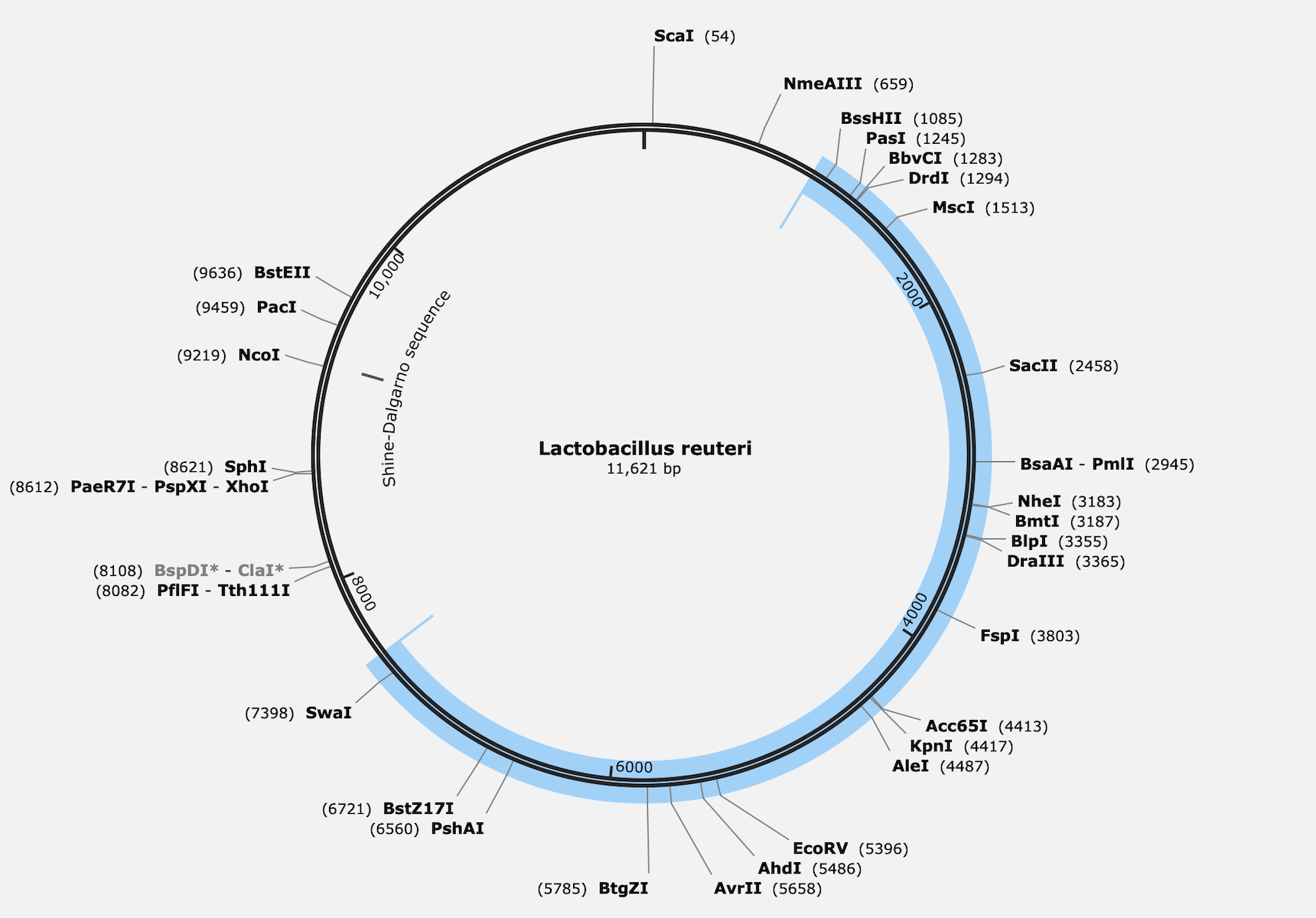 |
|---|
Promoters are essential regulatory DNA sequences that control transcription initiation and determine when and at what level genes are expressed. In this design, we have both inducible and constitutive promoters that serve distinct functions to achieve precise control over therapeutic output and biosafety.
The primary promoter is driven by an IL-6-responsive element, enabling inflammation-dependent gene expression. This promoter is activated by IL-6 elevation. By linking follistatin expression to IL-6 levels, the inducible promoter ensures that follistatin production is activated only when needed, thereby increasing specificity and reducing unnecessary metabolic activity in the host organism.
On the other hand, we have a constitutive promoter in the secondary promoter. This promoter is driven by continuous expression of regulatory components. Constitutive expression ensures that the safety mechanism remains active at all times, enabling rapid activation of the kill switch. This difference is the key between the inducible and constitutive promoters.
The devices also include double terminators to ensure that the device turns off the activation of certain proteins, so the host is no longer using metabolic energy. The double terminators are used to prevent “leaks” during transcription.
In Figure 5, you can see how the two would work together to build our device. Once the first promoter is activated, it produces follistatin, which is quickly detected by the constitutive promoter and activates the toxins, ultimately activating the kill switch.
| Figure 5: Synthetic gene circuit for inflammation-responsive follistatin delivery. This is an overview of the genetic system consisting of two modules. (Top) An IL-6-responsive sensing and therapeutic expression module + IL-6-sensitive promoter that activates the expression of Follisatin. | 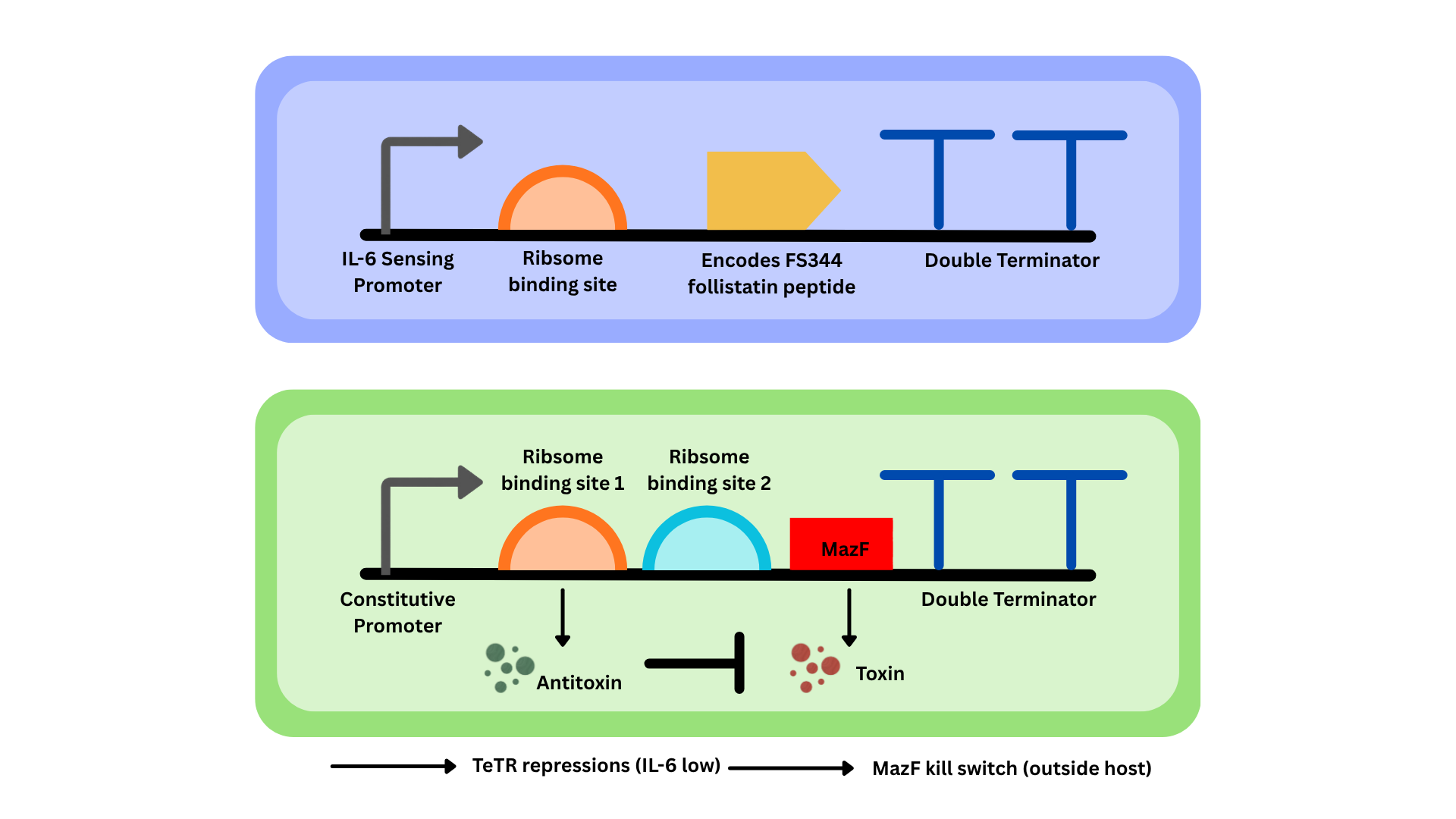 |
|---|
Limosilactobacillus reuteri is a probiotic bacterium commonly found in the human gastrointestinal tract and has been widely studied for its safety and therapeutic potential. Its designation as a Generally Recognized As Safe (GRAS) organism supports its use in engineered therapeutic systems, particularly those intended for oral delivery (Zhang et al., 2019). The organism’s ability to survive gastric transit and persist fleetingly within the gut environment makes it optimal for concentrated production of therapeutic biomolecules. In addition, L. reuteri and related Lactobacillus species have been successfully engineered to express heterologous proteins, showing compatibility with synthetic gene circuits and controlled expression systems (Mimee et al., 2016). These characteristics make it an effective chassis for probiotic-based delivery platforms that rely on non-invasive administration and host microbe interaction.
The IL-6-responsive promoter used in this system is derived from the SOCS3 regulatory region, which contains STAT3-binding elements activated by inflammatory signaling. IL-6 is a central cytokine in immune regulation and signals through the JAK/STAT3 pathway, where STAT3 phosphorylation and nuclear translocation drive transcription of downstream target genes (Rose-John, 2020). Incorporating STAT3-responsive elements into a synthetic promoter enables transcriptional activation in response to elevated IL-6 levels. These promoters have been widely studied for their ability to link extracellular inflammatory cues to gene expression, making them effective biosensors in engineered systems (Heinrich et al., 2003). This design allows the promoter to function as a precise input sensor, ensuring that downstream gene expression is tightly connected to disease-associated inflammatory states.
The therapeutic output of the system is the FST-344 isoform of follistatin, a secreted glycoprotein that inhibits myostatin and related TGF-β ligands, preventing their interaction with receptors, promoting muscle hypertrophy and regeneration (Lee & McPherron, 2001; McPherron & Lee, 1997). The FST-344 isoform is processed into an active, circulating form capable of exerting systemic effects, making it suitable for therapeutic applications targeting muscle-wasting diseases. Its efficacy in enhancing muscle mass and function has been demonstrated in many experimental models, supporting its selection as a therapeutic candidate (Pedersen & Febbraio, 2008). For use in a probiotic system, follistatin can be fused to a secretion signal peptide compatible with bacterial export pathways, enabling extracellular release and interaction with host tissues (Mimee et al., 2016). This ensures that the therapeutic protein reaches its target and performs its biological function effectively.
Safety
In humans, L. reuteri is found in many different body sites, including the gastrointestinal tract, mouth, and skin (Mu et al., 2018). Due to its multiple occurrences in the body, its presence supports its harmlessness for consumption and the environment. After leaving the body, it decomposes in the environment through natural processes, as it requires little oxygen. Additionally, the engineered probiotic is administered in a clinical setting as the patient needs, and studies on similar L. reuteri probiotic strains demonstrate that bacterial levels decline when administration stops and do not cause long-term ecological problems; they have even been shown to degrade organic waste, pollutants, and toxins rather than creating chemical pollution (Lee et al., 2025). After being generally recognized as safe (GRAS) by the U.S. Food and Drug Administration, L. reuteri probiotics are deemed to pose minimal risk to ecosystems under their intended use and therefore have little to no environmental impact (United States Environmental Protection Agency).
Experimental work involving the manipulation of these pathways would be conducted under appropriate biosafety conditions (BSL-1), due to the chassis, L. reuteri’s, status as GRAS (Generally Recognized As Safe) (Mu et al., 2018), using non-pathogenic laboratory strains within a controlled environment (Centers for Disease Control and Prevention [CDC], 2020; National Institutes of Health [NIH], 2019). The design assumes strict adherence to aseptic techniques during cloning, transformation, and expression procedures to prevent contamination and unintended spread of genetically modified material. Standard personal protective equipment, including gloves, lab coats, and eye protection, would be required, and all samples and reagents would be clearly labeled and handled to minimize exposure risk (CDC, 2020). Work surfaces and equipment would be routinely disinfected before and after use. All biological waste, including liquid cultures and contaminated consumables, would be decontaminated by chemical treatment or autoclaving prior to disposal, ensuring that no viable genetically modified organisms are released from the laboratory (Schmidt et al., 2009; NIH, 2019).
The use of a genetically modified organism to deliver follistatin presents many potential risks to the human body. This must be considered, as the overuse of probiotics may lead to gastrointestinal side effects such as bloating and gas (Cleveland Clinic, n.d.), which are common symptoms of rapid shifts in the gut microbiota. These symptoms can reflect a disturbance in the native microbiome when we introduce a new bacterium via a pill. Additionally, prolonged or excessive use of the probiotic can lead to overexpression of follistatin and unintentionally suppress myostatin signaling beyond necessary medical needs, thereby causing abnormal muscle growth or other physical effects. Therefore, there needs to be regulations of bacterial activity and robust biocontamination mechanisms to minimize these risks.
Further biosafety within our engineered L. reuteri probiotic incorporates two implemented regulatory mechanisms to control therapeutic output and ensure biosafety. A TetR repression loop functions as a transcriptional off-switch, suppressing follistatin overproduction once IL-6 levels normalize, maintaining homeostatic control and preventing unnecessary metabolic damage to the host (Ramos et al., 2005). When IL-6 drops below the 5 pg/mL threshold and TetR expression declines, the mazF kill switch is relieved of repression, triggering mRNA degradation and causing the bacteria to self-destruct before they can persist beyond the gut environment (Nariya & Inouye, 2008). Together, these mechanisms ensure that the probiotic remains active only during inflammatory episodes associated with DMD or sarcopenia, directly linking regulatory function to the conditions the therapy is designed to address.
The engineered L. reuteri employs a three-step safety procedure, but the system faces biological challenges of transcriptional leakiness and genetic instability. Even in the absence of elevated IL-6 levels, promoters still exhibit default activity, leading to leaky transcription of FST-344 (Cubillos-Ruiz et al., 2021). The implementation of double terminators will mitigate this and conserve host energy, but additional stability can be achieved by incorporating ribosome-binding or decoy sites to tightly regulate unintended protein production (Mimee et al., 2016). Additionally, pressure on the mazF-based kill switch could induce mutations that enable the bacteria to evade self-destruction (Nariya & Inouye, 2008). To counter this, utilizing redundant kill switches can ensure that any mutant is naturally eliminated. Essential gene coupling could also ensure elimination by linking the therapeutic plasmid to a gene necessary for survival in the gut environment (Ramos et al., 2005).
Discussions
The feasibility of this project is bolstered by the selection of Limosilactobacillus reuteri as a chassis, a bacterium naturally found in the body and generally recognized as safe. By leveraging synthetic biology components, such as IL-6-responsive promoters for damage sensing and TetR-regulated repression loops for homeostatic control, the design aligns with proven methods for engineering living therapeutics. Furthermore, integrating a mazF kill switch and double terminators addresses critical safety concerns and enables a noninvasive, scalable approach.
Since the genetically engineered probiotic will assist with muscle regeneration in individuals with Duchenne Muscular Dystrophy (DMD), there is a risk of misuse for purposes other than its intended use, such as unauthorized muscle enhancement or aesthetic modification. With this risk in mind, the probiotic would be distributed only in a clinical setting when needed and prescribed only to individuals with the disorder. The remedy is intended solely to relieve pain and promote muscle preservation, addressing the needs of these individuals, and will not be provided to those without DMD. The engineered bacteria are designed to stop activity once IL-6 levels return to normal, ensuring safety and specificity for patients with this condition.
Despite being among the most common approaches currently used to treat DMD and sarcopenia, myostatin inhibitors and gene therapy still face substantial delivery and economic challenges (Wagner, 2020; Elangkovan & Dickson, 2021). For instance, while myostatin inhibitors such as Trevogrumab and SRK-439 are designed to inhibit myostatin function, systemic delivery has yielded only modest results, requiring repetitive treatments (Wagner, 2020). Genetic treatment methods, such as exon skipping drugs and AAV gene therapy, are often too expensive, ranging from $300,000 to $3 million annually, or up to $3.5 million for one-time doses. Furthermore, their high mutation specificity limits their broader application, as evidenced by the recent failures of several late-stage clinical trials (Reuters, 2024).
The use of engineered L. reuteri as a probiotic vector can deliver follistatin orally, thereby avoiding the side effects and costly production processes associated with conventional therapies (Mu et al., 2018; Lee et al., 2025). With our unique combination of a tetR-regulated transcription circuit and a MazF-based kill switch, we achieve homeostasis in myostatin inhibition, ensuring that therapeutic FTS-344 is confined to the patient’s inflammatory levels (Ramos et al., 2005; Nariya & Inouye, 2008; Chan et al., 2015). This design enables active muscle repair while minimizing side effects and preventing the environmental release of engineered microbes.
There are several limitations associated with the production and large-scale implementation of probiotics. A key limitation is that their effects can vary widely among individuals who take them, depending on their gut microbiome. For example, even twins can have immensely different gut microbiome composition. Elevated IL-6 levels alone cannot distinguish between muscle degeneration and normal responses such as exercise or infection, because IL-6 is a general inflammatory cytokine. The difference is derived from the context: exercise causes a short spike in IL-6 levels that muscles can quickly resolve, while muscle degeneration produces a sustained elevation alongside markers such as tissue damage, elevated creatine kinases, and persistent inflammation. Engineering a probiotic to fit each individual is nearly impossible, making outcomes unpredictable (National Institutes of Health [NIH], 2023). Additionally, although probiotics are generally considered safe, there are documented risks of gastrointestinal side effects and infections in immunocompromised people (Cleveland Clinic, 2024). There is also a research gap that hinders our understanding of the long-term effects of both conventional and genetically modified probiotics (Hill et al., 2014). Probiotics face major limitations due to unpredictable individual responses, potential health risks in vulnerable populations, and limited research on their long-term effects.
Next steps
To move this design from a theoretical model to a functional therapeutic for DMD and sarcopenia, our research must proceed through phases of genetic optimization, component validation, and rigorous biosafety testing.
Our first priority is to refine the genetic circuit to address the inherent challenge of transcriptional leakiness. While our current design uses an IL-6-responsive promoter and a TetR-regulated repression loop, even minimal default promoter activity can lead to unintended production. To mitigate this, we will incorporate double terminators and optimize ribosome-binding or decoy sites to ensure tight control of protein expression (Cubillos-Ruiz et al., 2021; Mimee et al., 2016). We also intend to use AlphaFold to model the follistatin fragment, ensuring the peptide remains stable and biologically active upon secretion (Jumper et al., 2021).
Following construction, we must quantify the system’s responsiveness to IL-6, a keystone cytokine in inflammatory muscle degeneration (Hunter & Jones, 2015). We will conduct in vitro assays to measure the follistatin production levels in response to varying physiological concentrations of IL-6. These results will be calibrated against baseline myostatin-suppression metrics to verify therapeutic efficacy, relying on established understanding of myostatin regulation (Lee & McPherron, 2001).
Stability of our biosafety measures is critical. Because the mazF kill switch is susceptible to mutations that could allow the bacteria to evade self-destruction (Nariya & Inouye, 2008), we will develop redundant safety modules. The double terminators to prevent leakage, ribosome binding for tighter control over protein expression, and AlphaFold modelling to ensure our follistatin stays stable and active only when and where intended. Our future work will focus on integrating essential gene coupling, which links the therapeutic plasmid to genes critical for survival in the gut environment, ensuring that the strain cannot persist in the host should the regulatory system fail (Ramos et al., 2005). Finally, as L. reuteri is a GRAS-certified (generally recognized as safe) organism (Mu et al., 2018), we will continue to perform stress-testing in simulated gastrointestinal environments to verify that bacterial levels naturally decline when therapeutic administration ceases, thereby fulfilling the requirements for long-term ecological safety.
Author contributions
C.W. designed the original project idea. A.B, S.M, A.S, L.S, C.W. contributed to the writing of the Abstract, Background, Systems, Devices, Parts, Safety, and Discussion. A.B. wrote the next steps. S.M, A.S, and L.S. created the figures. A.B, S.M, A.S, L.S, and C.W. all compiled references.
Acknowledgements
We would like to thank Ms. Marmerstein for her valuable guidance, feedback, and support throughout our research process. We are also extremely grateful to our bio builder mentor, Dr. Kosuke Seki, for thoughtfully aiding our research, reviewing our work, and offering constructive criticism. Additionally, we would like to thank Western Reserve Academy for the opportunity to take CL Synthetic Biology 1 and for the numerous learning opportunities the course has provided. Finally, we acknowledge Biotreks for their support in making this design possible.
References
Barr, A., Heldt, F., Zhang, T., Bakal, C., & Novák, B. (2016). A dynamical framework for the all-or-none g1/s transition. Cell Systems, 2(1), 27-37. https://doi.org/10.1016/j.cels.2016.01.001
Biosafety in microbiological and biomedical laboratories (Center for Disease Control, Comp.). (n.d.). https://www.cdc.gov/labs/media/pdfs/2025/08/SF__19a_308133-A_BMBL6_00-BOOK-WEB-final-3.pdf
Birnkrant, D. J., Bushby, K., Bann, C. M., Alman, B. A., Apkon, S. D., Blackwell, A., Case, L. E., Cripe, L., Hadjiyannakis, S., Olson, A. K., Sheehan, D. W., Bolen, J., Weber, D. R., & Ward, L. M. (2018). Diagnosis and management of Duchenne muscular dystrophy, part 2: respiratory, cardiac, bone health, and orthopaedic management. The Lancet, 17(4), 347-361. https://www.thelancet.com/journals/laneur/article/PIIS1474-4422%2818%2930025-5/abstract
Bober, J. R., Beisel, C. L., & Nair, N. U. (2018). Synthetic biology approaches to engineer probiotics and members of the human microbiota for biomedical applications. Annual Review of Biomedical Engineering, 20(1), 277-300. https://doi.org/10.1146/annurev-bioeng-062117-121019
Chan, C. T. Y., Lee, J. W., Cameron, D. E., Bashor, C. J., & Collins, J. J. (2015). 'Deadman' and 'passcode' microbial kill switches for bacterial containment. Nature Chemical Biology, 12(2), 82-86. https://doi.org/10.1038/nchembio.1979
Cruz-Jentoft, A. J., Bahat, G., Bauer, J., Boirie, Y., Bruyere, O., Cederholm, T., Cooper, C., Landi, F., Rolland, Y., Sayer, A. A., Schneider, S. M., Sieber, C. C., Topinkova, E., Vandewoude, M., Visser, M., & Zamboni, M. (2019). Sarcopenia: revised European consensus on definition and diagnosis. Age and Aging, 48(1), 16-31. https://academic.oup.com/ageing/article/48/1/16/5126243?login=true
Cubillos-Ruiz, A., Guo, T., Sokolovska, A., Miller, P. F., Collins, J. J., Lu, T. K., & Lora, J. M. (2021). Engineering living therapeutics with synthetic biology. Nature Reviews Drug Discovery, 20, 941-960. https://www.nature.com/articles/s41573-021-00285-3
Dao, T., & Green, A. E. (2020, December 31). Sarcopenia and muscle aging: A brief overview. National Library of Medicine. Retrieved November 9, 2025, from https://pmc.ncbi.nlm.nih.gov/articles/PMC7803599/
Din, M. O., Danino, T., Prindle, A., Skalak, M., Selimkhanov, J., Allen, K., Julio, E., Atoll, E., Tsimring, L. S., Bhatia, S. N., & Hasty, J. (2016). Synchronized cycles of bacterial lysis for in vivo delivery. Nature, 536, 81-85. https://www.nature.com/articles/nature18930
Elangkovan, N., & Dickson, G. (2021). Gene therapy for Duchenne Muscular Dystrophy. Journal of Neuromuscular Dystrophy, 8(2), 303-316. https://pmc.ncbi.nlm.nih.gov/articles/PMC8673537/
Engstrom, M. D., & Pfleger, B. F. (2017). Transcription control engineering and applications in synthetic biology. Synthetic and Systems Biotechnology, 2(3), 176-191. https://doi.org/10.1016/j.synbio.2017.09.003
Fukuhara, S., Kobayashi, T., Hamaoka, M., Naruhiko, H., Oishi, K., Namba, Y., Oshita, K., Matsubara, K., Takei, D., Nakano, R., Sakai, H., Kuroda, S., Tahara, H., Ohira, M., & Ohdan, H. (2024). Sarcopenia's impact defined by grip strength and muscle mass on post-hepatectomy outcomes: A multicenter analysis. In Vivo, 38(6), 2827-2835. https://doi.org/10.21873/invivo.13763
Heinrich, P. C., Behrmann, I., Haan, S., Hermanns, H. M., MÜller-Newen, G., & Schaper, F. (2003). Principles of interleukin (IL)-6-type cytokine signaling and its regulation. Biochemical Journal, 374(1), 1-20. https://doi.org/10.1042/bj20030407
Hunter, C. A., & Jones, S. A. (2015). IL-6 as a keystone cytokine in health and disease. Nature Immunology, 16(5), 448-457. https://doi.org/10.1038/ni.3153
Johnson, D. E., O'Keefe, R. A., & Grandis, J. R. (n.d.). Targeting the IL-6/JAK/STAT3 signaling axis in cancer [Abstract]. Nature Reviews Clinical Oncology. https://www.nature.com/articles/nrclinonc.2018.8
Jumper, J., Evans, R., Pritzel, A., Green, T., Figurnov, M., Ronneberger, O., Tunyasuvunakool, K., Bates, R., Žídek, A., Potapenko, A., Bridgland, A., Meyer, C., Kohl, S. A. A., Ballard, A. J., Cowie, A., Romera-Paredes, B., Nikolov, S., Jain, R., Adler, J., . . . Silver, D. (2021). Highly accurate protein structure prediction with alphafold. Nature, 596(7873), 583-589. https://doi.org/10.1038/s41586-021-03819-2
Kim, A., Park, M., & Shin, H.-I. (2022). Pain characteristics among individuals with duchenne muscular dystrophy according to their clinical stage. BMC Musculoskeletal Disorders, 23(1). https://doi.org/10.1186/s12891-022-05504-5
Lee, A. H., Rodriguez Jimenez, D. M., & Meisel, M. (2025). Limosilactobacillus reuteri is a probiotic gut commensal with contextual impact on immunity. Gut Microbes, 17(1). https://doi.org/10.1080/19490976.2025.2451088
Lee, S.-J., & McPherron, A. C. (2001). Regulation of myostatin activity and muscle growth. Proceedings of the National Academy of Sciences, 98(16), 9306-9311. https://doi.org/10.1073/pnas.151270098
Leenhouts, K., Kok, J., & Venema, G. (n.d.). Lactococcal plasmid pWV01 as an integration vector for lactococci. PubMed Central. https://pmc.ncbi.nlm.nih.gov/articles/PMC183620/
Malatesta, M. (2012). Skeletal muscle features in myotonic dystrophy and sarcopenia: Do similar nuclear mechanisms lead to skeletal muscle wasting? European Journal of Histochemistry, 56(3).
Mimee, M., Citorik, R. J., & Lu, T. K. (2016). Microbiome therapeutics — advances and challenges. Advanced Drug Delivery Reviews, 105(A), 44-54. https://www.sciencedirect.com/science/article/abs/pii/S0169409X16301429?via%3Dihub
Mu, Q., Tavella, V. J., & Luo, X. M. (2018). Role of lactobacillus reuteri in human health and diseases. Frontiers in Microbiology, 9. https://doi.org/10.3389/fmicb.2018.00757
Nariya, H., & Inouye, M. (2008). MazF, an mRNA interferase, mediates programmed cell death during multicellular myxococcus development. Cell, 132(1), 55-66. https://doi.org/10.1016/j.cell.2007.11.044
NIH guidelines for research involving recombinant or synthetic nucleic acid molecules (National Institute of Health, Comp.). (n.d.). https://osp.od.nih.gov/wp-content/uploads/NIH_Guidelines.htm
Pedersen, B. K., & Febbraio, M. A. (2008). Muscle as an endocrine organ: Focus on muscle-derived interleukin-6. Physiological Reviews, 88(4), 1379-1406. https://doi.org/10.1152/physrev.90100.2007
Pelosi, L., Berardinelli, M. G., Forcina, L., Spelta, E., Rizzuto, E., Nicoletti, C., Camilli, C., Testa, E., Catizone, A., De Benedetti, F., & Musarò, A. (2015). Increased levels of interleukin-6 exacerbate the dystrophic phenotype in mdx mice. Human Molecular Genetics, 24(21), 6041-6053. https://doi.org/10.1093/hmg/ddv323
Pfizer's Duchenne gene therapy fails in late-stage study. (2024, June 12). Reuters. Retrieved November 6, 2025, from https://www.reuters.com/business/healthcare-pharmaceuticals/pfizers-gene-therapy-muscle-wasting-disorder-misses-main-goal-late-stage-study-2024-06-12/
Ramos, J. L., Martínez-Bueno, M., Molina-Henares, A. J., Terán, W., Watanabe, K., Zhang, X., Gallegos, M. T., Brennan, R., & Tobes, R. (2005). The tetr family of transcriptional repressors. Microbiology and Molecular Biology Reviews, 69(2), 326-356. https://doi.org/10.1128/mmbr.69.2.326-356.2005
Rose-John, S. (2020). Interleukin-6 signaling in health and disease. F1000Research, 9, 1013. https://doi.org/10.12688/f1000research.26058.1
Sarcopenia. (2022, June 3). Cleveland Clinic. Retrieved October 28, 2025, from https://my.clevelandclinic.org/health/diseases/23167-sarcopenia
Signaling pathway. (2025, August 25). BOC Sciences. Retrieved November 11, 2025, from https://www.bocsci.com/signaling-pathway.html?srsltid=AfmBOoqLlv2oS_gRq0GnGMgFuQWj3faZh35AcdLnlAVcG2VKuOhlxfzj
Translational Drug Development. (2023). Probiotics in focus: How does L. reuteri affect gut health? Translational Drug Development. https://blog.td2inc.com/probiotics-in-focus-how-does-l-reuteri-affect-gut-health?utm_source=chatgpt.com
Venugopal, V., & Pavlakis, S. (2023). Duchenne muscular dystrophy. National Library of Medicine.
Wagner, K. R. (2020). The elusive promise of myostatin inhibition for muscular dystrophy. Current Opinion in Neurology, 33(5). https://doi.org/10.1097/WCO.0000000000000853
Wen, T., Mao, C., & Gao, L. (2022). Analysis of the gut microbiota composition of myostatin mutant cattle prepared using crispr/cas9. PLOS ONE, 17(3), e0264849. https://doi.org/10.1371/journal.pone.0264849
Zahirović, A., & Berlec, A. (2022). Targeting IL-6 by engineered lactococcus lactis via surface-displayed affibody. Microbial Cell Factories, 21(1). https://doi.org/10.1186/s12934-022-01873-7
Zhang, L., Badgwell, D. B., Bevers, J. J., Schlessinger, K., Murray, P. J., Levy, D. E., & Watowich, S. S. (2006). IL-6 signaling via the stat3/socs3 pathway: Functional analysis of the conserved stat3 n-domain. Molecular and Cellular Biochemistry, 288(1-2), 179-189. https://doi.org/10.1007/s11010-006-9137-3
Zou, Z.-P., Du, Y., Fang, T.-T., Zhou, Y., & Ye, B.-C. (2023). Biomarker-responsive engineered probiotic diagnoses, records, and ameliorates inflammatory bowel disease in mice. Cell Host & Microbe, 31(2), 199-212.e5. https://doi.org/10.1016/j.chom.2022.12.004