
Nav Garg, Paul Stoica, and Nicolas Wong, Andover High School, Andover, Massachusetts, United States of America
Reviewed on 9 May 2026; Accepted on 22 June 2026; Published on 26 October 2026
With help from the 2026 BioTreks Production Team.
Egg allergies affect approximately 1.6-3.2% of children or around 2.3 million kids in the U.S., producing symptoms ranging from hives to life threatening anaphylaxis. This symptom is evoked by IgE-mediated mast cell degranulation and histamine release. Current treatments for this condition include allergen immunotherapy and antihistamines, which address the downstream allergic cascade– release of histamine in response to allergens binding onto IgE receptors. This paper delves into a solution to prevent the initiation of this cascade. It involves manufacturing a recombinant Fc-only IgE fragment consisting of the Cε2, Cε3, and Cε4 domains of the human IgE heavy chain. This will be used as a competitive inhibitor, inhibiting the FcεRI receptors on mast cells and basophils. By constructing these receptors without the Fab region, the fragment can block the allergen from binding and prevent the endogenous IgE antibody from binding to the FcεRI receptor, preventing the release of histamine and averting the allergic cascade. To cultivate this fragment, we propose to use the yeast Pichia pastoris to ensure glycosylation and high-affinity binding and purification using chromatography. Safety considerations include suppression of immune responses against parasites, risk of adverse immune responses to our modified antibody, and delivery system of this solution inside the bloodstream (Galli et al., 2012). The ethical and safety concerns center around the delivery system, biological sensitization, blocking the immune system, and high production costs, which could restrict this therapy among socioeconomic lines.
Keywords: Egg allergy, Immunoglobulin E (IgE), mast cells, histamine, competitive inhibition
Authors are listed in alphabetical order. Lindsey L'Ecuyer mentored the group. Please direct all correspondence to mailto:lindsey.lecuyer@andoverma.us.



Background
Egg allergy is an IgE-mediated hypersensitivity disorder affecting a significant portion of the population, particularly children. This immune hypersensitivity is caused by the body’s misidentification of egg proteins, which instead activate an immunological pathway meant to combat pathogens. This condition provokes the activation of mast cells and basophils, immune cells that release various chemical mediators that accumulate over time, ultimately producing a variety of mild to intense symptoms such as hives, itching, and more severe symptoms such as throat swelling, breathing difficulties, and a rapid pulse. These chemical mediators remain the culprit of an extensive range of conditions that encompass cutaneous, gastrointestinal, respiratory, cardiovascular, and even neurological symptoms. Discovering which immunological factors cause such aberrant responses is crucial for the development of allergen management to create alternative options that alleviate the effects of inflammation and discomfort. The culprits behind these harmful dysregulations are specific proteins found primarily in ovalbumin, which make up approximately 58% of the proteins in egg whites (Elsevier, n.d. (Ovalbumin)). Sensitization to these proteins can cause the immune system to produce specialized antibodies called immunoglobulin E (IgE). IgE is measured in allergen-specific IgE per liter (kUa/L); this unit is used in allergy testing to quantify the concentration of IgE antibodies produced due to contact with a certain allergen in a patient’s blood. For eggs, an IgE level of 7 is associated with a 95% predicted value of an allergic response (Bartnikas et al., 2015). These antibodies attach to specific spots, called epitopes, on proteins like ovomucoid (Gal d 1), ovalbumin (Gal d 2), ovotransferrin (Gal d 3), and lysozyme (Gal d 4). Among these proteins, ovomucoid and ovalbumin in particular are the most effective at stimulating IgE-mediated immune responses, primarily due to their epitopes being significantly more stable (Caubet & Wang, 2008). This stability allows them to resist denaturing that would otherwise occur during cooking and digestion, and remain identifiable to IgE antibodies and hence prolonging the erroneous immune reaction. Ovalbumin is less stable than ovomucoid, but comprises a majority of egg whites; its abundance increases the chance of the IgE antibodies recognizing the epitope versions that are still intact as shown in Figure 2.
| Figure 2: Flowchart of input and output levels Made with Canva | 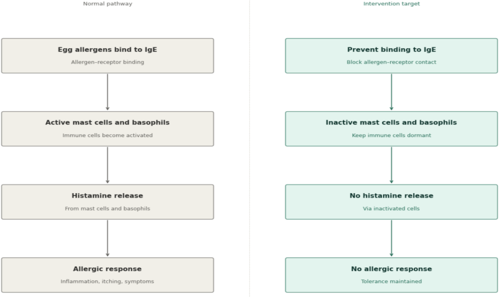 |
|---|
Systems level
The proposed system (most abstract level of the design) herein is aimed to develop targets to the biological cascade triggered by egg allergen exposure in individuals with egg allergies. At the broadest level, the input of the system is the presence of egg allergens that bind onto IgE receptors located on the surface of key immune cells– specifically mast cells and basophils. IgE receptors are antibodies produced by the immune system in response to allergen exposure, and their interaction with allergens serves as the primary trigger of the allergic cascade (Stone et al., 2010). Once egg allergens bind to these IgE receptors, mast cells and basophils become activated. This activation is a critical step in the allergic response, as it sets the downstream event of the allergic reaction.
Following activation, mast cells and basophils undergo degranulation– an immune process where cells release inflammatory mediators (Fong & Crane, 2023). The most significant of these mediators is histamine. Histamine is a bioactive molecule responsible for the symptoms of allergic reactions, including vasodilation– the widening of blood cells resulting in a severe decrease in blood pressure, increased vascular permeability, itching, and tissue inflammation (Stone et al., 2010). Histamine release is therefore the key effector event that produces the physiological harm associated with egg allergen exposure.
| Figure 1: The diagram illustrates a type 1 hypersensitivity reaction where IgE binds to the FcεRI receptor on mast cells, causing them to release chemical mediators such as histamine. | 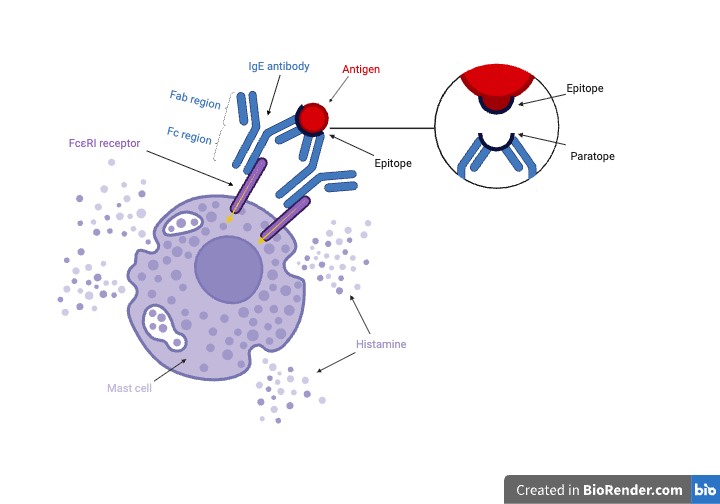 |
|---|
The Fc region of the IgE binds to the FcεRI receptor on the mast cell surface, facilitating an inflammatory immune response in the presence of an antigen (Leeson, 2023). After first exposure to an antigen, mast cells preload themselves with IgE, allowing it to instantly enable a reaction when re-exposed to the antigen (Galli et al., 2012). This mechanism triggers the release of inflammatory mediators such as histamine, which cause the symptoms during allergic reactions. The Fab region of the antibody is made up of six complementarity determining regions (CDRs), which are formed from heavy and light variable regions, ultimately coming together to form a paratope (Elsevier, n.d. (paratope)). Paratopes bind to their corresponding antigen receptor, known as the epitope, when in contact with an antigen. The binding is a highly specific one-to-one relationship and relies on non-covalent bonding, such as hydrogen bonding and van der Waals interactions (weak attractions between neutral atoms or molecules resulting in temporary imbalance of charges). However, the cross-linking of two IgE receptors to an antigen is required for a mast cell to induce a response (Janeway et al., 2001). Only then can mast cells undergo degranulation and release inflammatory molecules such as histamine through exocytosis (the active transport process of cell releasing materials) . Preventing the binding of IgE antibodies on these mast cells and basophils before they undergo degranulation would help eliminate the release of histamine.
By intervening at the level of allergen-receptor binding and cellular activation, the allergic response is suppressed at its origin rather than treating its symptoms. This upstream approach is significant because it addresses the root cause of histamine-mediated symptoms, rather than relying on downstream management, such as antihistamines, which only counteract the effects of histamine after it has already been released. Targeting the input– allergen binding to IgE receptors– could ultimately result in the full prevention of histamine release from inactivated mast cells and basophils.
Device level
| Figure 3: The diagram illustrates where the modified IgE binds to the FcεRI receptor on mast cells | 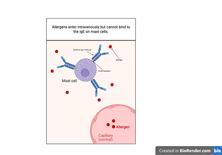 |
|---|
| Figure 4: Shows structure of Cε2 (blue), Cε3 (dark red), and Cε4 (green) domains of the IgE heavy chain which bind to FcεRI on mast cells | 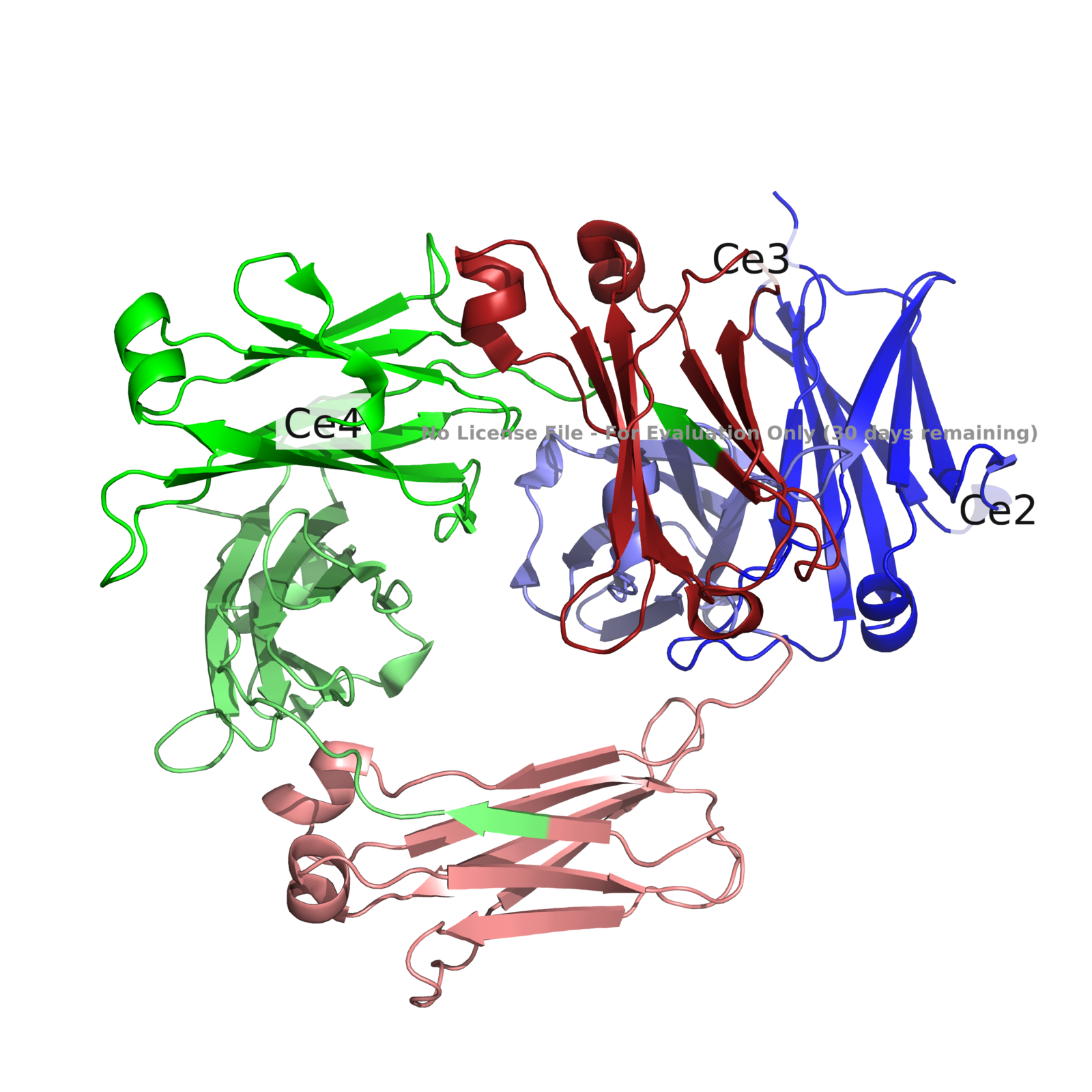 |
|---|
The immunotherapy proposed, is a modified IgE, specifically the Fc region of the antibody, which includes the Cε2, Cε3, and Cε4 domains of the IgE heavy chain. The IgE heavy chain will be modified to outcompete naturally produced IgE molecules for their IgE receptor (FcεRi) on mast cells and basophils. Unlike endogenous IgE antibodies, this fragment will only include the region that is responsible for binding to the receptor and is completely rid of the Fab region, which would bind to the allergen and initiate crosslinking, eventually releasing histamine. Since our fragment does not include the Fab region, it can only bind to the FcεRI, but cannot crosslink with the allergen, and will act as a competitive inhibitor, occupying the receptor but preventing IgE from activating an immune response. The fragment is produced in Pichia pastoris, a yeast that performs glycosylation. The Fc region is glycosylated, which would ensure correct protein folding that includes the high-affinity binding to FcεRI that endogenous IgE would have. Once expressed and secreted by the yeast, these modified fragments would be separated into individual components through chromatography, yielding purified protein with the same FcεRI binding affinity of IgE at Kd ~10-10 M (Eggel et al., 2014). The new purified protein would then be suspended in a liquid buffer at pH 7 to preserve structural integrity (Demarest et al., 2006). Since the fragments lack the Fab region, they do not carry a paratope and therefore cannot properly bind to allergens. This neither activates degranulation in mast cells, nor triggers subsequent sensitization. This is a distinction from typical allergen immunotherapy approaches, which often work by a buildup of dose in moderation, constantly exposing the individual to the allergen, which can provoke the high risk symptoms that the treatment aims to avoid.
Parts level
The main part of this proposed solution is the recombinant Fc-only region IgE fragment, which could act as a competitive inhibitor introduced via the bloodstream. This fragment will be produced by using a yeast cell to cultivate the specific DNA sequence. The gene encodes for only Cε2, Cε3, and Cε4 domains of the heavy chain of the human IgE antibody (Leeson, 2023). The sequence is retrieved from the NCBI GenBank database, a public dataset of mapped gene sequences. The gene DNA codes for the Fc region of IgE antibody, excluding the Fab region, so the antibody can bind to the FcεRI receptors on mast cells and basophils without the capability of recognizing any allergens. This gene construct is fundamental for the solution as the making of the modified IgE antibody is dependent on this sequence. (See NC_000014.9:c105601727-105600066 IGHE [organism=Homo sapiens] [GeneID=3497] [chromosome=14] DNA sequence of Fc region of IGE antibody; NCBI, 2025).
To cultivate this modified antibody we would use Pichia pastoris, which will serve as the host organism producing these antibodies. Once the DNA sequence is read by the yeast cell, we can produce Fc-Only fragments. Through this process the yeast cells also add sugar molecules to proteins through glycosylation, which is essential because naturally made Fc regions are naturally glycosylated and require these modifications to fold correctly and to bind to the FcεRI with sufficient affinity (Shade et al., 2015). After this, the yeast cells secrete the modified antibodies and are released, which will be collected and purified using chromatography. The purified Fc fragment is then prepared for introduction into the bloodstream, where it occupies FcεRI receptors on mast cells, blocking endogenous IgE from docking and preventing the allergic cascade from being initiated.
| Figure 5: The above image is of multiple Pichia pastoris | 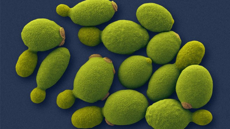 |
|---|
Safety & Ethical Consideration
A primary ethical concern of the proposed intervention is the potential compromise of the immune system's ability to respond to genuine biological threats. By occupying IgE receptors on mast cells and basophils with modified antibodies, the therapy may prevent naturally produced IgE antibodies from binding, which play a documented protective role against parasitic infections (Galli et al., 2012). If the modified antibodies successfully saturate the available receptors, patients— particularly those in regions where parasitic infections are endemic— may be left at elevated risk of reduced parasite clearance. Compounding this concern is the likelihood that natural IgE will continue to accumulate in the bloodstream without being able to engage its receptors, which may disrupt IgE's hypothesized role in tumor immune surveillance and raise long-term oncological risks that would require extensive investigation before the intervention could be considered clinically viable (Galli et al., 2012). A further concern involves the biological reliability of the modified antibodies themselves and the frequency of administration required to sustain the intervention. Yeast expression systems do not always replicate the precise glycosylation patterns found in human-derived antibodies, meaning the modified proteins may be recognized as foreign, triggering anti-drug antibody responses that reduce efficacy or cause hypersensitivity reactions over time (Shade et al., 2015). Additionally, IgE antibodies have a half-life of only approximately two to three days in circulation (Lawrence et al., 2017), meaning patients would require frequent injections to maintain sufficient receptor occupancy. This high dosing frequency increases cumulative exposure to the modified protein and raises the risk of adverse events associated with repeated administration, including injection site reactions and progressive immune sensitization.
The most immediate safety concern is the invasive nature of the delivery method combined with the significant unknowns surrounding the production process. Administration via repeated injection introduces risks including infection at the injection site and patient non-compliance over long treatment courses. More critically, while yeast cell systems are an established platform for recombinant protein expression, the precise behavior of the modified antibody once inside the human bloodstream—including how it folds, interacts with immune cells, and whether it produces off-target effects—cannot be fully predicted. Patients would be consenting to a treatment whose downstream biological interactions are not yet completely characterized, raising serious questions about the adequacy of informed consent and the ethical obligation of researchers to communicate this uncertainty transparently.
Finally, the intervention as currently conceived carries a significant risk of deepening existing health inequities. Biologic therapies derived from recombinant protein production are among the most expensive categories of medical treatment, owing to the costs of cell culture systems, purification, quality control, and cold-chain storage. If this intervention were to reach clinical use, access would likely be limited to patients with substantial financial resources or robust insurance coverage. Individuals from lower socioeconomic backgrounds—who are already disproportionately affected by limited access to allergy specialists and diagnostic testing — may be systematically excluded from benefiting, undermining the broader public health potential of the therapy.
Discussions
The treatments currently available are largely reactive rather than preventative. Antihistamines address symptoms after histamine has already been released, and allergen immunotherapy requires repeated allergen exposure, carrying the inherent risk of triggering the reaction it aims to prevent. The Fc-only IgE fragment proposed herein takes a different approach entirely—by occupying FcεRI receptors on mast cells and basophils before endogenous IgE can bind, the allergic cascade is interrupted at its origin. Because the Fab region is absent, the fragment cannot recognize allergens or initiate crosslinking, meaning it functions purely as a competitive inhibitor with no capacity to activate degradation on its own.
The selection of Pichia pastoris as the expression system is central to whether this design works in practice. Glycosylation is not a minor detail in this context—research has established that a single glycan on IgE is indispensable for initiation of anaphylaxis, demonstrating how structurally sensitive IgE-receptor interactions are and why correct protein folding matters for binding affinity (Shade et al., 2015). Hunt et al. further demonstrated that reducing IgE affinity for FcεRI dramatically reduces the allergic response in vitro and in vivo to humans, providing direct mechanistic support for the receptor competition strategy this design is built on. Whether the glycosylation produced by Pichia pastoris is sufficiently similar to human-derived patterns to achieve the binding affinity required remains the most important unresolved question, and one that can only be answered through laboratory work. If it can be confirmed, the mechanistic foundation of this intervention is strong,and it represents a meaningful step toward a preventative treatment for egg allergies that does not exist in this form today.
Next steps
The team plans to continue our research on current safety issues regarding the design. The most fundamental priority is the verification of receptor occupancy under experimental conditions, confirming that the modified antibodies can effectively and consistently bind onto mast cells and basophils as theorized. From there, further research into potential side effects of repeated treatment, and specifically, immune sensitization of the natural IgE antibodies. Additionally, the human body still needs its natural IgE to combat real threats, so more investigation is needed on how this intervention may impact the body’s ability to defend against parasitic infections, given that long-term receptor saturation may compromise natural IgE-mediated parasitic clearance.
As these safety questions are being addressed, further research into the optimal method of transporting the modified antibodies into the human body is needed, as minimizing invasiveness and maximizing delivery efficiency will be critical to patient compliance. Additionally, exploring how this intervention could be integrated with existing, complementary treatment strategies–such as omalizumab–may yield a more robust and comprehensive solution than any single intervention alone.
The theorized design of modified antibodies would be housed in a spring loaded autoinjector, similar to the delivery format of subcutaneous biologics such as omalizumab– an injectable biologic medication that reduces allergic inflammation. The subcutaneous route was selected because it will allow the fragment to enter the tissue and be distributed through blood vessels, where it diffuses into systemic circulation (Kim et al., 2017). Following the injection, the Fc fragment would gradually enter the bloodstream through capillary absorption, eventually reaching tissues where mast cells are densely populated, such as skin, lungs, and the gastrointestinal tract (Fong & Crane, 2023). The goal is to occupy the majority (~60%) of receptors, which would reduce the odds of allergen bound IgE molecules from crosslinking on the same mast cell cluster (Hunt et al., 2008). The device is designed for prophylactic administration every two to four weeks, with an initial dosage of ~150 mg to rapidly achieve receptor saturation, and maintenance doses of ~100 mg. Maintenance doses will be timed according to the fragment’s half life of two to three days when in serum; half life is prolonged after binding to FcεRi to 3 weeks (Lawrence et al., 2017). Ideally, the formulation will be refrigerated at 2-8°C to maintain protein stability (Huang et al., 2023).
A thorough cost analysis of the design, from yeast cell culture and protein purification to storage and clinical delivery, must also be undertaken to evaluate whether the therapy can realistically be made accessible beyond high-income populations. Ultimately, the most ambitious yet necessary long-term goal is the transition to hands-on laboratory work, where the modified antibody can be synthesized, tested in vitro, and evaluated in controlled preclinical models to validate the core assumptions of this intervention at the bench level.
Author contributions
All authors, Nav Garg, Paul Stoica, and Nicolas Wong, each contributed equally to the background research, and onto this paper.
Nav proposed the design solution, leading the research and development of it, and writing the parts-level analysis. Accompanied with Nav on the solution research is Paul. Paul contributed to the development of the design solution, authored the device-level section, and made all diagrams presented in the paper using BioRender. Finally, Nicolas conceived the original research idea and was responsible for the system-level design, as well as the safety and ethical considerations—in discussion with the team—portion of the paper.
Acknowledgements
Our team would like to sincerely thank Dr. Lindsey L’Ecuyer for her guidance and support throughout the course of our project. She played a critical role in assisting us with the direction of the project and provided consistent mentorship. We’d also like to thank Dr. Rebecca Crepeau for her expertise in immunology and her invaluable feedback that strengthened the background of the project.
References
Alexiou, A., Carreras-Kàtcheff, S., Hartmann, K., Treudler, R., Tassinari, P., Cardona, V., & Worm, M. (2025). Efficacy of omalizumab in food allergic adults: A retrospective analysis. World Allergy Organization Journal, 18(4), 101048. https://doi.org/10.1016/j.waojou.2025.101048
Bartnikas, L. M., Sheehan, W. J., Tuttle, K. L., Petty, C. R., Schneider, L. C., & Phipatanakul, W. (2015). Ovomucoid specific immunoglobulin E as a predictor of tolerance to cooked egg. Allergy and Rhinology, 6(3), 198–204. https://doi.org/10.2500/ar.2015.6.0135
Caubet, J.-C., & Wang, J. (2008). Recent advances in the understanding of egg allergens: Basic, industrial, and clinical perspectives. Journal of Agricultural and Food Chemistry, 56(13), 4874–4900. https://doi.org/10.1021/jf8001153
Demarest, S. J., Hopp, J., Chung, J., Hathaway, K., Mertsching, E., Cao, X., George, J., Miatkowski, K., LaBarre, M. J., Shields, M., & Kehry, M. R. (2006). An intermediate pH unfolding transition abrogates the ability of IgE to interact with its high affinity receptor FcepsilonRIalpha. The Journal of biological chemistry, 281(41), 30755–30767. https://doi.org/10.1074/jbc.M605190200
Eggel, A., Baravalle, G., Hobi, G., Kim, B., Buschor, P., Forrer, P., Shin, J. S., Vogel, M., Stadler, B. M., Dahinden, C. A., & Jardetzky, T. S. (2014). Accelerated dissociation of IgE-FcεRI complexes by disruptive inhibitors actively desensitizes allergic effector cells. The Journal of allergy and clinical immunology, 133(6), 1709–19.e8. https://doi.org/10.1016/j.jaci.2014.02.005
Elsevier. (n.d.). Ovalbumin. ScienceDirect Topics. https://www.sciencedirect.com/topics/biochemistry-genetics-and-molecular-biology/ovalbumin
Elsevier. (n.d.). Paratope. ScienceDirect Topics. https://www.sciencedirect.com/topics/immunology-and-microbiology/paratope
Fong M, Crane JS. Histology, Mast Cells. [Updated 2023 May 1]. In: StatPearls [Internet]. Treasure Island (FL): StatPearls Publishing; 2026 Jan-. Available from:
Galli, S. J., Tsai, M., & Piliponsky, A. M. (2012). IgE and mast cells in allergic disease. Nature Medicine, 18(5), 693–704. https://doi.org/10.1038/nm.2755
Huang, Z., Zhu, H., Xiao, L., Liu, T., Gan, H., Lin, R., Luo, W., & Sun, B. (2023). Allergy patient-specific IgE antibody shows significantly stability during 3 months of storage at multiple temperatures from -80 to 25°C. Frontiers in allergy, 4, 1239924. https://doi.org/10.3389/falgy.2023.1239924
Hunt, J., Bracher, M. G., Shi, J., Fleury, S., Dombrowicz, D., Gould, H. J., Sutton, B. J., & Beavil, A. J. (2008). Attenuation of IgE affinity for FcepsilonRI radically reduces the allergic response in vitro and in vivo. The Journal of biological chemistry, 283(44), 29882–29887. https://doi.org/10.1074/jbc.M804742200
Immunoglobulin Heavy Constant Epsilon. (2025). NCBI. https://www.ncbi.nlm.nih.gov/datasets/gene/3497/
Janeway, C. A., Jr., Travers, P., Walport, M., & Shlomchik, M. J. (2001). Effector mechanisms in allergic reactions. In Immunobiology: The immune system in health and disease (5th ed.). Garland Science. https://www.ncbi.nlm.nih.gov/books/NBKNBK27112/
Kim, H., Park, H., & Lee, S. J. (2017). Effective method for drug injection into subcutaneous tissue. Scientific reports, 7(1), 9613. https://doi.org/10.1038/s41598-017-10110-w
Lawrence, M. G., Woodfolk, J. A., Schuyler, A. J., Stillman, L. C., Chapman, M. D., & Platts-Mills, T. A. (2017). Half-life of IgE in serum and skin: Consequences for anti-IgE therapy in patients with allergic disease. The Journal of allergy and clinical immunology, 139(2), 422–428.e4. https://doi.org/10.1016/j.jaci.2016.04.056
Leeson, R. (2023, May 18). Antibodies 101: Fc effector functions. Addgene Blog. https://blog.addgene.org/antibodies-101-fc-effector-functions
Shade, K. T., Platzer, B., Washburn, N., Mani, V., Bartsch, Y. C., Conroy, M., Pagan, J. D., Bosques, C., Mempel, T. R., Fiebiger, E., & Anthony, R. M. (2015). A single glycan on IgE is indispensable for initiation of anaphylaxis. The Journal of experimental medicine, 212(4), 457–467. https://doi.org/10.1084/jem.20142182
Stone, K. D., Prussin, C., & Metcalfe, D. D. (2010). IgE, mast cells, basophils, and eosinophils. Journal of Allergy and Clinical Immunology, 125(2, Suppl. 2), S73–S80. https://doi.org/10.1016/j.jaci.2009.11.017