
Mahabubul Hasan Sowrav, Nafisha Karim, Yamileth Manzano-Ortiz, Lisbet Ortiz, Marta Reyes, and Warren Oliver, Cross Keys High School, Brookhaven, Georgia, United States
Reviewed on 9 May 2026; Accepted on 22 June 2026; Published on 26 October 2026
With help from the 2026 BioTreks Production Team.
Yogurt is a dairy product valued for its nutritional and probiotic benefits, and it is often favored for its fruity taste and soft texture. However, common industrial yogurt production often causes challenges that lead to inconsistent flavors and textures. These variations can alter the sensory profile of similar products, leading to discrepancies in consumer perception and enjoyment. To address this issue, our team aimed to engineer a probiotic yogurt that produces fruity flavors. Additionally, we included a purple chromoprotein for visual verification and aesthetic appeal. This project builds upon our previous year’s research, transitioning from a theoretical construct to an actual functioning biological system.
Keywords: Synthetic biology, flavor-producing bacteria, shuttle vector, food biotechnology, ATF1
Authors are listed in alphabetical order. Gitanshu Bhatia ■ LanzaTech mentored the group. Please direct all correspondence to mailto:ana_m_white@dekalbschoolsga.org.

Background
Yogurt is a widely consumed fermented dairy product that contributes significantly to human nutrition and digestive health. Benefits include its richness in proteins, calcium, phosphorus, and essential vitamins—including A, B, and B12—making it a valuable component of a balanced diet (Hadjimbei, 2022). Its fermentation relies on the probiotic starter cultures Lactobacillus bulgaricus and Streptococcus thermophilus, which help maintain the gut microbiota, enhance digestion, and support immune function (Popovic, 2020). These probiotics are also associated with increased disease resistance and potential anti-diabetic and anti-obesity effects, underscoring their importance in maintaining overall health (Wang, 2021). Moreover, the fermentation process naturally reduces lactose content, allowing individuals with lactose sensitivity to consume yogurt more comfortably (MedlinePlus, 2020).
Despite these benefits, yogurt production itself poses several challenges. Extended fermentation can lead to excessive acidity, negatively affecting taste, while fluctuations in temperature may reduce probiotic viability, compromising texture and nutritional value. Additionally, flavoring agents and thickening additives can alter yogurt’s sensory properties, occasionally causing excessive firmness or undesirable textures (Wajs et al., 2023).
To address these limitations, this project focuses on enhancing both the nutritional profile and sensory qualities of yogurt.
Our approach utilizes Lactobacillus paracasei, a probiotic species frequently used in fermented foods and associated with additional health benefits. The objective is to enhance probiotic content by enabling bacteria to biosynthesize natural flavor compounds. This strategy improves the yogurt’s nutritional profile while minimizing the reliance on additives that can negatively affect texture.
Due to the low transformation efficiency of L. paracasei, a dual-host shuttle vector was selected to enable expression of our ATF1-based flavor-producing pathway in both E. coli and L. paracasei. E. coli served as an intermediate host to validate plasmid assembly, transformation efficiency, and gene expression before introduction into L. paracasei (Hasan Sowrav et al., 2025).
| Figure 1: System-level overview of our design. The genetically engineered shuttle vector will first be tested in E. coli before being modified and transformed into L. paracasei (Hasan Sowrav, 2025). | 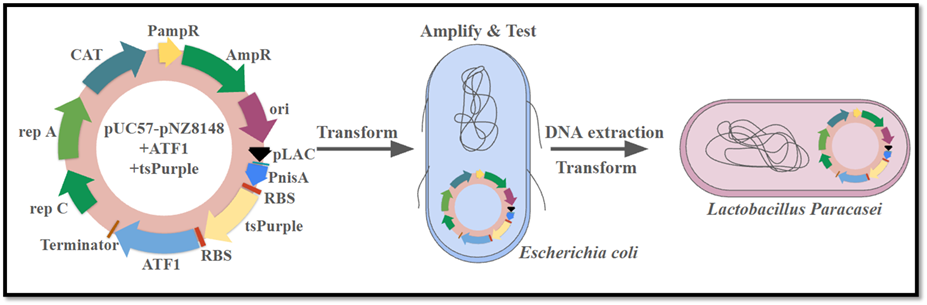 |
|---|
Our plasmid design incorporates two promoters to enable expression across both host organisms: a lac promoter for E. coli and the PnisA promoter for L. paracasei. To facilitate natural flavor production, we incorporated the ATF1 gene, which is naturally found in yeast and encodes the enzyme alcohol acetyltransferase (AATase). This enzyme catalyzes the synthesis of esters such as isoamyl acetate, the compound responsible for a distinct banana aroma. This approach utilizes synthetic biology to enable the in situ biosynthesis of natural flavors. ATF1. Isoamyl acetate is widely used in food and beverage fermentation and is recognized as safe for consumption (Verstrepen et al., 2003; FDA, 2023).
| Figure 2: This figure demonstrates the induction of AATase with Ethanol, Butyl, and Isoamyl to produce flavorful esters. (Verstrepen et al., 2003) | 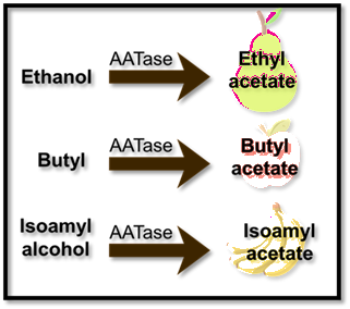 |
|---|
To complement ATF1, our construct includes the tsPurple gene, which encodes a chromoprotein derived from sea anemones. tsPurple serves as a visual marker of successful transformation and expression (Shih, 2015). Unlike antibiotic resistance markers, which are unsuitable for food-grade products, tsPurple provides a safe, non-antibiotic visual indicator. Its vivid purple coloration also offers potential for developing aesthetically appealing, naturally pigmented yogurt products (Sunico, 2024).
Ultimately, this study aims to engineer a dual-host plasmid that expresses both a flavor-producing enzyme (ATF1) and a chromoprotein reporter (tsPurple) in L. paracasei and E. coli. The system was first validated in E. coli, where IPTG induction successfully triggered gene expression, resulting in distinctly purple colonies that confirmed functional activity.
Protein production was further evaluated using SDS-PAGE under varying growth and induction conditions to optimize expression levels. These findings provide a successful proof of concept for using recombinant vectors to produce natural flavor esters such as isoamyl acetate. The work establishes a foundation for future development of food-grade engineered microorganisms capable of producing naturally flavored probiotic yogurt products (Hasan Sowrav et al., 2025).
To evaluate this system experimentally, we constructed and tested a dual-host recombinant plasmid in E. coli before future application in L. paracasei.
Materials and methods
Bacterial strains and plasmids
We selected L. paracasei as the host organism because it naturally ferments milk and offers recognized probiotic benefits. However, because of its low transformation efficiency, the shuttle vector pUC57-pNZ8148 was chosen. This vector carries the antibiotic resistance genes chloramphenicol acetyltransferase (CAT) and ampicillin resistance (AmpR) (Novopro, 2025). It can replicate in both E. coli and L. paracasei, enabling expression in both hosts. The vector also includes the lac promoter for E. coli and the nisin (PnisA) promoter for Lactobacillus (Novopro, 2025).
For visual and functional components, we used iGEM part BBa_K1033906, generously provided by the Frugal Science Academy at Georgia Tech, which contains the visual reporter gene tsPurple (Jamal, 2013). In addition, the pJL1 ATF1 plasmid (Addgene), which carries the ATF1 gene encoding the alcohol acetyltransferase (AATase) enzyme, was incorporated to enable the production of natural flavor esters such as isoamyl acetate, which is responsible for banana scent and flavor (Huang et al., 2018).
| Figure 3: Detailed maps of the three plasmids used in the experiments. To the left is the shuttle vector that carries the genes from the other plasmids. | 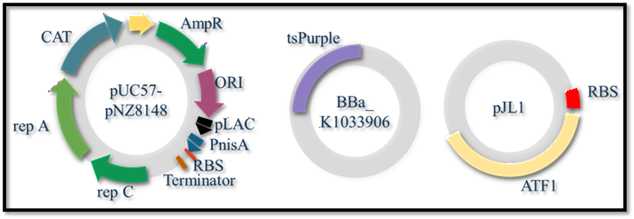 |
|---|
Plasmid design, initial transformations
Plasmid Construction Design: The intended order of gene assembly was Vector → tsPurple → ATF1 (inserted genes within the vector backbone). This arrangement allows tsPurple to utilize the ribosome binding site (RBS) already present in the vector, ensuring efficient translation initiation. The ATF1 gene was placed downstream to take advantage of an additional RBS introduced through a point mutation during amplification, supporting independent translation of both genes.
Initial Transformations and Plasmid Extraction: Due to limited initial quantities of tsPurple and ATF1 plasmids, E. coli transformations were performed using the New England Biolabs (NEB) transformation protocol. Transformed cells were cultured in LB medium to amplify the plasmids. Plasmid DNA was then isolated using the Monarch® Miniprep Kit (NEB) following the manufacturer’s protocol, yielding sufficient quantities for subsequent cloning and assembly experiments (New England Biolabs-Transformation protocol, 2025).
PCR amplification and vector preparation
For assembly, tsPurple and ATF1 fragments were amplified using polymerase chain reaction (PCR), while the shuttle vector was amplified and linearized to accept the inserts. Primer sequences were designed with NEBuilder software based on the following regions (New England Biolabs (Q5 PCR), 2025): the tsPurple fragment from BBa_K1033906 (2036–2736 bp of the coding region), the ATF1 region from pJL1 (666–2277 bp), and the vector linearization site from 2456–2443 bp. Primers were synthesized by Integrated DNA Technologies (IDT,2017).
The following is a chart of our primers for each plasmid:
| Name | Primer 5’ (overlap/spacer/Anneal) | Len. | %GC | 3’ %GC | 3’ Tm |
|---|---|---|---|---|---|
| Backbone_fwd | ggcatgcggtaccactag | 18 | 61 | 61 | 65.7 |
| Backbone_rev | ggtgagtgcctccttataatttattttg | 28 | 36 | 36 | 63.4 |
| ATF1 pJl1_fwd | ctttgagaccttttgttaactttaagaaggagg | 33 | 36 | 30 | 57.6 |
| ATF1 pJl1_rev | aactagtggtaccgcatgcccttattatggacccagcaac | 40 | 50 | 45 | 60.2 |
| tsPurple_fwd | attataaggaggcactcaccatggcgagcttggttaag | 38 | 47 | 50 | 62.3 |
| tsPurple_rev | gttaacaaaaggtctcaaagcttattacgtc | 31 | 36 | 43 | 60.2 |
Table 1: Color-coded chart of the primers designed for each plasmid, including calculated melting temperatures (Tm) and amplicon lengths. (The chart was based on the results from using NEBuilder Assembly Tools.)
Additionally, a point mutation was introduced in pJL1 at 682 bp, changing AGGAGA to AGGAGG to generate a functional ribosome-binding site (RBS) to enhance translation efficiency.
Methods summary: DNA assembly, transformation, and protein expression
PCR Cleanup: Before assembly, PCR cleanup was performed to maximize downstream efficiency. The Monarch® PCR & DNA Cleanup Kit (5 µg) from New England Biolabs (NEB) was used according to the manufacturer’s protocol to purify amplified DNA fragments and remove excess primers, nucleotides, and enzymes (New England Biolabs(Spin PCR & DNA Cleanup), 2025).
Plasmid Construction: The purified tsPurple and ATF1 fragments were assembled into the shuttle vector backbone using NEBuilder HiFi DNA Assembly. Based on the NEBuilder calculator, DNA fragments were diluted as instructed and combined with the assembly master mix and incubated according to the manufacturer’s instructions to facilitate seamless ligation (New England Biolabs (NEBuilder), 2025). The resulting recombinant plasmids were verified by gel electrophoresis. This assembly design enables independent translation of each gene, with individual ribosome-binding sites (RBS) ensuring consistent and balanced gene expression.
| Figure 4: Map of plasmid regions amplified by PCR, with color-coded fragments indicating targeted DNA segments. Also, a simple diagram illustrating the NEBuilder process for assembling the recombinant plasmid. | 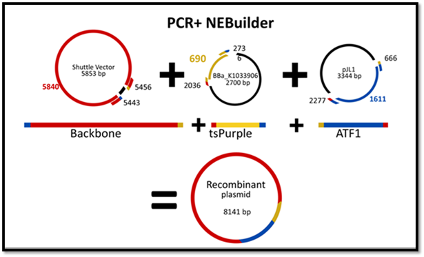 |
|---|
Transformation (Competent Cells): The recombinant plasmid was initially transformed into E. coli (NEB 5-alpha) due to its high transformation efficiency. Transformation followed the NEB heat-shock protocol: 2 µL of plasmid DNA was added to competent cells, incubated on ice, heat-shocked at 42°C for 30 seconds, then recovered in 950 µL of SOC medium at 37°C for 1 hour with shaking (New England Biolabs (Transformation), 2012).
Cells were plated on LB agar containing ampicillin and chloramphenicol and incubated overnight at 37°C. Ampicillin was used for final screening. Successful transformants were confirmed through colony growth and visual screening.
Protein Expression: Isopropyl β-D-1-thiogalactopyranoside (IPTG) was used to induce the lac promoter to confirm successful assembly of our plasmid. One plate was grown with no IPTG as a control. For testing, before spreading cells, 40 µL of 100 mM IPTG was spread on the agar surface and allowed to dry for 30 minutes. Subsequently, 50 µL of the transformed E. coli culture was spread on the IPTG-containing plates.
Induction of the lac promoter triggered the transcription and translation of tsPurple and ATF1. Successful expression was visually confirmed by the purple coloration of colonies, indicative of tsPurple expression.
Additionally, two liquid cultures were tested: one as a control and one induced with 25 µL of 100 mM IPTG, both incubated at 37°C for 2 days.
Flavor Expression: For testing the production of fruity esters in liquid culture, a 15 mL main culture of E. coli was grown at 37°C for two days. The culture was then aliquoted into eleven 1.5 mL microcentrifuge tubes, each containing 500 µL of culture. Two tubes were designated as controls, while the remaining tubes were divided into three groups corresponding to three different alcohol substrates (isoamyl alcohol, butyl alcohol, and ethyl alcohol), as shown in Figure 2.
Within each alcohol group, one tube was left uninduced, while two tubes were induced with IPTG. One of the two control tubes was also induced with IPTG. All tubes were incubated at room temperature in a biological safety hood for over 48 hours to allow expression of the ATF1 gene.
Following the expression, cultures were pelleted by centrifugation at 13,000 RPM for 5 minutes, and the supernatant was removed. Each pellet was resuspended in 200 µL of nuclease-free water. Alcohol substrates were then added directly to the resuspended cells: 2 µL of isoamyl alcohol (88.15 g/mol), 2 µL of butyl alcohol (74.12 g/mol), or 1 µL of ethyl alcohol, depending on the experimental group. The samples were incubated at room temperature for four hours to facilitate the enzymatic reaction between the alcohols and alcohol acetyltransferase (AATase). This process resulted in the synthesis of isoamyl acetate, butyl acetate, and ethyl acetate; the key esters responsible for fruity aromas.
After olfactory testing by surveying participants to see if they identified the scents, on the following day, a second round of alcohol was added to some tubes to observe whether the fragrance could become stronger. Each alcohol group was used to assess the effect of increased substrate concentration. This resulted in the following final conditions: one uninduced control, one IPTG-induced control, three tubes containing alcohol only, three tubes containing IPTG and alcohol, and three tubes containing IPTG with two sequential alcohol additions.
Protein Purification and SDS-PAGE Analysis: To assess protein expression levels under varying conditions, six liquid cultures of transformed E. coli were prepared. To start, one culture was grown for 16 hours. Next, 50 µL of the starting culture was added to the 6 LB medium tubes to mid-log phase (approximately 2 hours). Then, two cultures were kept as controls (no IPTG), two were induced with 25 µL of 100 mM IPTG, and two with 50 µL of 100 mM IPTG. Each set was tested at 37°C and at room temperature.
Cultures grown at 37°C were incubated overnight before pelletization and stored at 4°C, while those grown at room temperature were incubated for 24 hours. To prepare the samples and perform the SDS page, the standard protocol was followed. The samples were prepared using Laemmli buffer (with DTT added) and analyzed by SDS-PAGE to verify protein expression and compare the induction conditions (Team Fudan, 2022).
| Figure 5: A.Genetic circuit transformed in E. coli. ITPG was used to express our genes, tsPurple chromoprotein, and AATase. Isoamyl alcohol was later added to catalyze with AATase to produce isoamyl acetate, a molecule with a banana scent and flavor. B.Proposed transformed system for the recombinant plasmid in Lactobacillus, utilizing nisin to activate the PnisA promoter to start transcription. (Hasan Sowrav et al., 2024) | 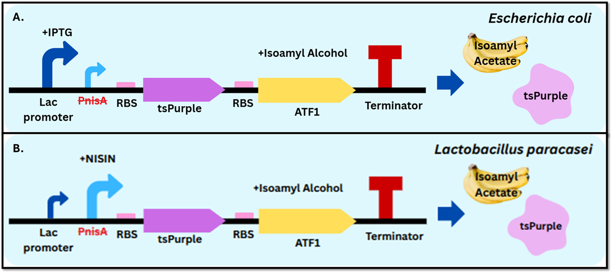 |
|---|
Results
PCR amplification and vector preparation
We successfully performed PCR on all our plasmids using NEB protocols and confirmed our results through gel electrophoresis, as seen in Figure 6. Additionally, a nano spectrophotometer was used to measure concentrations for the subsequent steps.
| Figure 6: Gel electrophoresis results after PCR confirming successful amplification of plasmids. DNA band sizes were compared to a 1 kb ladder as a reference. | 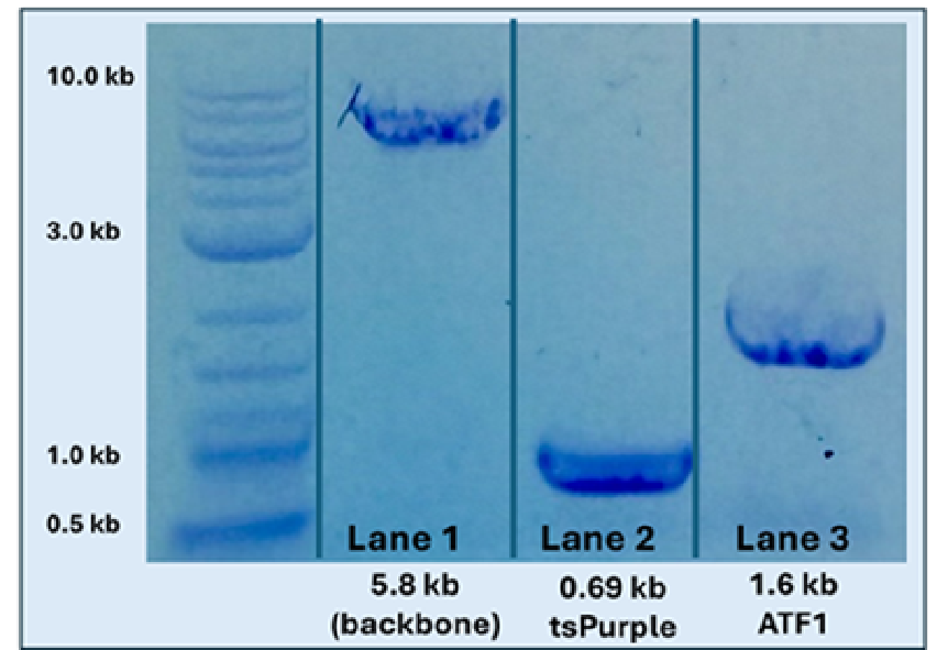 |
|---|
PCR cleanup
To address previous assembly issues, we purified our DNA using a NEB spin kit. As shown in Figure 6 below, there are clear bands for each amplified section, indicating suitable parts for assembly.
Plasmid construction
Following the cleanup, we assembled the plasmid using NEBuilder. Although it was not necessary to confirm successful assembly before transformation, we checked the products via gel electrophoresis.
| Figure 7: Gel of products after assembly. Our recombinant plasmid band (8.4 kb) in lane one was considerably higher than the positive control plasmid from NEB (5.2 kb) and aligned with the correct ladder segment. | 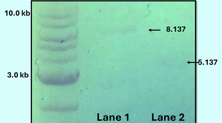 |
|---|
Transformations (competent cells)
We transformed our recombinant plasmid into DH5α cells using the heat shock method. A few purple colonies appeared before IPTG, indicating proper assembly, but also showed leaky expression of the lac promoter. Interestingly, the number of purple colonies increased when the plate was grown at 4°C, suggesting 4°C is the better fit for the optimal expression temperature of tsPurple.
| Figure 8: The first plate of our cells, and as seen on the bottom right side, there were a few purple colonies indicating the leaky nature of the lac promoter. | 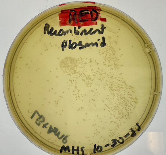 |
|---|
Protein expression
After growing control cells on a clean plate and on a plate layered with IPTG, we initially saw no purple colonies. However, we noticed the purple colonies after placing the plates at 4°C overnight. Additionally, as shown in Figure 9, tsPurple was observed on the control plates. Although the control plate has fewer purple colonies than the IPTG plate, purple colonies should not have been present in the control plates. This comparison shows the leaky nature of the Lac promoter.
| Figure 9: The right plate is the control for the experiment, and the left is the one induced with IPTG. The purple colonies represent cells that had successfully expressed tsPurple. | 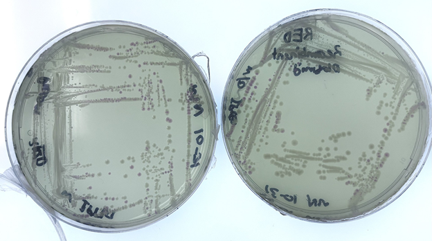 |
|---|
Flavor expression and sensory testing
On the first day of sensory testing, a survey was conducted with ten participants, and the liquid culture induction produced distinguishable results. Participants were able to differentiate between control samples, alcohol-only samples, and IPTG-induced samples across all alcohol conditions. Notably, a strong majority of participants (8 out of 10) reported that cultures induced with ethanol produced the fruitiest aroma.
On the second day, six additional participants were surveyed (n = 16). While participants continued to distinguish between samples, aroma preference shifted: butyl alcohol was preferred by 8 of 16 participants, ethyl alcohol by 6 of 16, and isoamyl alcohol by 2 of 16.
Although subjective, these results indicate ATF1 expression and ester production. The observed shift in aroma preference may reflect differences in the time required for each alcohol substrate to fully convert to its corresponding acetate ester.
| Figure 10: Percentage of participants preferring each ester aroma on Day 1 (n = 10) and Day 2 (n = 16). | 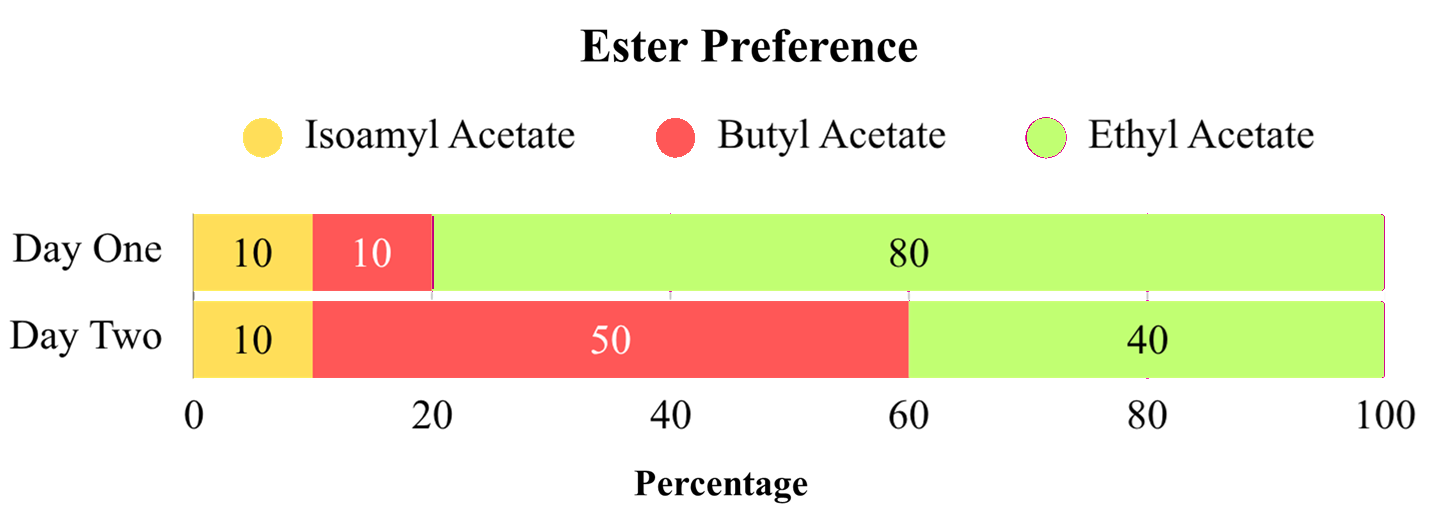 |
|---|
Protein expression observation
Basal expression (Figure 11A): Both uninduced (with IPTG) cultures exhibited visible purple pigmentation at room temperature, with color intensity increasing over time. This observation suggests significant basal expression ("leakiness") of the lac promoter, allowing for protein production even in the absence of the inducer(Figure 11A).
Aggregation and Temperature Sensitivity (Figure 11B): Protein aggregation occurred when cultures were incubated at 37°C over the weekend. This clumping likely indicates the overexpression of the recombinant protein (potentially AATase) or host strain (DH5α cells) intolerance to the metabolic burden. Notably, when the cultures were subsequently moved to refrigeration (4°C), the aggregated proteins appeared to resolubilize. This unusual reversibility suggests a temperature-dependent solubility characteristic of the expressed proteins (Figure 11B).
| Figure 11: Protein expression observations | 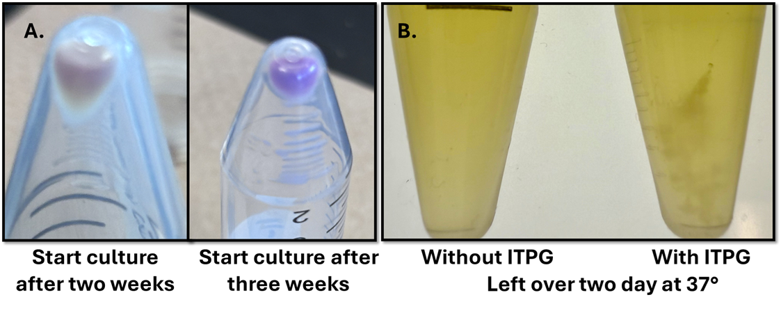 |
|---|
11A-Starter cultures were left at room temperature without IPTG for 2 and 3 weeks, respectively.
11B Cultures induced (right) and uninduced (left) that were left over the weekend at 37 degrees.
SDS-page
Although the successful assembly of our genes and plasmid was visually confirmed by tsPurple, the promoter's leakiness demonstrated that expression specificity was an issue, which could be detrimental to control. To determine the severity of the leaky Lac promoter, samples were induced with varying IPTG concentrations, and SDS-PAGE was used to objectively analyze expression.
As shown in the figure below, the bands for tsPurple (slightly under 37 kDa) were most apparent at room temperature (RT); the same was true for the AAtase enzyme (ATF1, under 75 kDa), although there were also bands for the other samples. Additionally, the bands for each protein were darker as the IPTG concentration increased. Taking these observations into account, we can conclude that the optimal expression temperature for the protein is approximately room temperature, and that although the lac promoter is leaky to some degree, it yields lower protein expression than regular IPTG induction.
| Figure 12: Labeled SDS-PAGE protein gel used to analyze the expression of proteins under differing conditions. The more apparent and thicker the band, the higher the level of expression. | 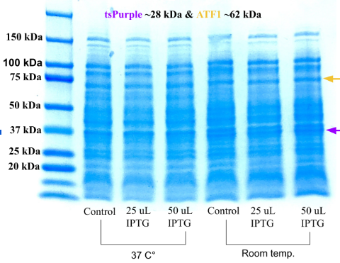 |
|---|
Ultimately, these results confirm that our recombinant plasmid was assembled correctly and expressed both tsPurple and ATF1 in E. coli, validating our idea and system before future testing in Lactobacillus.
Discussions
The designed construct and genetic system were confirmed to be functional, performing as expected. Successful assembly and expression were evidenced by the development of purple colonies and the detection of characteristic fruity aromas. Throughout the experimental process, a biological safety cabinet was utilized to ensure aseptic conditions and prevent contamination.
Several key insights emerged regarding the system’s performance. Notably, induction experiments revealed the "leaky" nature of the lac promoter, as purple pigmentation was observed even in uninduced samples. This suggests a lack of tight transcriptional control, which may have implications for the PnisA promoter intended for future Lactobacillus applications. Furthermore, protein expression appeared more robust at room temperature, suggesting temperature-dependent activity for tsPurple and potentially ATF1. At 37°C, induced cultures exhibited visible clumping, likely a result of metabolic burden or high levels of recombinant protein expression in the host cells.
This study had certain limitations, primarily that testing was restricted to E. coli due to time constraints, leaving the system’s performance in L. paracasei unverified. Additionally, we were unable to quantitatively confirm ester production using analytical methods such as gas chromatography. While qualitative data from participant surveys indicated fruity aroma production, these results do not constitute definitive chemical proof of ester synthesis.
Overall, these findings demonstrate the potential of this recombinant system to produce detectable flavor esters. This work supports the future development of engineered probiotics capable of generating natural flavors in situ during fermentation.
Next steps
As a successful expression of our pathway was confirmed in E.coli, our next task is to remove the antibiotic resistance genes from our plasmid before shuttling it into L. paracasei. This will be done in accordance with the rules of food-grade vectors, using PCR to remove the harmful genes. After removal and testing for safety with sequencing, we will be able to transform it into L. paracasei, which is the last step necessary to prepare for the fermentation of yogurt.
We also found that L. paracasei is used in many fermented products such as kefir, cheese, and sour beer. Being a probiotic, it is included in these products to increase the health benefits. With that in mind, we also plan to work with these products to enhance benefits and improve flavor profiles with many different fruity esters.
Overall, enabling yogurt cultures to generate their own natural flavors reduces reliance on external fruit additives, minimizes agricultural waste, and supports sustainable, food-safe production. Our project aligns with the growing interest in naturally derived ingredients and sustainable food technologies. Moreover, the use of genetically modified probiotics can enhance nutritional content while lowering the costs associated with flavoring and production (Maryville, 2022).
| Figure 13: Demonstrates our future idea for the fermentation process in yogurt; steps 1 through 4 occur before the fermentation process with the insertion of the starter cultures. | 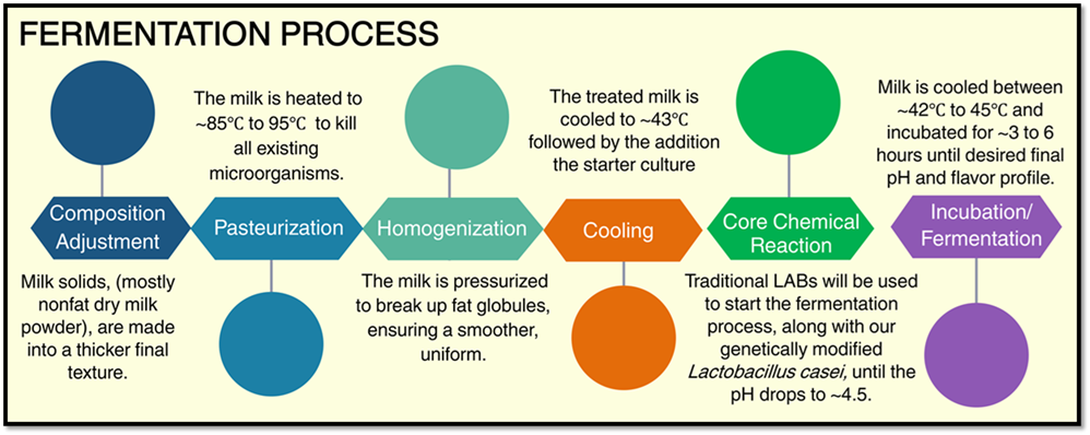 |
|---|
Author contributions
MHS, WO, LO, and YM developed the initial project concept in the previous year. MHS planned and performed the majority of the wet-lab experiments, with assistance from NK, who also documented experimental procedures and wrote the abstract. WO, LO, YM, and MR developed the background section, with WO additionally working on the next steps under guidance from MHS and NK. MHS wrote the materials and methods, results, and discussion sections and constructed the figures using Google Slides. MHS and NK coordinated and led manuscript editing. All authors contributed to formatting and final review of the manuscript.
Acknowledgements
This project was made achievable through the support of Ana White, who founded both the BioBuilder club and the research class at Cross Keys High School, where most of this work took place. We are deeply grateful for her dedication in providing us with the opportunity and continuous guidance in developing our ideas and carrying out this project.
We extend our appreciation to our mentor, Gitanshu Bhatia, for technical guidance in fermentation and for supporting the project’s development.
Finally, we would also like to greatly thank BioBuilder, Georgia Tech’s Frugal Science Academy, and the Bhamla Lab for providing time, guidance, and resources essential to completing our experiments.
References
Hadjimbei, E., Botsaris, G., & Chrysostomou, S. (2022). Beneficial Effects of Yoghurts and Probiotic Fermented Milks and Their Functional Food Potential. Foods, 11(17), 2691. https://doi.org/10.3390/foods11172691
Hasan Sowrav, M., et al. (2025). Flavor by design: Engineering flavored probiotics in yogurt. BioTreks, 10, 1–9. biotreks.org/e202541/
High Efficiency Transformation Protocol (C2987H/C2987I) | NEB. (n.d.). Www.neb.com. https://www.neb.com/en-us/protocols/0001/01/01/high-efficiency-transformation-protocol-c2987
Huang, A., et al. (2018). BioBitsTM explorer: A modular synthetic biology education kit. Science Advances, 4(8), eaat5105. https://doi.org/10.1126/sciadv.aat5105.
Jamal, S. (2013). Part:BBa_K1033906 – parts.igem.org. Parts.igem.org. parts.igem.org/Part:BBa_K1033906.
MARYVILLE UNIVERSITY. (2022, July 12). Why Is Agriculture Important? Benefits and Its Role. Maryville Online; Maryville University. https://online.maryville.edu/blog/why-is-agriculture-important/
MedlinePlus. (2020, August 18). Lactose intolerance: MedlinePlus Genetics. Medlineplus.gov. https://medlineplus.gov/genetics/condition/lactose-intolerance/
NEBuilder. (2025). Nebuilder.neb.com. https://nebuilder.neb.com/#
New England Biolabs. (2012, May 21). Transformation Protocol. Neb.com. https://www.neb.com/en-us/protocols/transformation-protocol
New England Biolabs. (2025a). Standard Cleanup Protocol using the Monarch® Spin PCR & DNA Cleanup Kit and Centrifugation (NEB #T1130). Neb.com. https://www.neb.com/en-us/protocols/standard-cleanup-protocol-using-the-monarch-spin-pcr-and-dna-cleanup-kit-and-centrifugation
News, Press Room, and Blogs | GreenChem Industries. (2025). Greenchemindustries.com. https://greenchemindustries.com/entry/150/isoamyl-acetate
PCR Using Q5® High-Fidelity DNA Polymerase (M0491) | NEB. (n.d.). Www.neb.com. https://www.neb.com/en-us/protocols/2013/12/13/pcr-using-q5-high-fidelity-dna-polymerase-m0491
Popović, N., Brdarić, E., Đokić, J., Dinić, M., Veljović, K., Golić, N., & Terzić-Vidojević, A. (2020). Yogurt Produced by Novel Natural Starter Cultures Improves Gut Epithelial Barrier In Vitro. Microorganisms, 8(10), 1586. https://doi.org/10.3390/microorganisms8101586
pUC57-pNZ8148 vector map and sequence. (2025). Novoprolabs.com. https://www.novoprolabs.com/vector/Vgeztsnrx
Shih, C.-H., Chen, H.-Y., Lee, H.-C., & Tsai, H.-J. (2015). Purple Chromoprotein Gene Serves as a New Selection Marker for Transgenesis of the Microalga Nannochloropsis oculata. PLOS ONE, 10(3), e0120780. https://doi.org/10.1371/journal.pone.0120780
Speicher, N. (2017, March 31). Expert tips for resuspending oligos and primers | IDT. IDT. https://www.idtdna.com/page/support-and-education/decoded-plus/tips-for-resuspending-and-diluting-your-oligonucleotides/
Súnico, V., Piunti, I., Bhattacharjee, M., Mezzetti, B., Caballero, J. L., Muñoz-Blanco, J., Ricci, A., & Sabbadini, S. (2024). Overview of Current Selectable Marker Systems and Novel Marker-Free Approaches in Fruit Tree Genetic Engineering. International Journal of Molecular Sciences, 25(22), 11902–11902. https://doi.org/10.3390/ijms252211902
Team Fudan iGEM. (2022, October 11). Preparation of bacterial cell lysate for SDS-PAGE (2022 iGEM). Protocols.io. https://www.protocols.io/view/preparation-of-bacterial-cell-lysate-for-sds-page-14egn2zwyg5d/v1?step=5
Verstrepen, K. J., Van Laere, S. D. M., Vanderhaegen, B. M. P., Derdelinckx, G., Dufour, J.-P., Pretorius, I. S., Winderickx, J., Thevelein, J. M., & Delvaux, F. R. (2003). Expression Levels of the Yeast Alcohol Acetyltransferase Genes ATF1, Lg-ATF1, and ATF2 Control the Formation of a Broad Range of Volatile Esters. Applied and Environmental Microbiology, 69(9), 5228–5237. https://doi.org/10.1128/aem.69.9.5228-5237.2003
Wajs, J., Brodziak, A., & Król, J. (2023). Shaping the Physicochemical, Functional, Microbiological, and Sensory Properties of Yoghurts Using Plant Additives. Foods, 12(6), 1275. https://doi.org/10.3390/foods12061275
Wang, X., Zhang, P., & Zhang, X. (2021). Probiotics Regulate Gut Microbiota: An Effective Method to Improve Immunity. Molecules, 26(19), 6076. https://doi.org/10.3390/molecules26196076