Badkundri Reva, Bharadwaj Skanda, Chennam Varsha, Kallu Eshawnvie, Krishnamoorthy Sarvesh, Murad Matthew, Raghunathan Aray, Rajuladevi Naga Sahithi, Song Yeryn, and Surange Swadha. ▫ BioBuildersClub, Lambert High School, Suwanee, Georgia, United States; Biobuilder Educational Foundation, Newton, Massachusetts, United States
Reviewed on 6 May 2023; Accepted on 13 July 2023; Published on 16 October 2023
With help from the 2023 BioTreks Production Team.
This year, the Lambert iGEM seminar team developed a project about the detection of the early onset of mast cell tumors, otherwise known as MCTs, in canids (dogs). Mast cells refer to specific types of white blood cells located in animals that are vital in the functioning of the immune system to fight off bacteria and parasites within the organism. However, if these cells begin to proliferate uncontrollably, they are capable of forming lumps on the skin known as mast cell tumors. These tumors have a negative impact on the health of animals, especially dogs, and this makes it crucial to create a novel detection system that can detect the presence of mast cell tumors before they negatively affect these mammals. This team’s inspiration came from Lambert iGEM’s competition team, which recently developed a novel detection method that utilizes miRNA biomarkers, small sections of RNA that serve to control mRNA gene expression, and two main processes known as rolling circle amplification (RCA) and fluorescence resonance energy transfer (FRET) to correlate the presence of certain diseases with decreased fluorescence (as shown in the diagram below). The fluorescence is then detected using the MicroQ sensor, which utilizes photoresistors to catch and quantify this fluorescence. The seminar team’s goal is to test the efficiency of this system using specific miRNAs that are related to the development of MCTs to analyze as well as making improvements such as changing the components of the MicroQ to better fit our project. The upregulation of a certain set of miRNAs, including miRNA-21, is directly correlated with mast cell tumors (MCTs) in canids. We hope this system improves early detection, providing the opportunity for treatment.
Keywords: miRNA, biomarker(s), canids, mast cell tumors
Authors are listed in alphabetical order. Chowdhury Sussmit, Kazman Maxwell, Matarrese Caroline, and Sharer Catherine mentored the group. Please direct all correspondence to .

Mast cell tumors, otherwise known as MCTs, are one of the most prevalent types of cancer present in canids. Studies show more than 20% of these animals obtain one at some point in their life. These tumors have the capability to release toxins into the body of the canids, leading to anaphylactic reactions that could become fatal (N. C. State Veterinary Hospital, n.d.). The Lambert iGEM seminar team hopes to provide a cost-effective and accurate way to detect the early onset of MCTs using microRNA (miRNA) biomarkers. miRNA refers to small sequences of RNA nucleotides that are responsible for regulating miRNA (set of RNA nucleotides) expression and can be used to show the presence of certain conditions, such as the coronary artery disease (the basis of Lambert iGEM’s project) (Lambert iGEM, 2022). In the recent year, the Lambert iGEM competition team utilized this concept along with an innovative biological process known as rolling circle amplification (RCA) and fluorescence detection (using a device called a MicroQ) to quantify the presence of their specific miRNA sequences (Lambert iGEM, 2022). mi-RNA 21, one of the key miRNAs involved in cell/tumor proliferation pathways in a variety of animals, has been shown to directly correlate with MCT growth and development. Currently, the best treatment available for the detection of mast cell tumors is expensive biopsies and examinations to find characteristics of the tumors (mi-RNA 21, one of the key miRNAs involved in cell/tumor proliferation pathways in a variety of animals, has been shown to directly correlate with miRNA. These processes cost a large amount of money, up to thousands of dollars, and do not always deliver the most accurate results (Gieger et al., 2005). By monitoring this condition using fluorescence, we hope that this project will help in monitoring the early onset and progression of MCTs in Canids to provide early treatment for dogs (Lambert iGEM, 2022).
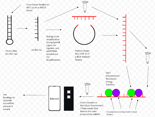
| Figure 1. Describes the process around our project. The first step would be to extract the miRNA, which is through the collection of saliva/blood. Then, it will be processed through RCA (described below) to produce the Rolling Circle Product After that, the process of FRET (described below) takes place, producing fluorescence output. Finally, the MicroQ detects this fluorescence output. |
|---|
Systems level
At a microscopic level, this system involves an intricate process that produces fluorescence used for quantification. The first main step in this process is what is known as RCA (Lambert iGEM, 2022). First, a padlock probe, which is a set of DNA nucleotides (30-150), has arms that directly complement specific miRNA sequences (Lambert iGEM, 2022). In a process called hybridization, the miRNA attaches to the arms of the padlock probe and the process of ligation, using the enzyme called SplintR ligase, to completely seal the full strand (Lambert iGEM, 2022). Then, a DNA polymerase called phi29 polymerase uses the miRNA as a template to transcribe/replicate a new strand known as the rolling circle product (RCP) (Lambert iGEM, 2022).

| Figure 2. The process of the miRNA binding to the DNA in Hybridization. Blue: Padlock Probe, Purple / Pink: miRNA (top strands) and Respective Complementary 5’ and 3’ arms (Lambert iGEM, 2022). Key: Blue: Padlock Probe Purple / Pink: miRNA (top strands) and Respective Complementary 5’ and 3’ arms |
|---|
After that, the process of luorescence resonance energy transfer (FRET) begins as the RCP. This RCP contains complementary regions for molecules known as linear probes and is rolled out into a linear strand (Lambert iGEM, 2022). Then, probe molecules known as fluorophores (molecules that enhance light) and quenchers (molecules that reduce light) are added to the process (Lambert iGEM, 2022). They bind together on the linear probe complementary DNA regions on the RoCR where the process of FRET takes place, which decreases fluorescent output (Lambert iGEM, 2022).
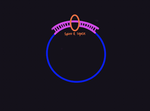
| Figure 3. The process of ligation in which the SplintR Ligase seals the gap in the structure (Lambert iGEM, 2022). |
|---|
The light in the sample decreases as the quantity of miRNA increases (Lambert iGEM, 2022).
Device level
Along with the additional devices listed above, which include the functional padlock probe nucleotide sequence, the process of RCA that amplifies the miRNA sequence, and the FRET process that involves the use of linear probes to quench fluorescence in quantifiable units, the device that is used to detect the fluorescence is known as the MicroQ sensor (Lambert iGEM, 2022). This sensor is equipped with some main functional parts, which include the blue light source, which shoots blue light waves (approximately 440 nm wavelength) through the sample present in the container. This is then converted into green light that passes through the emission filter, which directs the amount of the light to the photoresistor. This component collects and sends the light through wires to an ESP32, which calculates it in quantifiable units (schematics shown on competition team website and low) (Lambert iGEM, 2022).
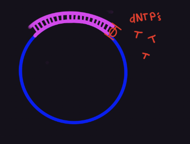
| Figure 4. The beginning of amplification where dNTPs are added using the phi29 polymerase enzyme (Lambert iGEM, 2022). |
|---|
In terms of engineering parts/organisms, the major engineered parts include the miRNA, the Padlock Probe, and the RCP (Lambert iGEM, 2022). As stated above, the padlock probe is vital to the replication, as it contains the complementary units to miRNA that are required to produce the RCP (Lambert iGEM, 2022). Then, the RCP is important in that it is where the linear probes bind to quench fluorescence, allowing for this process to work (Lambert iGEM, 2022).

| Figure 5. The rolling circle product from the RCA process (Lambert iGEM, 2022). |
|---|
Parts level
Overall, the parts that are required include some essential biological components and other engineering components (Lambert iGEM, 2022). The team requires the use of sequences of the specified miRNA, which is miRNA-21 (hsa-miR-21). This component serves as the fundamental part of the RCA process, as it is the main template that initiates hybridization in RCA (Lambert iGEM, 2022). Along with that, the team will also need to utilize the 5’ and 3’ arms for the miR-21 as well as the complementary padlock probes (5’ / 3’ arm for hsa-miR-21 and hsa-miR-21 padlock probe.) to execute the amplification process, as these parts will be involved in the hybridization and amplification process where the miRNA binds to the arms of the probe (described in depth above) Lastly, our team will need access to quencher and fluorophore probes, specifically the 6-carboxyl-fluorescein (FAM) Labeled DNA probe and the Black Hole Quencher 1 (BHQ1) DNA probe (Lambert iGEM, 2022). These probes allow the process of FRET, where the linear probes bind to the RCPs, to occur by serving as the linear probes.

| Figure 6. The rolling circle product is broken down into its consecutive parts (miRNA complements and linear probe complements). The circles represent linear probes known as quenchers (purple) and fluorophores (green) (Lambert iGEM, 2022). |
|---|
Safety
The team’s main safety concern for our lab is the execution of our proposed lab. In terms of the team’s development/manufacturing of the various parts/components needed for our experiment, the majority of the components will be accessible to us through the Lambert iGEM Competition Team, which have been proven safe and reliable to use after extensive studies with their project studying coronary artery disease (Lambert iGEM, 2022). The only part of the experiment that the team will have to worry about would be our sources for the cells we plan to extract. One of the most important ideas that we have to consider when running/constructing the experiment is to avoid contamination of the cells that were extracted, whether we use test bacteria, saliva samples, or blood samples. This contamination could proliferate into a contagion that can infect the lab, which also means that when handling these samples, PPE (personal protective equipment) is required, as well as cleaning equipment to maintain sanitary conditions. As stated before, the other materials, such as some of the miRNA molecules used can be used up without causing contamination, but it is still vital to use safe procedures to prevent any spills.

| Figure 7. The process of Fluorescent Resonance Energy Transfer (FRET) that decreases the fluorescence produced by the Fluorophores (Lambert iGEM, 2022). |
|---|
Discussions
Most certainly, the main benefits of the team’s experiment would be its implications. This experiment would reaffirm the Lambert iGEM competition team’s conclusions that it is possible to use miRNA biomarkers to detect and even quantify the propensity of certain conditions, including cancers like mast cell tumors. Obviously, more tests are going to have to be done to truly verify its results and what they actually mean at a large scale. The major challenges of this experiment would most likely be its feasibility for the team, as a lot of the parts and components the team needs are not simply obtained / have a high failure rate. Another challenge associated with the experiment would be being able to contact veterinarians and other doctors to bring/export our research to a higher level through human practices, as there is always the possibility of rejection due to certain aspects of our assay / the experiment. In terms of ethical concerns, there will be very little problems at the beginning of our trials, which would involve miRNA from non-pathogenic strains of bacteria such as Escherichia coli. However, if the team considers experimenting with canid cells, we will need to consider the threat the extraction of miRNA will have on these animals. The last major challenge would be effectively implementing the complex procedure designed by the competition team, which involves the use of processes that are prone to have a high failure rate, such as gel electrophoresis. This is due to the high amount of precision and accuracy required to produce visible separation of the DNA strands in the gel (as well as the processes of making the highest quality gel using TAE Buffer and Agarose), which may alter the team’s perception of the results / implications.
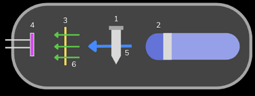
| Figure 8. The MicroQ involves the use of very high-level components that allow for fluorescence detection. Note: (1) The PCR tube that will be added to the device in order to detect fluorescence. (2) The blue laser 440 nm is used as a light source to excite the molecules in the sample. (3) The emission filter is used to direct the amount of light that reaches the photoresistor. (4) The photoresistor, which quantitatively measures the amount of light from the sample and sends it (with wires) to the ESP32, when it is then processes the amount and converts into the miRNA concentration. (5) Represents the blue light that passes through the sample. (6) Represents the green light that comes from the tube and goes through the emission filter. (Lambert iGEM, 2022) |
|---|
Next steps
We hope our team (or future teams) will continue this research by contacting local and global oncologists and veterinarians to understand more about the application and experimental design for this project. We also hope for other teams to conduct this experiment by looking into biosensor research to develop a novel sensor that they can use to detect their own specific miRNAs for their disorders and conditions (also study Lambert iGEM’s system). At the beginning of the next school year, we hope to be able to get access to the parts necessary to conduct experiments and produce verifiable results. This includes experiments such as purchasing miRNA samples, implementing the RCA and FRET processes above on bacterial samples and, eventually maybe animal cells (if permitted), and observing fluorescence output using our fluorescence sensor. This will have to be done through contact with our Lambert iGEM Competition Team to learn the major procedures necessary to successfully do the experiment.
Author contributions
A.R’s contributions include writing the abstract, keywords, article citations, background, systems level, device level, parts level, discussion, images, video, and safety of the article. N.S.R.’s contributions include writing the next steps, acknowledgments, and references S.B. and M.M: Original authors of the histamine detection system for mammary cancer, which was further altered as a team into miRNA biomarkers for mast cell tumors, a much more prevalent condition in dogs. for this article. V.S, S.K, S.S, S.B, Y.S, E.S, R.B, M.M, A.R, N.S.R – Contributed to the development of miRNA biomarkers proposal through research pertaining to KIT Proteins, miRNA biomarkers, histamine / mast cells, and related fields as part of a collaboration event for the Biobuilder Foundation.
Acknowledgements
Acknowledgements to our club mentors, Catherine Sharer and Caroline Matarrese, and our Georgia Tech mentors, Sussmit Chowdhury and Maxwell Kazman. They provided information and guided us through the process of figuring out the research topic and moving through with it. Regards to the Lambert iGem competition team for providing us the opportunity to build for our experiment and proposal. This includes running and analyzing tests produced from the MicroQ to show the frequency of the mi-RNAs in certain conditions that allows us to develop our own experiment.
References
Gieger, T., Northrup, N., & Wall, M. (2005). Clinical management of mast cell tumors in dogs. Compendium: Continuing Education for Veterinarians, 27(1), 56-67
Lambert IGEM (2022). CADlock. iGEM. Retrieved March 1, 2023, from https://2022.igem.wiki/lambert-ga/
N. C. State Veterinary Hospital. (n.d.). Medical oncology: Mast cell tumors in dogs. https://hospital.cvm.ncsu.edu/services/small-animals/cancer-oncology/oncology/mast-cell-tumors/ Zamarian, V., Stefanello, D., Ferrari, R., Chiti, L. E., Grieco, V., DallaCosta, E., Ceciliani, F., & Lecchi, C. (2023). Salivary miR-21 is a potential biomarker for canine mast cell tumors. Veterinary Pathology, 60(1), 47-51.