Frederick Suranyi and Abdullah Hashmi, Andover High School, Andover, Massachusetts, United States
Reviewed on 3 May 2025; Accepted on 9 June 2025; Published on 27 October 2025
With help from the 2025 BioTreks Production Team.
Skin cancer is one of the most common cancers globally, with melanoma being one of the most lethal subtypes due to its aggressive metastasis. Early detection is critical for improving a patient’s chance of survival (American Cancer Society, 2025). This project aims to propose a synthetic biological approach using engineered Escherichia coli (E. coli) to detect early-onset melanoma. Our system consists of E. coli modified to detect cancer biomarkers, by producing a visible color change. We arrange for people to schedule appointments for this as part of an annual routine. They would apply our foam-based solution evenly across their body, the foam approach allowing us to cover areas with hair, penetrating through hair to enable direct epidermal contact for reliable biomarker detection.
Keywords: skin cancer, melanoma detection, E. coli, synthetic biology, biosensor
Authors are listed in alphabetical order. Lindsey L'Ecuyer mentored the group. Please direct all correspondence to lindsey.lecuyer@andoverma.us.
Background
Cutaneous melanoma is one of humanity’s oldest documented malignancies, with evidence tracing UV-associated skin cancers back to early civilizations. Currently affecting 25 per 100,000 people annually in the U.S.Melanoma caused approximately 8,430 deaths this year (American Cancer Society, 2025). Despite these numbers dropping as more advanced treatments come out, melanoma is still the deadliest form of skin cancer out there, taking up a majority of skin cancer deaths while only accounting for about 1,4% of skin cancer cases (National Cancer Institute, 2025). This is because melanoma spreads much faster than other forms of skin cancer, not leaving much time for treatment and becoming fatal if it spreads to any vital organs. This is why early detection is so invaluable when treating melanoma.
We hypothesize that with our early-onset t cancer detection cream. Current detection methods are often invasive, expensive, or inaccessible. These include dermoscopy, full-body skin exams, biopsies, digital mole mapping, and AI-based imaging software used in clinical settings (American Cancer Society, 2025; Davis et al., 2019). In light of this, we aim to make detection more accessible using a biological system that can be applied at home. Using synthetic biology, we engineered E. coli to detect tyrosinase—an enzyme found at higher levels in melanoma cells—through a visible color change. Our method provides a non-invasive, cost-effective, and user-friendly alternative. The foam would be applied once per year, similar to an annual skin check, especially for individuals at higher risk for melanoma. It is spread across the body uniformly, left on for about 30–60 minutes, and then rinsed off. Any blue color change indicates potential melanoma biomarkers, telling the user to find professional help.
Systems Level
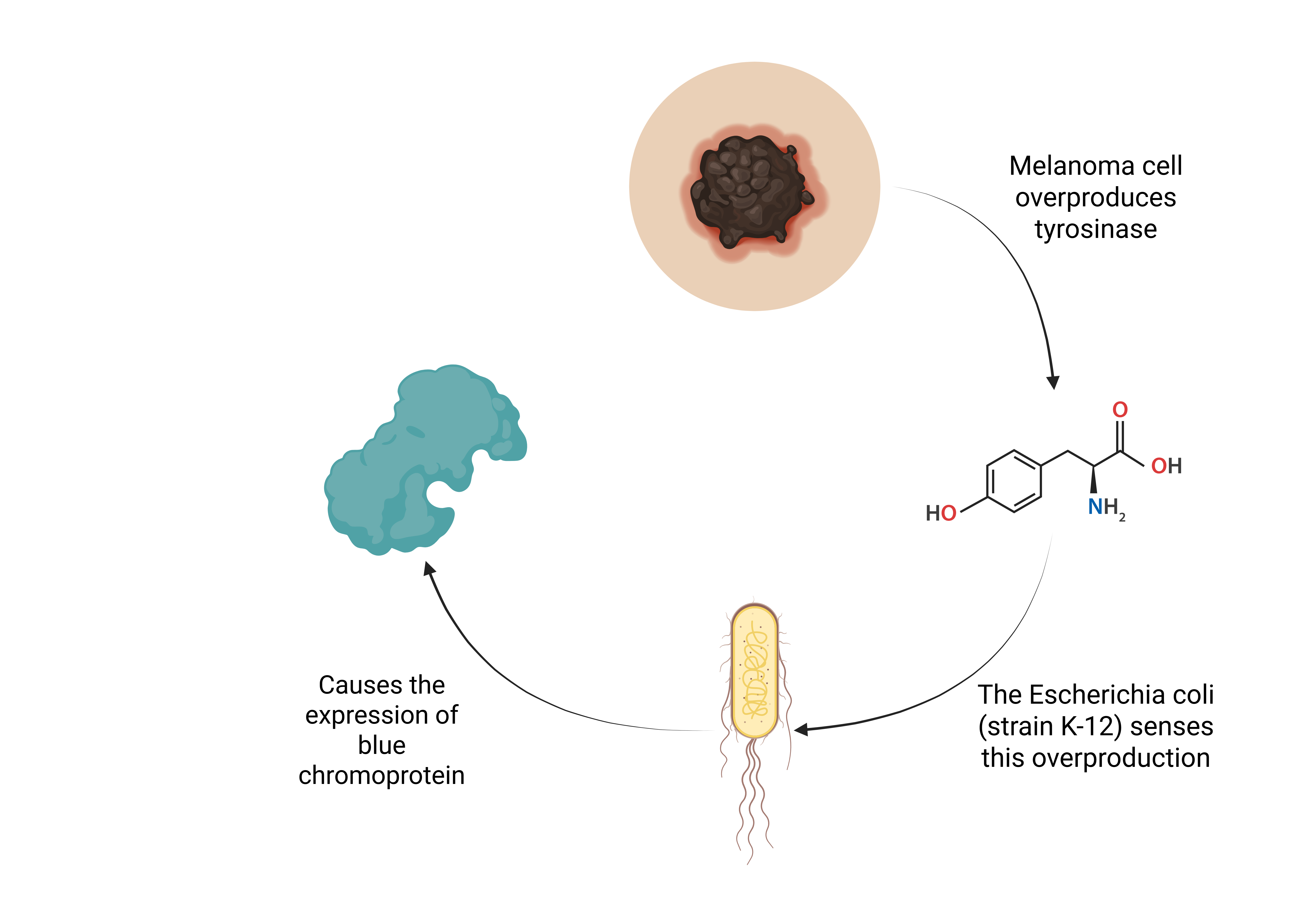
Our system is a topical foam designed to be applied across the entire surface of the skin. The foam contains genetically engineered E. coli DH5α, a derivative of the K-12 strain that is non-pathogenic and commonly used in molecular biology for its safety and high transformation efficiency (Thermo Fisher Scientific, 2025) (Figure 1).
DH5α is classified as a Biosafety level 1 (BSL – 1) organism, meaning that it is not known to cause disease in healthy humans and is considered safe for use. As a result, it is expected to have poor survivability outside a controlled environment and is unlikely to colonize or infect human tissue. Once applied, the engineered bacteria come into contact with the epidermis, interacting with the tyrosinase, a key melanoma biomarker. Tyrosinase is an enzyme produced by melanocytes, the cells responsible for melanin production. In the early stages of cutaneous melanoma, these melanocytes overproduce tyrosinase. Our bacteria are engineered to detect this unusually high concentration of tyrosinase and signal a potential melanoma spot (MedlinePlus Genetics, 2025). When this detection occurs, it triggers a genetic circuit that activates a reporter pathway, causing the bacteria to express blue chromoprotein (amilCP). This leads to a visible green skin discolorationunder UV or blue light, signaling that you should see a doctor for further medical evaluation (Tsien, 1998).To ensure control over the system, we also include a kill switch in the bacterial genome. This safety mechanism is designed to automatically destroy the bacteria after a set period or under specific environmental conditions, such as a lack of nutrients or exposure to certain temperatures or pH ranges. This helps ensure that the engineered E. coli does not persist or multiply uncontrollably on the skin.
| Figure 1. Mechanism of Detection in the Foam-Based E. coli Biosensor. Engineered E. coli in the foam detect 4-HPA, a metabolic byproduct of tyrosinase activity overexpressed in melanoma cells. Upon detection, the bacteria activate downstream modules that initiate a visible color change. |
|
Engineered E. coli in the foam detect 4-HPA, a metabolic byproduct of tyrosinase activity overexpressed in melanoma cells. Upon detection, the bacteria activate downstream modules that initiate a visible color change.
Device Level
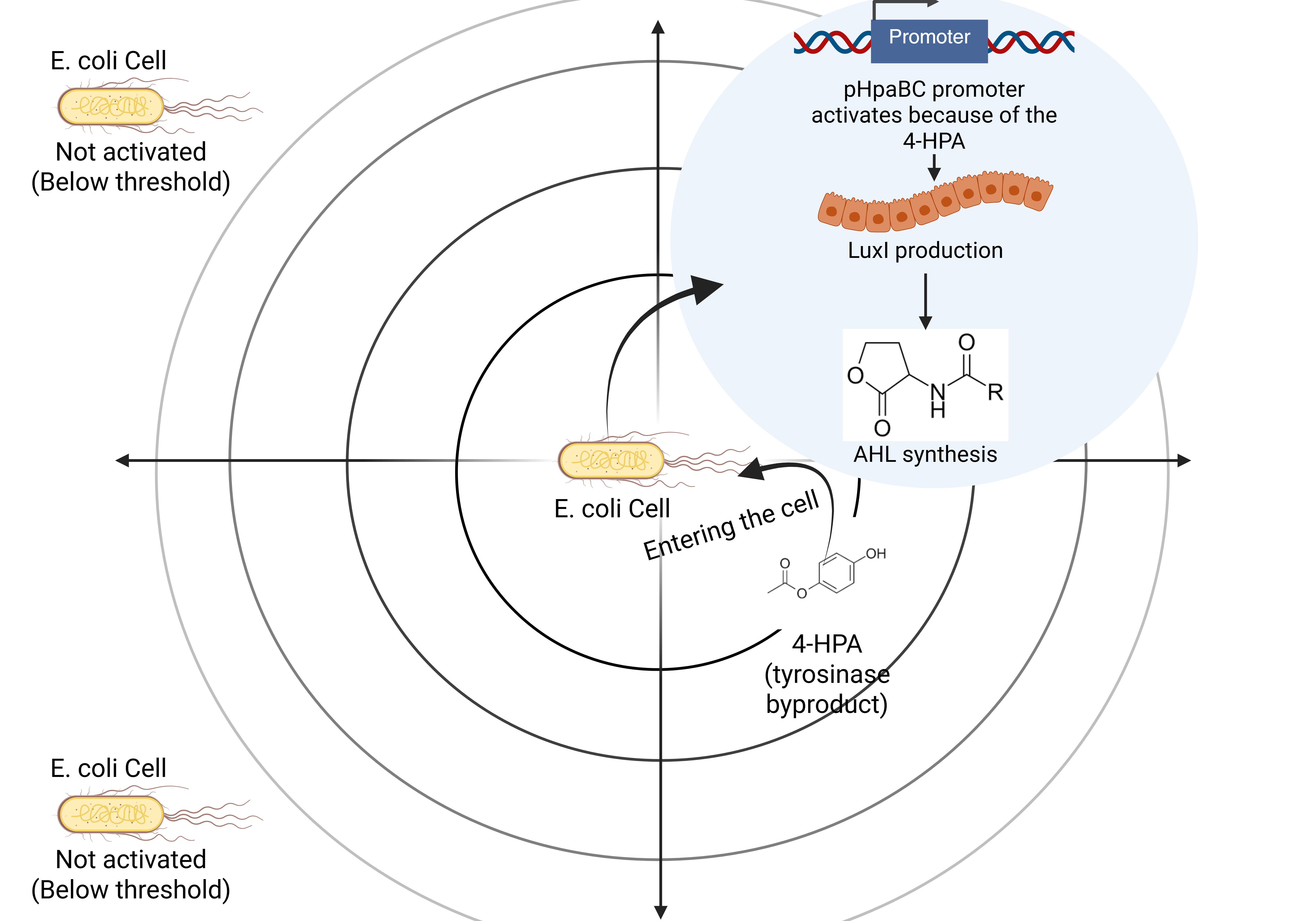
Our melanoma detection system employs genetically engineered Escherichia coli (strain K-12) as a living biosensor. The bacteria are programmed to detect elevated tyrosinase levels – an enzyme overproduced in melanoma cells – through a synthetic genetic circuit. When activated, this circuit triggers production of green fluorescent protein (GFP), causing affected skin areas to fluoresce under UV light. The device centers on a modular genetic circuit made of three main parts: a sensing module, a signal transduction cascade, and a reporter module.
Sensing module
This module uses a synthetic promoter that is responsive to catechol derivatives, which are a byproduct of tyrosinase activity on L-Dopa in the skin (Figure 2). To detect this, the E. coli is created with a pHpaBC promoter, which turns on in the presence of 4-HPA, a stable tyrosinase byproduct. This acts as our trigger (Teanphonkrang et al., 2021).
| Figure 2. Synthetic Genetic Circuit for Melanoma Sensing and Reporting. The circuit consists of three modules: (1) a sensing module that responds to 4-HPA; (2) a signaling module using quorum sensing to amplify the response across cells; and (3) a reporter module that produces blue pigment (amilCP) for visual output. |
|
Signal transduction module
The circuit consists of three modules: (1) a sensing module that responds to 4-HPA; (2) a signaling module using quorum sensing to amplify the response across cells; and (3) a reporter module that produces blue pigment (amilCP) for visual output.
Once activated, the sensing promoter drives transcription of a genetic amplifier system, making the system stronger. The core component here is a positive feedback loop using the LuxR & LuxI quorum sensing system. When one bacterium detects 4-HPA and activates the promoter, it also makes a molecule called AHL.(Saric-Bosanac et al., 2020). This molecule spreads to nearby E. coli and tells them to turn on too, even if they did not detect the 4-HPA themselves. This way, the whole area changes color, not just one or two cells. This loop activation increases the strength and persistence of the output signal, even with weak tyrosinase signaling. However, the spread of AHL is limited to a short range and only activates cells within the local area where the 4-HPA is, after the foam is removed or the AHL naturally diffuses away, the signal activation stops, preventing uncontrolled spread across the skin.
Reporter module
Our reporter is the amilCP gene, which makes blue chromoproteins. This protein changes color and is visible under natural light. Once the sensing and signaling systems are activated, amilCP gets made, and the blue color indicates where cancer might be. It is a clear, visible signal under normal light (iGEM Registry, 2025).
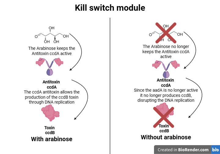
To prevent environmental release, the bacteria rely on arabinose to survive. Once the foam is washed off and arabinose is removed, the kill switch activates, leading to bacterial death within hours.
The kill switch uses a toxin-antitoxin system (ccdB/ccdA) to make sure the bacteria do not survive after use (Figure 3). During use, the foam contains an inducer (arabinose) that keeps the ccdA antitoxin gene active, allowing the bacteria to survive even while producing the ccdB toxin. Once the foam is washed off, the inducer is removed, and ccdA production stops. Without the antitoxin, the ccdB toxin disrupts DNA replication, killing the bacteria within hours. This prevents environmental contamination or long-term colonization of the skin (Fraikin et al., 2020).
| Figure 3. Kill Switch and Containment Mechanism in Engineered E. coli. To prevent environmental release, the bacteria rely on arabinose to survive. Once the foam is washed off and arabinose is removed, the kill switch activates, leading to bacterial death within hours. |
|
Parts Level
To makethis system work, we need to genetically engineer E. coli with three main sets of parts: the sensing module, the signal module, and the reporter module. Each module provides a specific genetic component, such aspromoters, coding sequences, and terminators, that work together in a pathway.
Sensing
We will use the hpaA gene from E. coli’s own genome, which naturally responds to 4-HPA (4-hydroxyphenylacetic acid). This gene works with a PBAD-like promoter that activates when 4-HPA is present. We will insert the hpaA gene under this promoter so the bacteria can recognize the 4-HPA biomarker produced near melanoma.
Signal (Quorum Sensing)
Once the 4-HPA has been detected, we then want the bacteria to start producing a quorum-sensing signal (AHL) that spreads to nearby E. coli. To do this, we use the luxI gene, which makes the AHL (N-acyl homoserine lactone), a small molecule used in bacterial communication.
Reporter (Expressing the amilCP)
The AHL travels to the cells next to it, where it binds to the LuxR protein. This then activates the Plux promoter, which triggers the amilCPexpression. The amilCP protein appears blue under visible light, revealing where cancer biomarkers were detected(IGEM registry of standard biological parts. 2025).
These genetic parts would be cloned into plasmids using tools like restriction enzymes and ligation, or Gibson assembly. Each plasmid would have a selection marker to ensure only the bacteria with our edited DNA survive in the culture. Then, we would transform them into lab-safe E. coli, K-12 to test the system.
Kill Switch
The kill switch consists of three core genetic parts: the pBAD arabinose-inducible promoter (BBa_I0500) controlling ccdA antitoxin expression, the ccdA antitoxin gene (BBa_K115001) that neutralizes the toxin, and the ccdB toxin gene (BBa_K115000) that kills cells when unopposed. During use, arabinose in the foam activates pBAD to produce ccdA, which counteracts the constitutively expressed ccdB toxin. When washed off, arabinose depletion stops ccdA production, allowing ccdB to kill the bacteria within hours. The system includes regulatory elements like araC and strong RBS sequences for proper control. All components are BioBrick-standard and optimized for reliable, safe operation in E. coli K-12 (VectorBuilder, 2025).
Safety
Before releasing our genetically engineered E. coli foam for public use, we must confirm that our product is completely safe for use on human skin and does not pose any health or environmental risks. To ensure this, our design includes built-in biological containment, and we follow an extensive safety testing plan. We focus on three key safety concerns:
- Confirming that that the E. coli is non-pathogenic and safe for topical use
- Preventing the the unintended spread or long-term build-up of bacteria
- Confirming that the blue chromoprotein amilCP and any chemical signals used are non-toxic and non-reactive with human skin
We are using a well-known strain of E. coli, DH5α, which is biosafety level 1 (BSL-1). This means it is not harmful to humans, does not produce toxins, and does not survive well outside a controlled lab. DH5α lacks key virulence factors and can not form colonies in the human body. It is commonly used in school and research labs for DNA cloning and protein production. Not only that, but according to the U.S. National Institutes of Health, it is genetically stable, meaning it does not mutate easily. It is also deficient in recombination (recA1) and restriction-modification systems (endA1), which improve safety and DNA stability (Thermo Fisher Scientific, 2025).
Not only this, but we are also implementing a kill switch system for biocontainment. We added a ccdB/ccdA toxin-antitoxin kill switch to ensure that our bacteria self-destruct after use. While in the foam, the bacteria make both the toxin (ccdB) and antitoxin (ccdA), so they are alive and functional. After the foam is rinsed off and the bacteria lose contact with the inducer, ccdA production stops. Only the toxin remains active, and the bacteria die within hours. This measure prevents the bacteria from colonizing the skin, being transmitted, or entering the environment.
Another measure we implement before human use is testing the foam in a lab-based setting using human skin cell cultures and synthetic skin models. These tests could include:
- Cytotoxicity assays, such as MTT (3-[4,5-dimethylthiazol-2-yl]-2,5 diphenyl tetrazolium bromide) or Lactate Dehydrogenase, to test if the bacteria or amilCPcause cell death
- Skin irritation assessments, using in vitro models like reconstructed human epidermis systems to measure inflammation, redness, and other irritation responses.
- Allergenic screenings on different skin types using immune response markers
- Wash-off trials to confirm that bacteria and amilCP can be easily removed with soap and water
We would also track how long bacteria survive on skin surfaces to ensure the kill switch works properly and nothing is left behind after rinse.
In addition, AmilCP is nontoxic, nonimmunogenic, and widely used in biotech and medicine. It does not penetrate the skin or enter the bloodstream, and the chemical signal AHL breaks down quickly in air and light. Itis also used in many microbial systems without reported harmful effects.
After extensive preclinical validation with positive safety and efficacy results, we will initiate a staged clinical testing protocol. The first phase will involve controlled epidermal patch testing on healthy volunteers to evaluate: (1) local skin reactivity using standardized irritation scoring systems, and (2) potential allergic responses through dermatological assessment. Following the successful demonstration of cutaneous safety, we will progress to clinical validation studies in populations with elevated melanoma risk, incorporating performance comparisons against established diagnostic reference standards.
Discussions
One of the primary benefits of this project is its potential to provide an accessible, non-invasive method for early melanoma detection, which is critical for treatment and the survival rate. Using our topical foam containing engineered E. coli that detects overproduction of tyrosinase, our design allows for simple visual identification of melanoma in specific areas. This decreases the dependence on expensive imaging equipment or the need for specialized dermatological evaluations and creates a cheap, time-effective treatment.
Additionally, our system’s use of amilCP provides a clear visualization of the target area under UV or blue light, flagging only melanoma-suspected. This targeted detection approach reduces the likelihood of misdiagnosis and unnecessary treatment, supporting more personalized healthcare. Furthermore, this system could be used forother forms of skin disease.
A significant advantage also lies in the project’s biocontainment features. The inclusion of the ccdB/ccdA kill switch limits the survival of genetically modified E. coli beyond the application time. This is important for public and environmental safety, ensuring that this product does not colonize the skin or persist in nature after its needed use. However, we pride ourselves on this project’s safety, as E. coli DH5α is widely recognized as safe.
However, despite all our successes’, we faced many challenges that need to be addressed before this system can be safely implemented in the real world. Firstly, while the pHpaBC promoter responds to catechol derivatives of tyrosinase activity, it may lack specificity, leading to false positives in inflammation or non-malignant pigmentation areas. Further refinement of the sensor module or replacement with melanoma-specific promoter systems will improve selection.
Another issue that arises is the delivery and consistency of the foam-based application. Users may spread the foam unevenly or fail to leave it on long enough for adequate detection. This variation could reduce the product’s reliability and make tests more uncertain. Additionally, the visibility of the amilCP signal under natural light could lead to some users missing spots if they are not thorough enough. Exploring options for visibility under normal lighting conditions will be optimal going forward.
While E. coli DH5α and amilCP are considered safe, the potential for skin irritation, immune responses, or long-term exposure risks must be carefully tested extensively. Although amilCP is not toxic and is used widely in biomedical settings, its long-term presence on the skin and possible interaction with the skin remains largely unstudied in the context of consumer-based products.
Next Steps
To move this project forward, the next step would be doing a controlled lab experiment to test if our engineered E. coli DH5α can detect tyrosinase actively and change color, as we hypothesize it would. This would help us establish that our biosensor circuit actually works. We would start by transforming E.coli DH5a cells with our designed plasmid. This plasmid would have a special promoter called pHpaBC (which turns in response to 4-HPA, a chemical made when tyrosinase is active), along with an RBS (ribosome binding site), the amilCP gene, and a terminator. These parts would be assembled into a plasmid like pSB1C3, commonly used in synthetic biology because it has a high copy and BioBrick.
After transformation (using a heat shock method), the bacteria would be spread on LB agar plates with chloramphenicol to select the ones that took the plasmid. After incubating overnight at 37 °C, we would grow the colonies in liquid LB media and add different amounts of 4-HPA (0,50 and 100 μM) to see if the bacteria change color more with high tyrosinase byproduct levels (Alberts et al. 2002).
We would then observe the blue coloration directly meaning that our detection system is working.
We also want to test our kill switch, which is designed to make the bacteria die off after a while. For that, we would use another plasmid with a toxic gene like ccdB under a pBAD promoter only active with arabinose. By growing the bacteria with and without arabinose, we can see if they survive only under the condition when the antitoxin is present.
Basically, this experiment would allowus to test both our detection system and safety system. If this works, our next step would be applying the bacteria in a foam format and testing it on synthetic skin models to mimic real skin reactions.
Author Contributions
F.S. developed the initial concept for the project and designed diagrams to illustrate key aspects of the work. A.H. conducted research and compiled the necessary sources. Both authors worked closely together throughout the process, collaboratively drafting, refining, and revising the manuscript to ensure the scientific content was clear, accurate, and cohesive.
Acknowledgements
We would like to express our sincere gratitude to our mentor, Dr. Lindsey L’Ecuyer, for her continuous support and invaluable guidance throughout our project. Dr. L’Ecuyer provided us with essential resources, fact-checked our work, and clarified complex concepts, which helped strengthen our understanding of the subject. Her feedback and suggestions kept us on track, ensuring that our project stayed focused and progressed smoothly. We are incredibly grateful for her time, patience, and expertise, which played a key role in shaping our work. Without her help, this project would not have been possible.
References
AIM at Melanoma Foundation. (2025). Understanding melanoma.
Alberts, B., Johnson, A., Lewis, J., Raff, M., Roberts, K., & Walter, P. (2002). From DNA to RNA. In Molecular biology of the cell (4th ed.). NCBI Bookshelf. https://www.ncbi.nlm.nih.gov/books/NBK26887/
American Cancer Society. (2025). Key statistics for melanoma skin cancer. American Cancer Society. (2025). Tests for melanoma skin cancer.
Davis, L. E., Shalin, S. C., & Tackett, A. J. (2019). Current state of melanoma diagnosis and treatment. Cancer Biology & Therapy, 20(11), 1366-1379. https://doi.org/10.1080/15384047.2019.1640032
Durland, J., & Ahmadian-Moghadam, H. (2022). Genetics, mutagenesis. In StatPearls. StatPearls Publishing. https://www.ncbi.nlm.nih.gov/books/NBK560519/
Fraikin, N., Goormaghtigh, F., & Van Melderen, L. (2020). Type II toxin-antitoxin systems: Evolution and revolutions. Journal of Bacteriology, 202(7), e00763-19. https://doi.org/10.1128/JB.00763-19
Havloujan, J. (2024, May 31). Why is melanoma so dangerous? Skin Analytics.
iGEM Registry of Standard Biological Parts. (2025). Part: BBa_K082010.
iGEM Registry of Standard Biological Parts. (2025). Part: BBa_K592009.
Long, W. T. (2025). Why melanoma is more dangerous than other types of skin cancer. Manhattan Dermatology. https://www.dermatologistnewyork.org/blog/why-melanoma-is-more-dangerous-than-other-types-of-skin-cancer
Mayo Clinic. (2025). Melanoma – Symptoms and causes.
Mayo Clinic. (2025). Skin cancer – Diagnosis and treatment.
MedlinePlus Genetics. (2025). TYR gene.
National Cancer Institute. (2025). Melanoma of the skin – Cancer Stat Facts.SEER. https://seer.cancer.gov/statfacts/html/melan.html
Saric-Bosanac, S., Clark, A. K., Sivamani, R. K., & Shi, V. Y. (2020). The role of hypothalamus-pituitary-adrenal (HPA)-like axis in inflammatory pilosebaceous disorders. PubMed. https://pubmed.ncbi.nlm.nih.gov/32239884/
ScienceDirect. (2025). Gene amplification. ScienceDirect Topics. https://www.sciencedirect.com/topics/agricultural-and-biological-sciences/gene-amplification
Skin Analytics. (2025). Understanding melanoma.
Teanphonkrang, S., Suginta, W., Sucharitakul, J., Fukamizo, T., Chaiyen, P., & Schulte, A. (2021). An electrochemical method for detecting the biomarker 4-HPA by allosteric activation of Acinetobacter baumannii reductase C1 subunit. Journal of Biological Chemistry, 296, 100467. https://doi.org/10.1016/j.jbc.2021.100467
Thermo Fisher Scientific. (2025). DH5α competent cells.
Tsien, R. Y. (1998). The green fluorescent protein. Annual Review of Biochemistry, 67(1), 509-544. https://doi.org/10.1146/annurev.biochem.67.1.509
VectorBuilder Inc. (2025). pBAD bacterial recombinant protein vector.
Zhang, H., Tao, S., Chen, H., Fang, Y., Xu, Y., Chen, L., Ma, F., & Liang, W. (2020). The biological function of the type II toxin-antitoxin system ccdAB in recurrent urinary tract infections. PubMed Central. https://pmc.ncbi.nlm.nih.gov/articles/PMC11059956/