Sihyang Baek, Qianyi Guo, Claire Hua, Isabella Haslinger-Johnson, Sofia Kovacevic, Ruihan Qin, Binita Shaw, Aparajita Shimpi, Lingzhen Wang, Wen Xiao, and Huichen Yang, Western Reserve Academy, Hudson, Ohio, United States
Reviewed on 3 May 2025; Accepted on 9 June 2025; Published on 27 October 2025
With help from the 2025 BioTreks Production Team.
Radioactive metals are natural or synthetic elements that release alpha (α), beta (β), or gamma (γ) rays harmful to both humans and the environment. The growth of the uranium mining industry has led to increased amounts of contaminated radioactive wastewater being released into the atmosphere, leading to ecological and public health threats. Our design is a cost-effective and sustainable alternative to traditional wastewater treatment methods. It also offers increased versatility by targeting U(VI) and U(IV). Deinococcus radiodurans—known for its robust genetic repair mechanisms, which allow it to resist extremely high radiation levels—will be engineered as a heavy-metal uranium remediator. Our design introduces a fusion protein construct combining D. radiodurans’ S-layer protein with PhoN. This acid phosphatase enzyme releases inorganic phosphate (Pi), which reacts with uranium (U) to form a solid uranium-phosphate compound. We will express multiheme c-type cytochromes that reduce uranium from its hexavalent state U(VI) to a less soluble and less toxic tetravalent state U(IV). To enable U(IV) binding, we will also introduce Pelosinus sp. strain UFO1’s uranium binding complex (UBC) encoded by UFO_4202 and UFO_4203. This construct combines bioprecipitation, bioreduction, and sequestration to enhance the immobilization of uranium in two oxidation states. It provides a solution for sustainable uranium detoxification and prevents the spread in contaminated environments. If successful, this construct could be deployed at uranium-contaminated sites, offering an effective, low-cost approach to environmental cleanup and ecosystem protection.
Keywords: Uranium, bioremediation, mineralization, Deinococcus radiodurans, heavy metal
Authors are listed in alphabetical order. Jason Boock and Beth Pethel mentored the group. Please direct all correspondence to pethelb@wra.net.

Background
Uranium is a naturally occurring element known for its radioactive properties. Often extracted through mining and milling, uranium is obtained from the Earth, crushed, and then chemically processed to separate and concentrate the element (“Radioactive Waste”, 2025). It is commonly used in nuclear research and energy production to initiate and control nuclear fission, providing a source of electricity. Nuclear power plants generate over 10% of the world’s electricity (What Is Uranium?, 2024).
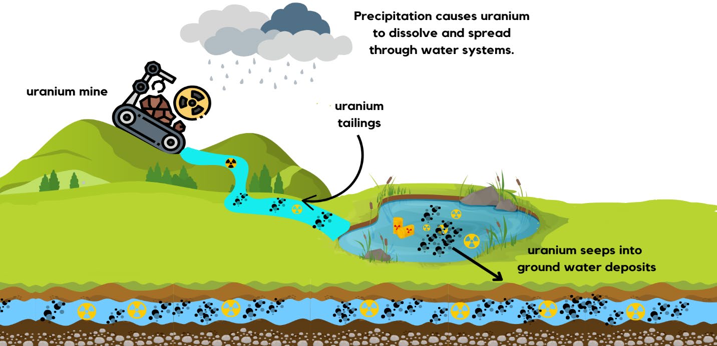
Though it is useful, uranium has toxic properties that contaminate soil and water resources. Recently, the mining and milling industries have become leading contributors to uranium contamination, releasing uraniferous tailings and waste. Contamination dispenses uranium into surrounding groundwater, surface water, soil, and air (Huang et al., 2022; Figure 1). This poses a significant health concern, as uranium affects vital organs such as the liver, kidneys, brain, bones, and lungs (Huang et al., 2022). Uranyl ions, a predominant form of U(VI) in the environment, enter the body through inhalation, ingestion, and absorption through the skin (Gallois et al., 2021). This can cause issues in the central nervous system, resulting in disruptions of the sleep-wake cycle, poor memory, and elevated anxiety (Vellingiri, 2023). Additionally, exposure to alpha particles through uranium exposure can damage the DNA, resulting in lung cancer (“Uranium-235”, 2024).
| Figure 1. Pathway of uranium contamination from mining activities into water systems. Rain dissolves uranium from exposed ore and tailings, causing surface runoff into nearby bodies of water. Contaminants accumulate in ponds before seeping into groundwater deposits, posing risks to surface and subsurface water quality. |
|
Current remediation solutions for uranium contamination include reverse osmosis, which requires membranes to separate U(VI), among other heavy metals and contaminants, from aqueous solutions. Another solution is ion resins, which attract and bind uranium ions, isolating them from the contaminated water (Ighalo et al., 2024). While effective, these methods are high-maintenance and costly. In reverse osmosis, membranes can foul over time and generate concentrated brine waste streams that contain uranium. Furthermore, additional wastewater ions such as calcium and magnesium often compete with uranium for absorption sites on ion resins, preventing ion resins from functioning effectively (DeSilva, 2005).
Though all forms of uranium are considered toxic due to their chemical and radioactive properties, certain oxidation states are less harmful than others. The two most common oxidation states in the environment are hexavalent and tetravalent uranium, abbreviatedU(VI) and U(IV) (Romanchuk et al., 2020). U(VI) forms highly-soluble uranyl ions [U02]2+, allowing it to spread quickly through water and soil and increasing environmental contamination risks (Liu et al., 2024). Its mobility makes it particularly dangerous; plants can absorb it, allowing it to enter the food chain and bioaccumulate within organisms (Li et al., 2024). In contrast, U(IV) typically forms as a tetravalent solid, which limits its mobility in the environment (Liu et al., 2024). Because of its reduced solubility, U(IV) is less likely to contaminate water sources or be absorbed by plants and animals.
Current physical and chemical uranium remediation methods often incur high costs or sanitary waste. Reverse osmosis, a common physical method, relies on a semi-permeable membrane and a high raw-water quality; ion-exchange method selectively binds and removes uranium from water but are energy intensive; chemical precipitation produces large volumes of sludge; and absorption onto carbon or mineral sorbents have limited capacity (United States Environmental Protection Agency, n.d.; Ighalo et al., 2024). Conventional bioremediation techniques address only one removal pathway and often fail when redox conditions shift or radiation levels are high (You et al., 2021). In contrast, our approach combines mineralization, reduction, and accumulation within a single radiation- tolerant construct. This eliminates the need for expensive infrastructures, limits secondary waste, and enables deployment with high specificity and long term stability.
Our project aims to engineer Deinococcus radiodurans—a bacterium known for its remarkable genetic repair mechanisms and ability to withstand high radiation levels—as a uranium remediator. By constructing two recombinant plasmids, pHPI-PhoN and pUBC, for transformation into this organism, we will deliver a genetically-enhanced bacterium that can reduce uranium burden through the three-pronged process of reduction, accumulation, and mineralization. Compared to current solutions, this remediation method will not require expensive energy and high maintenance; additionally, its targeted pathway will allow it to optimize uranium remediation specifically (Xiong et al., 2025).
Systems Level
This project aims to construct a bacterium capable of uranium remediation that can be introduced into environments with high levels of heavy metal waste. Remediation consists of three simultaneous processes: mineralization, where uranium ions in the environment combine with other chemical groups to form solid minerals; reduction, which transforms uranium from its hexavalent state U(VI) to its tetravalent state U(IV); and accumulation, which collects the transformed uranium outside of the bacteria (Zhang et al., 2020; You et al., 2021). The chassis D. radiodurans already exhibits effective resistance to radioactive environments via accelerated DNA repair mechanisms and S-layer proteins, and it can absorb uranyl ions from wastewater naturally (Gerber et al., 2015). To maintain optimal growth for D. radiodurans, it will be cultured in TGY medium (1% Bacto Tryptone, 0.5% yeast extract, 0.1% glucose, pH 7.0) and incubated at 30°C under aerobic conditions (Venkateswaran et al., 2000). Ampicillin (2 μg/mL) and chloramphenicol (0.1 μg/mL) will be added to the medium for plasmid maintenance and selection (Xu et al., 2013).
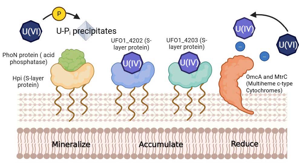
To further enhance D. radiodurans’ uranium remediation abilities, genes encoding factors that increase mineralization, reduction, and accumulation rates will be introduced on y two recombinant plasmids, pHPI-PhoN and pUBC. The phoN gene encodes an enzyme that removes phosphate groups from other molecules (Vu & Downs, 2021). By binding the PhoN enzyme with the S-layer of D. radiodurans, this fusion protein construct will be located at the S-layer of the transformed cells , which should improve D. radiodurans’ efficacy in biomineralizing uranium (Misra et al., 2021). The system also incorporates multi-heme c-type cytochromes, which reduce uranium from its hexavalent state to its tetravalent state in the form of uranium phosphate (Almeida et al., 2025).
To promote the extracellular accumulation of uranium phosphate, a second recombinant plasmid, pUBC, will be constructed using the plasmid vector pDEINO1 as its backbone (Brumwell et al., 2022). This plasmid will introduce the UFO_4202 and UFO_4203 genes, which encode a uranium-binding complex (UBC) (Thorgersen et al., 2017). The expression of these genes is designed to enhance the bacterium’s ability to bind and precipitate uranium outside the cell, thereby minimizing intracellular toxicity and facilitating the easier recovery of uranium phosphate. The UFO_4202 and UFO_4203 come initially from the bacterium Pelosinus sp., and it is hoped that they can transfer to D. radiodurans via our constructed plasmid (Thorgersen et al., 2017).
UBC binds uranium through noncovalent interactions with S-layer proteins, making the binding reversible under conditions that disrupt these interactions (e.g., changes in pH or competing ligands). Once the UBC sites on the bacterial surface become saturated, we plan to remove the bacteria to prevent the potential re-release of uranium upon cell death and lysis.
As shown in Figure 2, the expression of these proteins involved in uranium mineralization, reduction, and accumulation should enhance the remediation efficiency of D. radiodurans.
| Figure 2. Uranium mineralization, accumulation, and reduction pathways for engineered D. radiodurans. The Hpi-PhoN fusion protein releases inorganic phosphate that binds with soluble U(VI), forming insoluble uranium phosphate precipitates on or near the bacterial surface. The UFO_4202 and UFO_4203 genes encode a uranium-binding complex (UBC) on the outer membrane that captures and accumulates U(VI). Additionally, multiheme c-type cytochromes transfer electrons to U(VI), reducing it to U(IV), a less soluble form that enhances uranium immobilization. Together, these pathways facilitate efficient uranium bioremediation. |
|
Device Level
Mineralization and Reduction
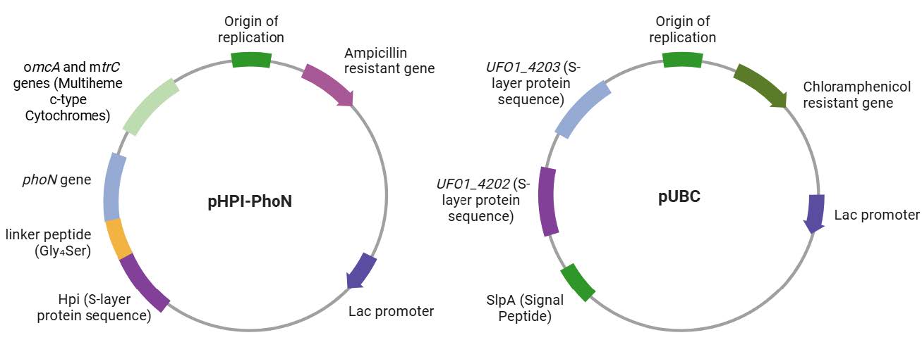
The bioengineering of D. radiodurans involves assembling two recombinant plasmids through restriction enzyme digestion and ligation. With the plasmid backbone pRAD1, we plan to express the multiheme c-type cytochrome genes omcA and mtrC from Shewanella spp. and the fusion protein targeting mineralization (Jing et al., 2020; Marshall et al., 2006). To construct the fusion protein, we will clone the hpi and phoN genes within the open reading frame, with the hpi gene placed at the N-terminal end to facilitate proper surface localization in D. radiodurans (Misra et al., 2021). To maintain structural flexibility and functional independence of each domain, we will insert a flexible linker peptide (Gly₄Ser)₃ between the hpi and phoN genes to allow for proper folding and activity of the PhoN enzyme, as depicted in Figure 3.
The fusion construct will be cloned into pRAD1 downstream of the lac promoter (Chen et al., 2013). This configuration ensures that the Hpi-PhoN fusion protein can self-assemble at the S-layer while retaining full acid phosphatase activity, facilitating efficient metal biomineralization. The PhoN protein functions by releasing phosphate ions, which react with heavy metals to create stable mineral deposits to support biomineralization of uranium phosphate (Rogiers et al., 2022). Following the fusion protein gene, inserting omcA and mtrC genes will enable uranium reduction. Multiheme c-type cytochromes function as terminal reductases in extracellular electron transfer pathways, enabling the reduction of insoluble metals, including uranium U(VI) (Zarei et al., 2023). They transfer electrons directly to soluble uranyl ions (UO₂²⁺) in the environment and reduce them to U(IV), which precipitates as insoluble uraninite (UO₂) or other U(IV) minerals (You et al., 2021).
Accumulation
For the second plasmid backbone, pDEINO1, two uranium-binding protein genes, UFO1_4203 and UFO1_4202, will be inserted downstream of the lac promoter (Thorgersen et al., 2017), as illustrated in Figure 3. To ensure the successful expression of heterologous S-layer proteins, a signal sequence will be added upstream of the gene to direct the protein to the cell surface, facilitating proper folding and localization. SlpA peptide will be incorporated since it’s another S-layer protein’s N-terminal signal peptide from D. radiodurans that directs surface localization (Rothfuss et al., 2006; Owji et al., 2018).
Amino acid sequences of the proteins in this design will be reverse-translated to obtain their corresponding DNA sequences for gene synthesis (Wicaksono et al., 2023), since their specific complete DNA sequences are not found on Genbank and other credible databases. Codon optimization methods, such as usage bias matching and mRNA secondary structure minimization, will be applied to match the preferences of the host organism.
To ensure the propagation of transformed bacteria, we will use ampicillin resistance, whose gene is already present in the backbone of pRAD1, and chloramphenicol resistance, present in pDEINO1, as selectable markers. Additionally, the lac selectable marker allows blue/white screening by disrupting the β-galactosidase gene when an insert is present, so colonies harboring plasmids with the desired insert remain white on X-gal plates. Antibiotic resistance indicates the presence of recombinant plasmids in the bacteria, whereas, and a broken lactose gene (beta galactosidase that doesn’t cleave X-gal) indicates the presence of inserted genes in our plasmids.
| Figure 3. Plasmid construction. pHPI-PhoN carries genes responsible for uranium mineralization and reduction, while pUBC carries genes involved in accumulation. |
|
Parts Level
Plasmid pRAD1 is a convenient general-purpose cloning vector that can replicate autonomously in both E. coli and D. radiodurans (Meima & Lidstrom, 2000). pRAD1 contains the lac operon regulatory elements, ampicillin-resistant gene, and M13 reverse primer site that is helpful when verifying the promoter-gene fusions and the reading frame near the insertion site. The other plasmid vector, pDEINO1, is engineered with D. radiodurans’ origin of replication and a codon-optimized chloramphenicol resistance gene (Brumwell et al., 2022).
These two plasmid vectors will enable the creation of a fusion protein targeting uranium mineralization by the linkage of the phoN gene from Salmonella typhimurium and the natural S-layer protein gene sequence hpi (Makde et al., 2007; Peters et al., 1987; UniProt Consortium, n.d.). The (Gly₄Ser)₃ flexible linker is designed to connect the two proteins due to its resistance to proteolytic cleavage, which enhances the stability of the fusion protein in diverse environments (Chen, 2019). PhoN expresses a periplasmic acid phosphatase, which breaks down organic phosphate compounds, releasing inorganic phosphate that binds with uranium to form stable mineral precipitates. Its periplasmic location limits its exposure to environmental contaminants and poses a cytotoxicity risk. By fusing PhoN with S layer proteins, the enzyme will be displayed directly on the cell surface, increasing accessibility of phosphatase active sites and protecting cell organelles from heavy metal damage. Multiheme c-type cytochromes encoded by genes omcA and mtrC from Shewanella spp will be expressed on the outer membrane of the bacterium and facilitate the transfer of electrons to uranium, reducing U(VI) to U(IV) (SO_RS08155 OmcA/MtrC, n.d.). Lastly, this design will employ the Uranium Binding Complex (UBC) from Pelosinus sp. Strain UFO 1 by inserting genes UFO_4202 and UFO_4203 (Brown et al., 2014). UBC contains an S-layer domain that sequesters U(IV). In contrast, most others only bind to U(VI), with UFO1_4202 protein containing an OprB or porin superfamily domain and UFO1_4203 protein containing an outer membrane channel superfamily domain.
Safety
Working with U(IV) and U(VI) requires strict safety protocols due to their chemical toxicities and radiological hazards. Given the associated dangers, we can construct our design, but it cannot be tested in the setting of a high school laboratory. Natural uranium’s chemical toxicity, particularly to the kidneys, is a significant concern in laboratory settings (Anderson et al., 2025). If not handled properly, radioactive chemicals emitted by uranium can enter the body through inhalation, ingestion, and skin absorption (Hazardous Substance, 2001). It can irritate the skin, cause coughing and shortness of breath, and damage essential organs like the kidneys and liver (Hazardous Substance, 2001). Due to its carcinogenic nature, prolonged exposure to uranium also poses the risks of cancer in the lung, larynx, and bone from (Hazardous Substance, 2001).
Therefore, addressing these health concerns requires careful laboratory preparation. Specifically, while personal protective equipment, including lab coats, gloves, and eye protection, is of fundamental importance, this approach should be complemented by engineering controls to ensure proper ventilation, negative pressure enclosure, and isolation of contaminated areas (Guide for Radiological, 2012). Moreover, radiological monitoring is crucial for recording and regulating radiation levels in the laboratory to prevent inhalation and ingestion exposures and minimize other potential threats to the well-being of the kidneys and liver. Routinely conducting evaluations for uranium exposure in measurable ways, including collecting air samples, is essential (Hazardous Substance, 2001). Strategically, the reduction of hexavalent to tetravalent uranium, which allows its removal, also requires that the environment remains reductive. Exposure to oxygen or any other unfavorable oxidizing agent can lead to the reoxidation of U(IV) back to the more toxic, hexavalent state (Anderson et al., 2003). Thus, this underscores the importance of monitoring the reduction potential and conducting chemical assessments periodically.
Wild type Deinococcus radiodurans has been associated with skin inflammation diseases (Chen et al., 2021). While handling this organism, wearing gloves is essential to avoid contact with bare skin. Since this bacterium does not possess any pathogenic traits, it falls under the Biosafety Level 1 category. It requires minimal safety precautions such as wearing gloves, safety goggles, and a lab coat (University of Texas Rio Grande Valley, n.d.). Similarly, Pelosinus spp is nonpathogenic and follows the same safety protocols as Deinococcus radiodurans (Schober et al., 2024).
A thorough investigation of the potential environmental risks is necessary to address the potential hazards associated with introducing a genetically-modified organism into the environment. Before field testing, we strongly recommend an evaluation of three areas: 1) the potential for engineered D. radiodurans to initiate horizontal gene transfer into bacteria native to the environment, 2) the potential for engineered D. radiodurans to produce toxins, and 3) the impact of this introduced species on the ecological community. Additionally, assessing the consequences of such effects on humans following the US Environmental Protection Agency’s (EPA) Human Health Risk Assessment guidelines is vital (Human Health, 2025). Such evaluations are necessary to minimize the potential harm to the environment and the lab personnel, ensuring the project’s safe conduct.
Discussions
This project engineers D. radiodurans to perform uranium remediation by integrating mineralization, reduction, and accumulation. Unlike traditional methods such as reverse osmosis and ion resins, this cell–based construct offers a more sustainable, self-replicating solution that uses
multiple bioremediation pathways to prevent the spread and reduce the toxicity of uranium water pollution (Ighalo et al.). By combining several genes and proteins identified by previous research, this design introduces a more comprehensive approach to heavy metal detoxification.
One key challenge in implementing this system is obtaining government approval to work with uranium, which is classified as both radioactive and hazardous material. Working with uranium in laboratory and field settings requires specialized facilities, a license, and adhering to strict safety protocols enforced by agencies such as the Nuclear Regulatory Commission (NRC) (Uranium Recovery, 2025). Additionally, evaluating the efficacy of each remediation module—mineralization, reduction, and accumulation—may pose technical difficulties. These processes often occur simultaneously or interact with one another, making it difficult to isolate their contributions. Only the overall uranium removal can typically be quantified, while detecting the exact remediation mechanism requires specialized technologies and analysis of system-generated byproducts (Ighalo et al.). Moreover, uranium speciation is highly complicated in environmental samples, as uranium often binds to other elements in the surrounding matrix (Huang et al., 2024). This can significantly influence remediation efficacy in ways that are difficult to predict or replicate under controlled laboratory conditions.
This design primarily proposes the use of chemical transformation to introduce heterologous genes into D. radiodurans. In circumstances where direct transformation may hinder genes’ functionality, conjugation-based transformation methods should be considered, in which a donor strain of E. coli harboring a mobilizable plasmid equipped with an origin of transfer, along with a helper plasmid, will be transferred to the chassis by cell-to-cell contact (Brumwell et al). Given D. radioduran’s robust restriction-modification system, conjugation offers the advantage of minimizing DNA degradation while promoting stable genomic integration or episomal expression of targeted genes. The conjugation method also utilizes the fact that E. coli is typically easier to bioengineer than D. radiodurans. If direct transformation into D. radiodurans proves inefficient or if its restriction-modification system degrades incoming DNA, we will attempt conjugation to bioengineer the chassis by cell-to-cell DNA transfer.
If the proposed D. radiodurans-based construct fulfills the need to control uranium contamination, future advancements of this design may include using biochar or installing a membrane filtration system. For example, we plan to integrate this D. radiodurans-based construct into the biochar system that Parikh’s group built in 2020, creating an adsorbent by embedding Fe₃O₄ particles into a porous biochar matrix and using iron-treated cedar sawdust through thermal pyrolysis (Wan et al., 2020). When applied to contaminated soil slurry, the material effectively removed 20–30% of arsenic, cadmium, and lead in just 24 hours due to a reduction of metal(loid) binding across multiple sites. We also plan to apply a filtration membrane that retains the D. radiodurans and uranium particles, while allowing treated water to pass through. This approach of using a filtration membrane minimizes costs, reduces energy consumption, and avoids introducing chemicals into the environment in comparison to other filtration methods. A polyethersulfone membrane with a pore size of 0.2 µm will block bacteria and capture uranium precipitates generated through the organism’s mineralization, reduction, and accumulation pathways. Together, these advancements highlight the strong potential for a safer, more efficient, and environmentally adaptable solution to uranium bioremediation.
Next Steps
Target gene sequences will be retrieved from source organisms and codon-optimized for expression in D. radiodurans. Synthetic DNA fragments and plasmid vector backbones will be ordered from Twist Bioscience and Thermo Fisher, and restriction digestion will be used to generate sticky ends for gene insertion. The digested vector and insert DNA will be ligated using T4 DNA ligase, forming a recombinant plasmid containing the target gene under an appropriate promoter. The recombinant plasmid will be introduced into D. radiodurans via chemical transformation.
To confirm successful plasmid construction and transformation following ligation, antibiotic marker selection and blue/white screening will be conducted to select successfully transformed chassis. Individual colonies will then be screened using PCR to verify the presence and size of the inserted genes. Restriction digestion and gel electrophoresis will be performed to confirm the expected fragment sizes corresponding to both the plasmid backbone and the insert. Finally, bidirectional Sanger sequencing will be used to determine the full sequence integrity of the plasmid and verify the absence of mutations.
Following successful plasmid assembly, protein expression and pathway efficiency of Hpi-PhoN, OmcA/MtrC, and UBC will be validated. Protein location and function are assessed through Western blotting and immunofluorescence microscopy (Im et al., 2018). Mineralization via phoN will be assessed by measuring the amount of phosphate released, specifically filtering out the precipitate, centrifuging the sample, and weighing the pellet. Reduction of U(VI) to U(IV) will be monitored using X-ray Absorption Near Edge Structure (XANES) to determine uranium’s oxidation state (Guda et al., 2021). Accumulation through the expression of UFO_4202 and UFO_4203 proteins will be quantified using Inductively Coupled Plasma Mass Spectrometry (ICP-MS), an instrument that detects the resulting ions based on their mass-to-charge ratio (Baghaliannejad et al., 2021). These assays will ensure the effectiveness of the three remediation modules when functioning in combination.
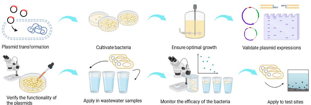
Furthermore, the bioengineered D. radiodurans will be tested with uranium-contaminated water samples, as illustrated in Figure 4. Upon approval from the relevant government agencies, water samples collected from polluted sites will be used to evaluate the bacteria’s remediation capabilities. Tanks containing uranium-contaminated water will initially be analyzed using alpha spectrometry to establish baseline levels of environmental radioactivity. Following the introduction of the engineered bacteria, uranium concentrations will be measured again to evaluate the effectiveness of the bioremediation process. Variables such as uranium concentration, water pH, and temperature will be systematically controlled and varied to generate a comprehensive dataset, ensuring cell viability and functional performance under different levels of radioactivity. If remediation proves effective, we will conduct field trials in controlled real-world environments and evaluate the outcomes at test sites compared to negative controls lacking the bioengineered bacterial remediator. Simultaneously, any environmental side effects associated with using the engineered bacteria and their byproducts will be closely monitored and assessed. If the results are successful, further steps towards implementing the product may be considered.
| Figure 4. Building and testing procedures. This figure outlines the workflow of the project, from plasmid transformation to bacterial culturing, construct validation, and final application. |
|
Author Contributions
L.W. conceived the original idea and initiated introductory research. I.H., L.W., and W.X. conducted the early research process. S.B., Q.G., C.H., I.H., S.K., R.Q., B.S., A.S., L.W., W.X., and H.Y. contributed to the writing and proofreading of the paper. S.B., I.H., C.H., and A.S. animated the video. S.K., I.H., L.W., and W.X. designed the images and graphics for this project.
Acknowledgements
We sincerely thank Western Reserve Academy for offering the resources and opportunities that made this project possible. We are especially grateful to Dr. Pethel and Dr. Boock for their guidance and advice throughout the challenges of our work. Their support and insight have played a crucial role in the development of this design.
References
Almeida, A., Turner, D. L., Silva, M. A., & Salgueiro, C. A. (2025). New insights in uranium bioremediation by cytochromes of the bacterium I. Journal of Biological Chemistry, 301(2), 108090. https://doi.org/10.1016/j.jbc.2024.108090
Anderson, R. T., Vrionis, H. A., Ortiz-Bernad, I., Resch, C. T., Long, P. E., Dayvault, R., Karp, K., Marutzky, S., Metzler, D. R., Peacock, A., White, D. C., Lowe, M., & Lovley, D. R. (2003). Stimulating the in situ activity of Geobacter species to remove uranium from the groundwater of a uranium-contaminated aquifer. Applied and Environmental Microbiology, 69(10), 5884-5891. https://doi.org/10.1128/aem.69.10.5884-5891.2003
Anderson, W. A., Domingo-Relloso, A., Galvez-Fernandez, M., Schilling, K., Glabonjat, R. A., Basu, A., Nigra, A. E., Gutierrez, O. M., Scherzer, R., Goldsmith, J., Sarnak, M. J., Bonventre, J. V., Kimmel, P. L., Vasan, R. S., Ix, J. H., Shlipak, M. G., & Navas-Acien, A. (2025). Uranium exposure and kidney tubule biomarkers in the multi-ethnic study of atherosclerosis (MESA). Environmental Research, 271, 121060. https://doi.org/10.1016/j.envres.2025.121060
Baghaliannejad, R., Aghahoseini, M., & Amini, M. K. (2021). Determination of rare earth elements in uranium materials by ICP-MS and ICP-OES after matrix separation by solvent extraction with TEHP. Talanta, 222, 121509. https://doi.org/10.1016/j.talanta.2020.121509
Brown, S. D., Utturkar, S. M., Magnuson, T. S., Ray, A. E., Poole, F. L., Lancaster, W. A., Thorgersen, M. P., Adams, M. W. W., & Elias, D. A. (2014). Complete genome sequence of Pelosinus sp. strain UFO1 assembled using single-molecule real-time DNA sequencing technology. Genome Announcements, 2,:10.1128/genomea.00881-14. https://doi.org/10.1128/genomea.00881-14
Brumwell, S. L., Van Belois, K. D., Giguere, D. J., Edgell, D. R., & Karas, B. J. (2022). Conjugation-Based genome engineering in Deinococcus radiodurans. ACS Synthetic Biology, 11(3), 1068-1076. https://doi.org/10.1021/acssynbio.1c00524
Chen, F., Zhang, J., Ji, H. J., Kim, M.-K., Kim, K. W., Choi, J.-I., Han, S. H., Lim, S., Seo, H. S., & Ahn, K. B. (2021). Deinococcus radiodurans exopolysaccharide inhibits Staphylococcus aureus biofilm formation. Frontiers in Microbiology, 12:712086https://doi.org/10.3389/fmicb.2021.712086
Chen, J. (2019, August 4). Part:BBa_K2976011. Registry of Standard Biological Parts. Retrieved April 14, 2025, from https://parts.igem.org/Part:BBa_K2976011
Chen, X., Zaro, J. L., & Shen, W.-C. (2013). Fusion protein linkers: Property, design and functionality. Advanced Drug Delivery Reviews, 65(10), 1357-1369. https://doi.org/10.1016/j.addr.2012.09.039
Consortium, U. (n.d.). Q9RRB6 – Tumor necrosis factor receptor superfamily member 1A (tnfrsf1a) [Rattus norvegicus]. UniProt. Retrieved April 14, 2025, from https://www.uniprot.org/uniprotkb/Q9RRB6/entry
DeSilva, F. (2005, April 6). Uranium in water removal by iron exchange. WaterWorld. Retrieved April 14, 2025, from https://www.waterworld.com/residential-commercial/article/14307975/uranium-in-water-removal-by-ion-exchange
University of Texas Rio Grande Valley. (n.d.). Environmental health, safety &
risk management. Retrieved May 5, 2025, from https://www.utrgv.edu/ehsrm/
programs/lab-safety/biological-safety-program/biosafety-levels/index.htm
Gallois, N., Alpha-Bazin, B., Bremond, N., Ortet, P., Barakat, M., Piette, L., Mohamad Ali, A., Lemaire, D., Legrand, P., Theodorakopoulos, N., Floriani, M., Février, L., Den Auwer, C., Arnoux, P., Berthomieu, C., Armengaud, J., & Chapon, V. (2021). Discovery and characterization of UipA, a uranium- and iron-binding PepSY protein involved in uranium tolerance by soil bacteria. The ISME Journal, 16(3), 705-716. https://doi.org/10.1038/s41396-021-01113-7
Gerber, E., Bernard, R., Castang, S., Chabot, N., Coze, F., Dreux‐Zigha, A., Hauser, E., Hivin, P., Joseph, P., Lazarelli, C., Letellier, G., Olive, J., & Leonetti, J. (2015). Deinococcus as new chassis for industrial biotechnology: Biology, physiology and tools. Journal of Applied Microbiology, 119(1), 1-10. https://doi.org/10.1111/jam.12808
Guda, A. A., Guda, S. A., Martini, A., Kravtsova, A. N., Algasov, A., Bugaev, A., Kubrin, S. P., Guda, L. V., Šot, P., van Bokhoven, J. A., Copéret, C., & Soldatov, A. V. (2021). Understanding X-ray absorption spectra by means of descriptors and machine learning algorithms. npj Computational Materials, 7, 203.https://doi.org/10.1038/s41524-021-00664-9
Guide for radiological laboratories for the control of radioactive contamination and radiation exposure. (2012). United States Environmental Protection Agency #EPA 402-R-12-005.https://www.epa.gov/sites/default/files/2015-05/documents/402-r-12-005_contamination_guide_aug_2012.pdf
Hazardous substance fact sheet. (2001, December). New Jersey Department of Health and Senior Services. Retrieved April 14, 2025, from https://nj.gov/health/eoh/rtkweb/documents/fs/1969.pdf
Huang, F., Dong, F., Chen, L., Zeng, Y., Zhou, L., Sun, S., Wang, Z., Lai, J., & Fang, L. (2024). Biochar-mediated remediation of uranium-contaminated soils: Evidence, mechanisms, and perspectives. Biochar, 6:16. https://doi.org/10.1007/s42773-024-00308-3
Huang, L., Li, S., Zhou, W., Gao, J., Yin, J., Wang, Z., & Li, J. (2022). Cellular transport of uranium and its cytotoxicity effects on CHO-k1 cells. Ecotoxicology and Environmental Safety, 246, 114166. https://doi.org/10.1016/j.ecoenv.2022.114166
Human health risk assessment. (2025, January 31). U.S. Environmental Protection Agency. Retrieved April 14, 2025, from https://www.epa.gov/risk/human-health-risk-assessment
Ighalo, J. O., Chen, Z., Ohoro, C. R., Oniye, M., Igwegbe, C. A., Elimhingbovo, I., Khongthaw, B., Dulta, K., Yap, P.-S., & Anastopoulos, I. (2024). A review of remediation technologies for uranium-contaminated water. Chemosphere, 352, 141322. https://doi.org/10.1016/j.chemosphere.2024.141322
Im, K., Mareninov, S., Diaz, M. F. P., & Yong, W. H. (2018). An introduction to performing immunofluorescence staining. In: Yong, W. (eds) Biobanking. Methods in Molecular Biology, vol 1897. Humana Press, New York, NY. https://doi.org/10.1007/978-1-4939-8935-5_26
Jing, X., Wu, Y., Shi, L., Peacock, C. L., Ashry, N. M., Gao, C., Huang, Q., & Cai, P. (2020). Outer membrane c-type cytochromes OmcA and MtrC play distinct roles in enhancing the attachment of Shewanella oneidensis MR-1 cells to goethite. Appl Environ Microbiol, 86:e01941-20. https://doi.org/10.1128/aem.01941-20
Li, Z., Sun, P., Zhang, C., Zhu, N., Xu, N., Li, D., Gao, Y., & Zhao, J. (2024). Translocation and transformation of uranium along the aquatic food chain: New insights into uranium risks to the environment. Journal of Hazardous Materials, 478, 135499. https://doi.org/10.1016/j.jhazmat.2024.135499
Liu, X., Xie, Y., Hao, M., Li, Y., Chen, Z., Yang, H., Waterhouse, G. I. N., Wang, X., & Ma, S. (2024). Secondary metal ion-induced electrochemical reduction of U (VI) to U (IV) solids. Nature Communications, 15, 7736. https://doi.org/10.1038/s41467-024-52083-1
Makde, R. D., Mahajan, S. K., & Kumar, V. (2007). Structure and mutational analysis of the PhoN protein of Salmonella typhimurium provide insight into mechanistic details. Biochemistry, 46(8), 2079-2090. https://doi.org/10.1021/bi062180g
Marshall, M. J., Beliaev, A. S., Dohnalkova, A. C., Kennedy, D. W., Shi, L., Wang, Z., Boyanov, M. I., Lai, B., Kemner, K. M., McLean, J. S., Reed, S. B., Culley, D. E., Bailey, V. L., Simonson, C. J., Saffarini, D. A., Romine, M. F., Zachara, J. M., & Fredrickson, J. K. (2006). C-Type cytochrome-dependent formation of U(IV) nanoparticles by Shewanella oneidensis. PLoS Biology, 4(8), e268. https://doi.org/10.1371/journal.pbio.0040268
Meima, R., & Lidstrom, M. E. (2000). Characterization of the minimal replicon of a cryptic Deinococcus radiodurans SARK plasmid and development of versatile Escherichia coli-D. radiodurans shuttle vectors. Applied and Environmental Microbiology, 66(9), 3856-3867. https://doi.org/10.1128/aem.66.9.3856-3867.2000
Misra, C. S., Sounderajan, S., & Apte, S. K. (2021). Metal removal by metallothionein and an acid phosphatase PhoN, surface-displayed on the cells of the extremophile, Deinococcus radiodurans. Journal of Hazardous Materials, 419, 126477. https://doi.org/10.1016/j.jhazmat.2021.126477
Owji, H., Nezafat, N., Negahdaripour, M., Hajiebrahimi, A., & Ghasemi, Y. (2018). A comprehensive review of signal peptides: Structure, roles, and applications. European Journal of Cell Biology, 97(6), 422-441. https://doi.org/10.1016/j.ejcb.2018.06.003
Peters, J., Peters, M., Lottspeich, F., Schäfer, W., & Baumeister, W. (1987). Nucleotide sequence analysis of the gene encoding the Deinococcus radiodurans surface protein, derived amino acid sequence, and complementary protein chemical studies. Journal of Bacteriology, 169(11), 5216-5223. https://doi.org/10.1128/jb.169.11.5216-5223.1987
Radioactive waste from uranium mining and milling. (2025, January 29). United States Environmental Protection Agency. Retrieved April 14, 2025, from https://www.epa.gov/radtown/radioactive-waste-uranium-mining-and-milling#
Rogiers, T., Van Houdt, R., Williamson, A., Leys, N., Boon, N., & Mijnendonckx, K. (2022). Molecular mechanisms underlying bacterial uranium resistance. Frontiers in Microbiology, 13, 822197. https://doi.org/10.3389/fmicb.2022.822197
Romanchuk, A. Y., Vlasova, I. E., & Kalmykov, S. N. (2020). Speciation of uranium and plutonium from nuclear legacy sites to the environment: A mini review. Frontiers in Chemistry, 8, 630. https://doi.org/10.3389/fchem.2020.00630
Rothfuss, H., Lara, J. C., Schmid, A. K., & Lidstrom, M. E. (2006). Involvement of the s-layer proteins Hpi and SlpA in the maintenance of cell envelope integrity in Deinococcus radiodurans R1. Microbiology, 152(9), 2779-2787. https://doi.org/10.1099/mic.0.28971-0
Schober, I., Koblitz, J., Sardà Carbasse, J., Ebeling, C., Schmidt, M. L., Podstawka, A., Gupta, R., Ilangovan, V., Chamanara, J., Overmann, J., & Reimer, L. C. (2024). BacDive in 2025: The core database for prokaryotic strain data. Nucleic Acids Research, 53(D1), D748-D756. https://doi.org/10.1093/nar/gkae959
SO_RS08155 OmcA/MtrC family decaheme c-type cytochrome [Shewanella oneidensis MR-1]. (n.d.). Retrieved April 14, 2025, from https://www.ncbi.nlm.nih.gov/gene?Db=gene&Cmd=DetailsSearch&Term=1169553
Thorgersen, M. P., Lancaster, W. A., Rajeev, L., Ge, X., Vaccaro, B. J., Poole, F. L., Arkin, A. P., Mukhopadhyay, A., & Adams, M. W. W. (2017). A highly expressed high-molecular-weight S-layer complex of Pelosinus sp. strain UFO1 binds uranium. Applied and Environmental Microbiology, 83:e03044-16.https://doi.org/10.1128/aem.03044-16
United States Environmental Protection Agency. (n.d.). Point-of-use reverse osmosis systems. Retrieved May 20, 2025, from https://www.epa.gov/watersense/point-use-reverse-osmosis-systems#Uranium-235 (U-235) and Uranium-238 (U-238). (2024, April 17). CDC Radiation Emergencies. Retrieved April 14, 2025, from https://www.cdc.gov/radiation-emergencies/hcp/isotopes/uranium-235-238.html#
Uranium Recovery Regulations, Guidance, and Communications. (2025, February 20). Retrieved April 16, 2025, from https://www.nrc.gov/materials/uranium-recovery/regs-guides-comm.html
Vellingiri, B. (2023). A deeper understanding about the role of uranium toxicity in neurodegeneration. Environmental Research, 233, 116430. https://doi.org/10.1016/j.envres.2023.116430
Venkateswaran, A., McFarlan, S. C., Ghosal, D., Minton, K. W., Vasilenko, A., Makarova, K., Wackett, L. P., & Daly, M. J. (2000). Physiologic determinants of radiation resistance in Deinococcus radiodurans. Applied and Environmental Microbiology, 66(6), 2620-2626. https://doi.org/10.1128/aem.66.6.2620-2626.2000
Vu, H. N., & Downs, D. M. (2021). An unexpected role for the periplasmic phosphatase PhoN
in the salvage of B6 vitamers in Salmonella enterica. Applied and Environmental Microbiology, 87:e02300-20. https://doi.org/10.1128/aem.02300-20
Wan, X., Li, C., & Parikh, S. J. (2020). Simultaneous removal of arsenic, cadmium, and lead from soil by iron-modified magnetic biochar. Environmental Pollution, 261, 114157. https://doi.org/10.1016/j.envpol.2020.114157
What is uranium? How does it work? (2024, May 16). World Nuclear Association. Retrieved April 14, 2025, from https://world-nuclear.org/information-library/nuclear-fuel-cycle/introduction/what-is-uranium-how-does-it-work
Wicaksono, A., Kharisma, V. D., & Parikesit, A. A. (2023). New perspectives on reverse translation: Brief history and updates. Universitas Scientiarum, 28(1), 1-20. https://doi.org/10.11144/javeriana.sc281.npor
Xiong, X., Liu, J., Xiao, T., Lin, K., Huang, Y., Deng, P., Hu, H., & Wang, J. (2025). Remediation of uranium-contaminated water and soil by biochar-based materials: A review. Biochar, 7, 41. https://doi.org/10.1007/s42773-025-00438-2
Xu, S., Battaglia, L., Bao, X., & Fan, H. (2013). Chloramphenicol acetyltransferase as a selection marker for chlamydial transformation. BMC Research Notes, 6, 377. https://doi.org/10.1186/1756-0500-6-377
You, W., Peng, W., Tian, Z., & Zheng, M. (2021). Uranium bioremediation with U (VI)-reducing bacteria. Science of the Total Environment, 798, 149107. https://doi.org/10.1016/j.scitotenv.2021.149107
Zarei, M., Fatemi, F., Ghasemi, R., Mir-Derikvand, M., Hosseinpour, H., & Samani, T. R. (2023). The effect of non-anaerobicization and discolored bacteria on uranium reduction by Shewanella sp. 7. Applied Radiation and Isotopes, 192, 110551. https://doi.org/10.1016/j.apradiso.2022.110551
Zhang, D., Chen, X., Larson, S. L., Ballard, J. H., Knotek-Smith, H. M., Ding, D., Hu, N., & Han, F. X. (2020). Uranium biomineralization with phosphate—biogeochemical process and its application. ACS Earth and Space Chemistry, 4(12), 2205-2214. https://doi.org/10.1021/acsearthspacechem.0c00252