Daryn Amos, Yunwoo Choi, Phalak Dhingra, Suzanna Gebhart, Dongyeon Kim, and Xochi Nandi, Western Reserve Academy, Hudson, Ohio, United States
Reviewed on 3 May 2025; Accepted on 9 June 2025; Published on 27 October 2025
With help from the 2025 BioTreks Production Team.
Cataracts are a common disease in dogs, causing vision impairment and extreme pain. Cataracts are especially prevalent in diabetic dogs, with 75 and 80 percent of diabetic dogs developing diabetic cataracts within 370 to 470 days of diagnosis. Cataracts develop when the crystallin proteins that make up the lens of the eye clump together, leading to decreased visual quality and often blindness. Risk factors for cataracts include old age, genetic predisposition, and certain diseases, such as diabetes at advanced stages. Current treatments include daily lanosterol eye drops, phacoemulsification surgery, which removes just the cataract, and enucleation to remove the entire eye. Phacoemulsification surgery is effective but costly, ranging from $2,700 to $4,000, making it inaccessible to most pet owners. While current lanosterol eye drops come at a relatively low price, the multiple daily applications are burdensome for both the pet and the owner, and cataract recurrence is possible. Lanosterol, a naturally occurring tetracyclic triterpenoid, has shown great potential in reversing the aggregation of proteins within the eye and thus restoring lens transparency. Our innovative solution for lifelong cataract prevention in dogs utilizes an engineered yeast, part of the normal flora of dogs’ eyes, to produce lanosterol, ensuring long-term drug delivery within the eye. This treatment would be administered shortly after birth, and the yeast, Candida albicans, would live in the eye, producing lanosterol consistently. The sustained production of lanosterol offers a non-surgical alternative that not only eliminates the risks and recovery associated with cataract surgery, but may also prevent cataract formation and lifelong vision loss, improving quality of life.
Keywords: lanosterol, Candida albicans, cataracts, eye
Authors are listed in alphabetical order. Beth Pethel and Kissaou Tchedre mentored the group. Please direct all correspondence to pethelb@wra.net.


Background
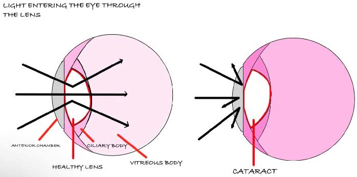
In a healthy eye, the lens focuses incoming light onto the retina, producing clear and precise vision. A cloudy lens scatters rather than directs light, resulting in blurred vision and even blindness. Cataracts develop when proteins in the eye lens clump together, leading to a cloudy appearance and progressive vision loss, as shown in Figure 1 (Cornell University College of Veterinary Medicine, n.d.). A clear lens allows light to pass through and focus on the retina, enabling sharp vision. However, when the lens becomes cloudy, as pictured in Figure 2, it reflects incoming light off the lens instead of directing it into it, resulting in blindness.
Symptoms of cataracts include clouding, changes in the color of the eye, scratches, and signs of vision impairment (Jones, 2023). Advanced cataracts can cause extreme pain and discomfort to dogs.
Cataracts can develop due to different factors, most often related to aging. A genetic predisposition (certain conditions and mutations) also increases risk. Some breeds, including the American cocker spaniel, Labrador retriever, French poodle, Boston terrier, and Welsh springer spaniel, are more susceptible to cataracts due to genetic predispositions (Barnes et al., 2024). Beyond genetics, other diseases, such as diabetes, trauma to the eye, nutrition imbalance, and chronic uveitis also contribute to cataract formation (Cornell University College of Veterinary Medicine, n.d.). Among diabetic dogs, approximately 50% will develop cataracts within five months of diagnosis, 75% within one year, and 80% within 16 months (Veterinary Vision Center, 2022).
| Figure 1. A dog with cataracts, notice the milky cloudiness of the lens. |

|
| Figure 2. Normal lens and lens with cataracts while light travels through a normal lens, a lens with cataracts reflects the light, causing blurry vision and blindness. |
|
Some long-term effects include increased anxiety and discomfort, as well as an increased risk of injury. Cataracts ultimately diminish their quality of life as activities and playing become more challenging.
Treatment depends on the severity of the disease. In the most severe cases, the only course of action is enucleation, a surgical procedure in which the eye is completely removed to alleviate pain and prevent further complications (Cornell University College of Veterinary Medicine, n.d.). Phacoemulsification surgery is the only reliable method for vision restoration. During the procedure,the cataract is emulsified using ultrasonic vibrations, allowing its removal via a hollow needle and subsequent replacement with an artificial intraocular lens. The success rate is approximately 90%, allowing most dogs to regain functional vision if performed early and followed by diligent postoperative care (Cornell University College of Veterinary Medicine, n.d.). However, recovery can take several weeks, and complications such as infection, posterior capsule opacification, or inflammation may occur, highlighting the need for less invasive and more accessible alternatives. The cost of this surgery for dogs is also significant, typically ranging from $2,700 to $4,000 (Jones, 2023). Complications are uncommon but can be grave. A small portion of the cataract can return, likely from scar tissue (Beers, 2017.).
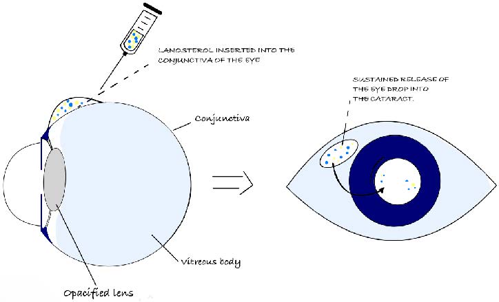
Alternative non-surgical treatments, such as anti-inflammatory medications, antioxidant supplements, and specially formulated eye drops can delay the progression of cataracts but cannot reverse the condition. Lanosterol eye drops degrade the protein clumps which cause opacity within the lens. As demonstrated in Figure 3, these drops dissolve protein aggregates by directly delivering the naturally occurring compound lanosterol to the conjunctiva (the thin, transparent membrane that covers the inside of the eyelids) of the eye. This then gradually travels into the lens, restoring the lens’s transparency and improving vision (Lam, 2023.). Lanomax®, a form of lanosterol eye drops, is only implemented after cataracts have formed in the eye and requires consistent application. Pet owners should be prepared for an extended treatment period, where improvement may be seen within six weeks of starting the drops at the earliest, but this can vary between individual dogs (Lam, 2023). Advanced cataracts may not respond to lanosterol alone (Lam, 2023). In some cases, longer, consistent usage is required, not to mention the struggle of putting the eyedrop in, which could present challenges with application consistency. Lanosterol eye drops offer a hopeful solution for pet owners considering alternatives to surgery to manage cataracts in their dogs (Innovative Veterinary Care Journal, 2023). The cost of lanosterol drops can range from $70 to $160 (Lumen Pro, n.d.).
However, while initial results are promising, lanosterol therapy remains in its early stages, and the effectiveness may vary depending on the cataract type and stage of the cataract involved. It may be more effective in earlier stages of cataract development. It may not be as effective in advanced cases or for cataracts caused by underlying medical conditions (including diabetes mellitus, chronic uveitis, and hereditary cataracts) (Lam, 2023).
Thus, the only proven method for vision restoration in cataract-affected dogs, phacoemulsification, is highly effective but comes at a substantial cost. This high cost causes significant drawbacks, making pet owners reluctant to treat their pets’ cataracts. Non-surgical alternatives, such as medicated eye drops and antioxidant supplements, may delay cataract progression but cannot reverse vision loss.. The dilemma underscores the need for an accessible, long-term preventative solution that can help preserve vision without the current options’ financial and logistical constraints.
| Figure 3. Mechanism of lanosterol eye drops. Lanosterol cataract preventative eye drops insert the medication into the conjunctiva of the eye. Eventually, the lanosterol travels into the lens and releases the solution directly into the cataract, leading to the breakdown of aggregate proteins. |
|
This project aims to provide a novel solution for the ongoing prevention of cataracts in dogs using an enzyme-based approach. By creating a more affordable option that does not require frequent application, our product overcomes the limitations of the products currently on the market. We propose genetically designing yeast to continually generate lanosterol in the eye, enhancing natural lens function and avoiding protein aggregation that leads to cataracts. The genetically designed yeast will make enzymes responsible for producing lanosterol, which generates a protective substance when mixed into a controlled eye solution. The yeast will be introduced into a predetermined eye solution, where it continually synthesizes lanosterol to keep the lenses clear. Although this method minimizes frequent replenishment of the solution, occasional dosing every 4-6 weeks may still be needed to maintain therapeutic concentrations.
The yeast chosen is part of the normal flora of canine eyes. The treatment is intended to prevent canine cataracts by addressing the underlying problem (protein build-up in the lens) before the condition occurs. The solution is cost-effective because there’s no need for expensive surgeries or long-term therapy. Once established, the yeast reproduces itself, which offers ongoing protection and reduces ongoing costs for pet owners compared to traditional interventions. The lanosterol eye drops integrate synthetic biology to support a natural, anti-aging effect on dog vision. Using lanosterol eye drops as an intervention could eliminate the need for cataract surgery while improving life expectancy and quality of life.
Systems Level
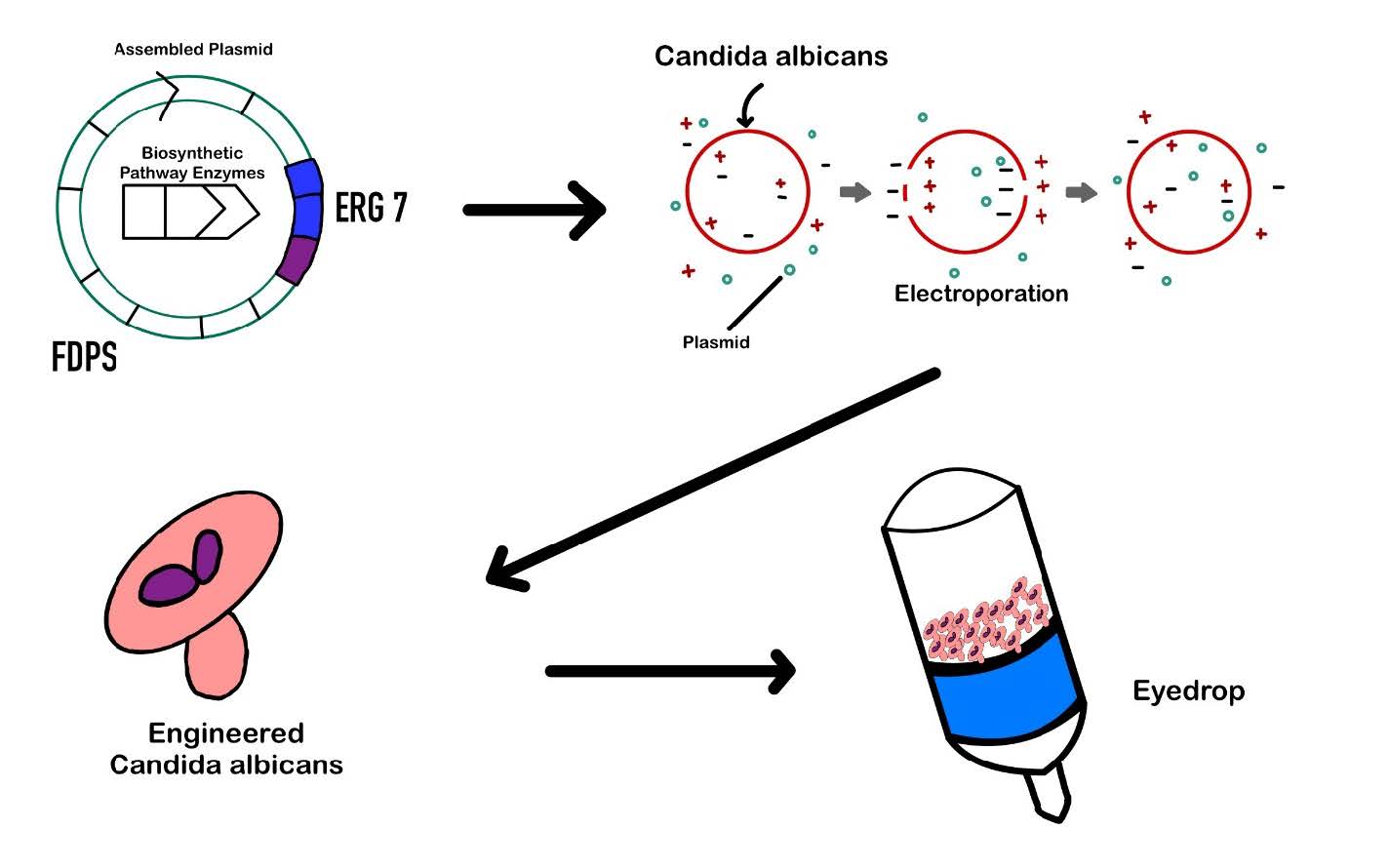
We will engineer a genetic circuit to express and release a copy of the lanosterol analog within C. albicans for our mock eyedrop solution experiments. In wild-type C. albicans, lanosterol is typically converted into ergosterol as part of the sterol biosynthesis pathway. However, our engineered strain is designed to accumulate and secrete lanosterol instead of converting it, making it suitable for therapeutic use. Since C. albicans is a naturally occurring yeast and non-pathogenic in specific microbiomes, it is considered safe for veterinary ophthalmic applications. To achieve this, we will construct a plasmid encoding the first nine enzymes of the lanosterol biosynthetic pathway, assembled using Start-Stop Assembly to ensure seamless integration and efficient gene expression.
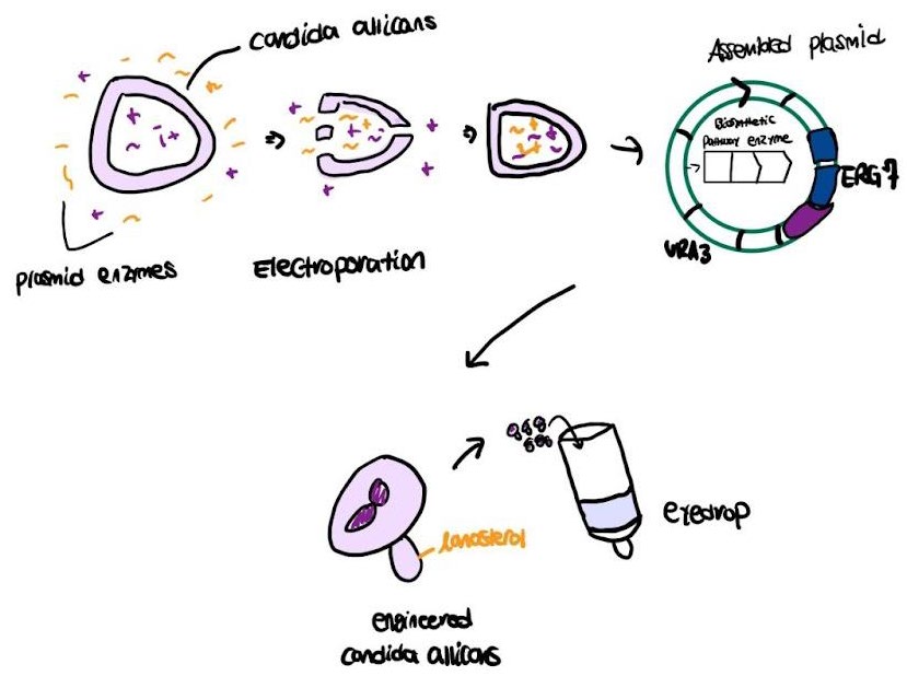
| Figure 4. Yeast transforming into an eyedrop. The plasmids are transformed into C. albicans through electroporation— a technique that uses electrical pulses to create temporary pores in cell membranes, allowing substances like nucleic acids to enter and be delivered into cells. Then, the engineered C. albicans are mixed into sterile saline to form the eyedrop. |
|
|
The assembled plasmid will then be introduced into C. albicans via electroporation or the lithium acetate method. Electroporation is a technique that uses an electrical pulse to disrupt the yeast cell membrane, allowing plasmid uptake temporarily. Post-electroporation incubation in a recovery medium allows yeast cells to reseal their membranes while initiating plasmid expression. The C. albicans cells then undergo treatment with lithium acetate to enhance their membrane permeability in this method. The combination of carrier DNA and polyethylene glycol (PEG) is used to introduce the plasmid, which promotes its uptake into cells. The application of heat shock helps to maximize transformation efficiency. The transformed yeast will be grown on selective media to verify successful plasmid integration, followed by functional validation through protein expression analysis and high-performance liquid chromatography (HPLC) to confirm secretion.
Device Level
The engineered plasmid is designed to enable C. albicans to express and secrete a lanosterol analog, a key compound in sterol biosynthesis. The plasmid contains a carefully assembled genetic circuit, incorporating essential regulatory elements that ensure efficient transcription, translation, and secretion of the target molecule. We chose to use the start-stop method because it ensures that each enzyme is translated properly by optimizing start and stop codons within the construct. It reduces the risk of unwanted frameshift mutations or incorrect gene order, which could disrupt lanosterol biosynthesis, and it allows for the simultaneous integration of multiple genes without the need for iterative cloning steps.
To achieve tissue-specific expression, we will utilize the keratocan promoter (KERA), which is naturally active in corneal tissue and plays a role in maintaining corneal transparency. KERA has demonstrated activity in murine models, and we aim to extend its application to canine ocular tissues (Allgoewer et al., 2010). The modular design of the plasmid ensures efficient and reliable expression of the mock lanosterol pathway. Start-stop assembly creates the construct by enabling the scarless integration of multiple genetic elements. The biosynthetic pathway enzymes undergo PCR amplification and subsequent ligation with restriction enzymes like BsaI during Golden Gate Assembly to achieve precise gene assembly (Engler et al., 2008). E. coli cells first amplify the plasmid, which researchers then extract and purify for transformation into C. albicans. Researchers will cultivate C. albicans cells that express the initial nine enzymes of the lanosterol pathway in regulated conditions after transformation. Researchers will then induce expression and quantify secretion efficiency through biochemical assays and protein measurement techniques. The final product is collected from the culture medium to ensure enough yield for mock eyedrop solution preparation.
Parts Level
We are targeting ERG7, a key enzyme in sterol biosynthesis, which encodes lanosterol synthase (LSS). Enhancing ERG7 expression increases lanosterol production, leading to its intracellular accumulation. To construct our plasmid, we plan to use the start-stop assembly method to combine nine genes into one single construct. This method is the most efficient way to put such a large number of genes into a plasmid without major complications. Each genetic component undergoes PCR amplification, restriction enzyme digestion, and ligation to form the assembled plasmid. Once transformed into C. albicans, the plasmid enables lanosterol synthesis and facilitates its extracellular secretion via the α-Mating factor signal (Taylor et al., 2019).
The collected product will be contained in a veterinary sterile saline solution (0.9% NaCl) designed for the safety of the canine eye. Adjustments to the solution’s pH will occur only when required to maintain biocompatibility with the ocular environment. Regular serum electrolyte measurements of sodium (Na⁺), potassium (K⁺), calcium (Ca²⁺), chloride (Cl⁻), and bicarbonate (HCO₃⁻) are crucial for evaluating fluid balance and metabolic stability during treatment when the therapy alters systemic physiology. These electrolytes play a fundamental role in keeping osmotic balance and the proper functioning of nerves and muscles when treatments are administered inside living organisms. The solution passes through a 0.22 μm filter, which removes bacterial contaminants to ensure sterility before stability and safety testing (Lemp et al., 2007). Gene expression in ocular tissues is regulated by KERA, while the initial nine enzymes of the lanosterol biosynthetic pathway facilitate biosynthesis. An α-Mating factor secretion signal is included to promote extracellular secretion of pathway enzymes, rather than the small molecule itself, to support efficient delivery and processing. Proper transcription termination is achieved through the CYC1 terminator. For selection, URA3 and NAT1 serve as selectable markers, with URA3 playing a crucial role in RNA synthesis and NAT1 aiding in the detoxification of harmful compounds. The vector backbone integrates an assembly cassette, resistance marker, and replicon, allowing seamless plasmid replication and fusion site integration.
Safety
Safety and contamination control are paramount when developing an ophthalmic treatment for canines. Improper application of eye drops can contaminate the bottle or applicator, potentially introducing harmful pathogens into the ocular environment of patients. Overusing specific formulations may also exacerbate dry eye symptoms, while preservatives within the ocular solution may cause irritation or discomfort. We will utilize sterile saline in production to mitigate these risks and implement rigorous sanitation protocols for all dispensing equipment. This includes autoclaving containers and maintaining an aseptic technique during formulation and administration.
Particular attention must also be paid to C. albicans overgrowth, which can result in opportunistic infections, also known as candidiasis. Reports show that candidiasis occurs infrequently in dogs and cats but has been observed in connection with oral, respiratory, intestinal, urinary, and ocular disorders (Cleveland Clinic, n.d.). The bloodstream invasion of C. albicans, known as invasive candidiasis, leads to spread throughout critical organs like the eyes, which creates serious health threats. The condition is known to affect human ICU patients more prevalently, but also occurs in dogs, especially when they are immunocompromised or suffering from systemic illnesses while undergoing long-term antibiotic treatment. Working with immunocompromised or vulnerable animal populations requires additional safety precautions to manage these risks.
Before making the product, we must effectively test our solution on other organisms and dogs to ensure it is safe for use. To conduct safe scientific testing on dogs and other animals, we will adhere to strict regulations and work in conjunction with the Institutional Animal Care and Use Committee (IACUC), following the Biosafety Level 2 precautions and prioritizing the 3Rs (Replacement, Reduction, and Refinement) to ensure proper animal welfare, including but not limited to appropriate housing, care, and pain management when testing (Russell & Burch, 1992). To safely test eyes, we will use a combination of ophthalmoscopy, fluorescein staining, behavioral tests like the menace response, visual placing reaction, and other animal-safe processes to assess vision and identify any other potential issues.
Our eye treatment’s proper handling and use are critical to deliver safety and effectiveness. The product should not be placed into open wounds; minor stinging upon application may occur. The most common issues with eye drops are contamination, causing infection or blindness, overuse, exacerbation of dry eye symptoms, and preservatives causing irritation. Misapplying the solution by touching the applicator to the eye or its vicinity can bring pathogens into the bottle and onto the eye surface, leading to infection risks and ocular damage. We will reduce risks by supplementing the formulation with sterile saline while following strict sanitation procedures, including autoclaving equipment and maintaining aseptic techniques throughout production and administration.
Discussions
The prevalence of cataracts among dogs represents a considerable veterinary challenge, as they are common, progressive, and painful for the dogs. Though surgery is available, the high cost and time-consuming post-operative treatments may deter owners. A different solution that is biologically suitable is therefore necessary.
We need to address several challenges before proceeding with the design. First, to secrete and accumulate lanosterol, we need to take into account the feasibility of genetically modifying C. albicans. Saccharomyces cerevisiae is the microbe most widely used for applications in synthetic biology. C. albicans is more difficult to use for this purpose because of its diploid genome and a less characterized suite of genetic tools. The diploid genome, which carries two copies of each chromosome, exhibits a high degree of genome plasticity, with variations in chromosome numbers, structure, and gene content, making it challenging to establish a reference genome for accurate genetic studies. Although electroporation is a routine method for the transfer of plasmids into yeast cells (Gietz & Schiestl, 2007), it is rather inefficient for C. albicans; other methods, such as lithium acetate transformation, may be employed.
Our solution—genetically modified lanosterol-producing yeast—offers a novel intervention grounded in both synthetic biology and microbial ecology (Guo et al., 2016). While current treatments aim to reverse damage after cataracts form, our approach seeks to prevent protein aggregation in the lens altogether. This preventative model aligns with strategies in human medicine, such as using probiotics and gene therapy to maintain long-term health and function. However, applying this to the ocular environment introduces a new frontier: reengineering the eye’s microbiome for therapeutic benefit.
Though often overlooked, the canine eye hosts its microbiota—a delicate and relatively under-researched ecosystem. Recent veterinary microbiome studies suggest that ocular flora plays a role in both immune defense and homeostasis (Veterinary Vision Center, 2022). Our project leverages this fact by selecting a yeast strain already present in the eye, thus minimizing the likelihood of adverse immune reactions or ecological disruption (Bedinghaus, 2024).
Next Steps
Our initial step involves choosing the right enzyme from the lanosterol biosynthesis pathway to start the Start-Stop Assembly process. The production of lanosterol begins with acetyl-CoA, which takes part in multiple enzyme-driven reactions to generate lanosterol, which serves as an essential intermediate in sterol biosynthesis. The enzyme HMG-CoA reductase (HMGR) leads the pathway by converting HMG-CoA into mevalonate, which initiates the formation of isoprenoid precursors (Goldstein & Brown, 1990). The Start-Stop Assembly process starts with LSS because the ERG7 gene encodes this enzyme, transforming squalene to lanosterol. We can precisely target lanosterol synthase by choosing ERG7 as our starting point and efficiently integrate downstream enzymes in the biosynthetic pathway (Lai et al., 2022). ERG7 is ideal as a starting point because it encodes lanosterol synthase, which catalyzes the key step in lanosterol biosynthesis and anchors the downstream pathway.
To build the genetic pathway for lanosterol production, we are using a method called Start-Stop Assembly. This technique helps us combine multiple genes into one long sequence that can be inserted into yeast, C. albicans, so the yeast can produce the enzymes needed to make lanosterol. Start-Stop Assembly is special because it connects the genes without adding any extra DNA. Although this has not yet been tested in C. albicans, this method has been successfully utilized in other related yeast species, suggesting potential for adaptation. This is important because it helps each enzyme work correctly and reduces the chance of errors like frameshift mutations or misplaced genes. It’s also faster and more reliable than traditional cloning methods, which often require assembling one gene at a time with more manual steps.
After selecting the correct enzyme, we will construct a plasmid that includes all essential enzymes needed for lanosterol biosynthesis. The Start-Stop Assembly method enables us to build the plasmid by integrating each of the nine pathway enzymes into their correct positions (Taylor et al., 2019). The Start-Stop Assembly method proves advantageous because it allows for the flawless merging of multiple genes without creating unwanted scars or additional sequences between the coding regions. The Start-Stop Assembly method is critical for achieving the correct enzyme expression order and quantity needed to drive efficient lanosterol biosynthesis in C. albicans (Taylor et al., 2019).
Since we are working with nine different enzymes, trying to assemble them all at once could get complicated and increase the risk of mistakes. So, instead of building everything in one step, we aim to divide the enzymes into three smaller groups, with three enzymes in each group. This allows us to test each group as we go, making it easier to troubleshoot and confirm that everything is working properly. After assembling and testing each group in yeast to see if lanosterol (or its precursors) is being produced, we will combine all three groups into a final construct. This step-by-step strategy will help us stay organized and make the project more manageable, especially in a high school lab setting.
The C. albicans will receive the plasmid construct using electroporation or lithium acetate transformation techniques. These transformation techniques are well-established for yeast and enable high-efficiency DNA uptake when paired with carrier DNA and polyethylene glycol (Gietz & Schiestl, 2007; Thompson et al., 1998). After transformation, we will grow the yeast in selective media to confirm plasmid incorporation and enzyme production. Transformants will be selected using the MET15 gene (Viaene et al., 2000).
We will employ KERA to achieve tissue-specific enzyme expression within ocular tissues, localizing lanosterol production to our targeted site (Lai et al., 2022).
Author Contributions
PD wrote the introduction and system devices sections, contributed to the video editing, and conducted primary research. YC was responsible for compiling citations and sourcing relevant images. DK authored the parts-level section, assisted with video editing, and revised the background. SG managed the overall project, contributed to the introduction, and authored the next steps section. DA created the figures, participated in video editing, helped write the device-level section, and contributed to the discussion section. XN recorded the voiceovers and wrote the safety section as well as the discussion.
Acknowledgements
Dr. Pethel has shown unwavering support to Precision Vision throughout the process of writing and researching this paper. With her vast experience in the science field, she was able to provide thoughtful insights ranging from helping us choose the correct enzyme to complete this paper to showing us how to properly format a scientific paper. We couldn’t have asked for a more dedicated mentor, and we sincerely thank her for the time, effort, and care she invested in us and this paper. We would also like to thank Dr. Kissaou Tchedre for lending us his time through a consultation with our group.
We would also like to thank all of the authors whose work we referenced throughout this paper. Their research provided a well-established foundation and intellectual framework that guided our understanding of the topic and informed the development of our own ideas. Without their contributions to the scientific community, this project would not have been possible.
References
Allgoewer, I., McLellan, G. J., & Agarwal, S. (2010). A keratoprosthesis prototype for the dog. Veterinary Ophthalmology, 13(1), 47-52. https://doi.org/10.1111/j.1463-5224.2009.00759.x
Barnes, C., Weir, M., & Ward, E. (2024). Cataracts in dogs. VCA Animal Hospital. Retrieved November 12, 2024, from https://vcahospitals.com/know-your-pet/cataracts-in-dogsBedinghaus, T., OD. (2024, September 30). Eye drops for cataracts, A future alternative to cataract surgery? Very Well Health. Retrieved April 14, 2025, from https://www.verywellhealth.com/eye-drops-for-cataracts-3421711#citation-10
Beers, H. (2017, August 7). Cataract surgery restores dogs’ sision. University of Illinois Urbana-Champaign College of Veterinary Medicine. https://vetmed.illinois.edu/pet-health-columns/cataract-surgery-dogs/
Cleveland Clinic. (n.d.). Invasive Candidiasis. Retrieved April 8, 2025, from https://my.clevelandclinic.org/health/diseases/22308-invasive-candidiasis
Cornell University College of Veterinary Medicine. (n.d.). Canine cataracts. Retrieved November 12, 2024, from https://www.vet.cornell.edu/departments-centers-and-institutes/riney-canine-health-center/canine-health-information/canine-cataracts
Engler, C., Kandzia, R., & Marillonnet, S. (2008). A one pot, one step, precision cloning method with high throughput capability. PLOS One. https://doi.org/10.1371/journal.pone.0003647
Gietz, R. D., & Schiestl, R. H. (2007). High-efficiency yeast transformation using the LiAc/SS carrier DNA/PEG method. Nature protocols, 2(1), 31–34. https://doi.org/10.1038/nprot.2007.13
Goldstein, J. L., & Brown, M. S. (1990). Regulation of the mevalonate pathway. Nature, 343(6257), 425-430. https://doi.org/10.1038/343425a0
Guo, S., Qiu, B.-L., Zhu, C.-Q., & Yang, Y.-Y. G. (2016). Effects of comprehensive function of factors on retention behavior of microparticles in gravitational field-flow fractionation. Journal of Chromatography B, 1031, 1-7. https://www.sciencedirect.com/journal/journal-of-chromatography-b/vol/1031/suppl/C?
Innovative Veterinary Care Journal. (2023, June 12). Non-surgical option looks promising for treating cataracts in dogs. Retrieved November 12, 2024, from https://ivcjournal.com/eye-drops-for-cataracts/
Jones, S. (2023, September 1). How much does dog cataract surgery cost? Canine Journal. https://www.caninejournal.com/dog-cataract-surgery-cost/
Lai, B., Li, D., Meng, C., & Weaver, P. (2022). Decreasing the incidence of presbyopia through oxidative stress reduction with flavonoid-producing gut bacteria. BioTreks, 7(1). https://biotreks.org/wp-content/uploads/2022/10/e202209.pdf
Lam, S.(2023, May 6). Lanomax for dogs cataracts: A comprehensive guide. RehabVet. Retrieved November 12, 2024, from https://rehabvet.com/blog/lanomax-for-dogs-cataracts-a-comprehensive-guide/
Lemp, M. A., Baudouin, C., Baum, J., Dogru, M., Foulks, G. N., & Kinoshita, S. (2007). The definition and classification of dry eye disease: Report of the definition and classification subcommittee of the international Dry Eye WorkShop (2007). Ocular Surface, 5(2), 75-92. https://doi.org/10.1016/s1542-0124(12)70081-2
Lumen Pro. (n.d.). Pet Vision Products [Advertisement]. LumenPRO. Retrieved December 4, 2024, from https://lumenpro.com/products/
Russell, W.M.S., & Burch, R.L. (1992). The principles of humane experimental technique. Methuen, London. The Center for Alternatives to Animal Testing. Retrieved April 22, 2025, from https://caat.jhsph.edu/the-principles-of-humane-experimental-technique/
Taylor, G. M., Mordaka, P., & Heap, J. T. (2019). Start-Stop assembly: A functionally scarless DNA assembly system optimized for metabolic engineering. Nucleic Acids Research, 47(3), e17. https://doi.org/10.1093/nar/gky1182
Thompson, J. R., Register, E., Curotto, J., Kurtz, M., & Kelly, R. (1998). An improved protocol for the preparation of yeast cells for transformation by electroporation. Yeast (Chichester, England), 14(6), 565–571.
Veterinary Vision Center. (2022, November 3). Diabetic cataracts and retinopathy in dogs. Retrieved January 14, 2025, from https://veterinaryvisioncenter.com/diabetic-cataracts-and-retinopathy-in-dogs/
Viaene, J., Tiels, P., Logghe, M., Dewaele, S., Martinet, W., & Contreras, R. (2000). MET15 as a visual selection marker for Candida albicans. Yeast, 16(13), 1205-1215.