Mia Chen, Isabelle Lee, Yi-An Loh, Megan Su, Caden Tan, Florence Wu, Taipei American School, Department of Scientific Research, Taipei, Taipei, Taiwan
Reviewed on 3 May 2025; Accepted on 9 June 2025; Published on 27 October 2025
With help from the 2025 BioTreks Production Team.
Chitin, a naturally occurring biopolymer with high tensile strength, has the potential to serve as a sustainable alternative to Kevlar in protective gear. Kevlar armor is effective but heavy, bulky, and heat-retaining, limiting comfort and mobility. With rising gun violence, there’s a need for lighter, eco-friendly alternatives. This project proposes biosynthetic chitin as a sustainable, comfortable substitute for Kevlar. Our research focuses on the synthetic fabrication of chitosan fibers derived from chitin and their potential application in protective gear. We utilized chitin powder to develop downstream methods for turning chitin into a usable biomaterial. To this end, we dissolved the chitin and re-solidified it into a functional form in two ways. First, we combined chitin with polyethylene oxide (PEO) and electrospun the solution into nanofibers. Using scanning electron microscopy (SEM), we compared our synthesized chitin with naturally derived shrimp shell chitin. Analysis revealed similar elemental compositions, primarily consisting of carbon and nitrogen, though the shrimp shell contained additional elements such as calcium and phosphorus. Second, we mixed chitin with formic acid and air-dried it to fabricate fibers and thin films, whose structural strength we quantitatively measured through stress tests. Ultimately, we hope to incorporate these fibers to produce stronger impact-resistant body armor or other biodegradable materials.
Keywords: Escherichia coli, chitin, chitosan fibers, electrospinning
Authors are listed in alphabetical order. Jonathan Hsu mentored the group. Please direct all correspondence to hsujo@tas.edu.tw.
Background
Traditional Kevlar armor is recognized for its capability to absorb kinetic energy but comes with substantial drawbacks. Its weight (1.44 g/cm3 vs chitin’s 1.37 g/cm3), bulkiness, and retention of heat limit wearer mobility and can negatively affect health, particularly in high-temperature conditions (Wang, Xue, et al. Textile Research Journal, 2024; Otex, 2022). These concerns are exacerbated by the increasing prevalence of gun violence globally, necessitating exploration into alternative materials that offer a combination of lightweight properties, flexibility, and biodegradability (Riley et al., 2017). Our project proposes synthetic chitin, produced through bacterial biosynthesis, as a sustainable substitute for Kevlar, leveraging chitin’s unique mechanical properties to develop protective armor that prioritizes environmental sustainability and wearer comfort (Fernandez & Ingber, 2013; Schmitz et al., 2019).
Chitin, a biopolymer found abundantly in nature, like in the exoskeletons of arthropods and crustaceans, as well as the cell wall of fungi, exhibits exceptional tensile strength and flexibility attributable to its hierarchical nanofibrillar structure optimized through evolutionary processes (Ibitoye et al. 2018). The tensile strength of chitin is 80 MPa, and the tensile strength of kevlar is 23.36 MPa (Zhang et al., 2019; Gadgey, 2017). The nanostructure facilitates effective load distribution and energy dissipation, ensuring that chitin fibers remain robust yet pliable. Moreover, through chemical modifications such as deacetylation to form chitosan, researchers can enhance the material’s bonding capabilities and structural integrity (Sacco & Masotti, 2010; Cui et al., 2016). This has been applied in creating advanced layers for body armor that effectively absorb high-energy impacts, as highlighted in recent studies (Duan et al., 2013; Petroni et al., 2023).
In contrast to Kevlar, which is a synthetic and non-biodegradable polymer, chitin is a natural polymer that can decompose without leaving toxic residues (Casadidio et al., 2019). This biodegradability aligns with the increasing demand for eco-friendly materials and helps mitigate environmental issues associated with disposal. The shift towards chitin-based armor represents not only a functional advancement but also reflects a commitment to sustainable development within protective gear (Wachsmann et al., 2024; Huang et al., 2021). This study aims to develop methods for evaluating the mechanical strength of chitin derived from commercially available raw sources and to investigate its potential for conversion into a functional fiber suitable for use as a biomaterial, to serve as an alternative to Kevlar in body armor applications. The resulting process will involve purifying chitin via centrifugation and chromatography, followed by fabricating fibers and mats utilizing electrospinning and spin coating techniques to fine-tune the mechanical properties of the material (Ding et al., 2012).
Conducting systematic mechanical testing, including assessments of tensile strength and impact absorption, will allow us to refine chitin formulations. By experimenting with different concentrations and processing techniques of chitin, we aim to identify the optimal configurations that ensure effectiveness and sustainability in armor applications. This comprehensive research approach positions our work as having the potential to advance protective gear suitable for military, law enforcement, and diverse civilian contexts (Radwan et al., 2011; Xiong et al., 2023).
Materials and methods
Electrospinning
Materials:
In the electrospinning segment of our experiment, we employed chitosan supplied by Sigma-Aldrich, which had a low molecular weight ranging from 5–19 kDa, and polyethylene oxide (PEO) with an average molecular weight of 900 kDa. The acetic acid used, with a purity of 99.81%, was sourced from Choneye Pure Chemicals. This specific combination is consistent with established methods in electrospinning, where chitosan and PEO offer advantageous properties such as enhanced electrospinnability and improved fiber morphology (Ohkawa et al., 2004; Spasova et al., 2004; Wardhani et al., 2019).
Electrospinning apparatus:
The electrospinning apparatus was acquired from Inovenso, which included essential components such as a tapered tip nozzle, ferrule, and tubing connector. We configured the apparatus horizontally, utilizing a 10 mL Luer lock syringe filled with our mixed polymer solution. This setup was complemented by a programmable syringe pump (NE-300 from New Era Pump Systems) and a high-voltage power supply from Inovenso. The chosen configuration ensured stability and uniformity in the electrospinning process, critical for achieving the desired structural properties in the fibers (Spasova et al., 2004; Muerza-Cascante et al., 2015; Zhang et al., 2007).
Methods:
Following the protocol developed by Klossner et al. (Klossner et al., 2008), separate solutions of chitosan and PEO were prepared before blending them to achieve proper homogeneity. The chitosan solution was created by dissolving 4% (by weight) of low molecular weight chitosan in an 80% acetic acid solution. This concentration was achieved by diluting pure acetic acid by a factor of 1.25 with distilled water. Similarly, a PEO solution was made by dissolving 3% (by weight) PEO in deionized water, which is a standard method for ensuring adequate polymer dissolution (Chen et al., 2011; Desai et al., 2008). We meticulously prepared 100 mL batches of the blended solution, ensuring both components were completely stirred for 48 hours at 650 RPM to achieve a homogenous mixture prior to electrospinning.
Electrospinning:
During the electrospinning process, we maintained consistent parameters: a flow rate of 15 µL/min, applied voltage of 25 kV, and a needle-to-collector distance of 10 cm. All procedures were conducted at room temperature, adhering to the recommended conditions for optimal fiber formation from polymer solutions (Li & Hsieh, 2006; Pierpaoli et al., 2021). Trapped air was effectively removed from the syringe to prevent disruptions in the flow of the solution. Once the solution commenced leaking from the nozzle, the electrospinning began and continued for a total duration of 45 minutes, resulting in a collection of fibers on a stationary collector plate prepared with aluminum foil for easy future characterization (Yang, 2022; Klumdoung & Pankaew, 2017).
Through these carefully outlined methodologies, we aim to achieve a stable production of chitosan/PEO nanofibers, which will be analyzed post-collection for their properties and potential applications in various fields (Marklein & Burdick, 2009; Jin et al., 2002).
Sublimation of Chitin Gel
Materials:
In the fabrication of chitin gel sheets, we used chitin powder (Sigma-Aldrich, ≥95% purity) and formic acid (Emperor, 94% purity). To measure the tensile strength of the sheets, we utilized Logger Pro Dual Sensor Force Meter (Vernier), ballistic gelatin powder (ScholAR Chemistry, 10% purity), a 50 gram hooked steel weight (Carolina Scales Inc.), 3-1 polyoxypropylenediamine – epoxy resin (Emperor Chemical Co.).
Gel formation:
Following the procedure outlined in Tokura et. al, we dissolved 5 g of chitin in 100 mL of formic acid under continuous magnetic stirring for 24 hours at room temperature (22°C ± 2°C), yielding a 5% (w/v) chitin-formic acid solution.
The solution underwent at least 10 freeze-thaw cycles to induce gelation. Each cycle consisted of freezing at -20°C for 3 hours followed by thawing at room temperature for 3 hours. After the final cycle, the mixture transformed into a turbid, semi-solid gel.
Sheet fabrication:
The gel was transferred in-between two pieces of oven paper and compressed under a weight of around 3.5 kg total for 48 hours at 25°C, producing dried sheets (0.35 ± 0.05 mm thickness).
Mechanical testing:
Standard tensile testing
Utilizing the Vernier dual force sensor, we looped the fiber around the hook on the apparatus. Then, we evaluated tensile properties using a Logger Pro Dual Sensor Force Meter following ASTM D638 standards. Following this procedure, we produced five trials.
DIY tensile validation
We followed instructions in a DIY experiment on determining tensile strength of fabrics (Alex Lab). We constructed 2 layers of chitin sheets (25cm x 5 cm x 1.4mm) using a commercial epoxy resin (Epikote 828, Hexion) at a 1:3 epoxy-resin weight ratio. For comparison, composites of epoxy only, epoxy and kevlar fiber (Japan), and epoxy with chitin powder (Sigma-Aldrich) were also created. All epoxy setups were cured at 60°C for 48 hours. This produced rigid composites with improved crosslinking density through epoxy-chitin interactions.
A vertical tensile stand with digital scale (5 kg capacity) and motorized crosshead (2 mm/min) was used to validate results. Epoxy-reinforced specimens demonstrated fracture forces exceeding 35 N in all trials.
Impact resistance
Using 10% ballistic gelatin, the 50 g weight dropped from a height of 2.8 m created 0.4 ± 0.1 cm indentations in gelatin beneath chitin sheets. Additionally, the chitin sheets did not crack under pressure of the weight. We require more trials under similar conditions before approximating the tensile strength.
Scanning electron microscopy (SEM):
We placed carbon tape on the SEM stub and placed the sample on the other side of the tape. We placed the stub, containing the tape and the sample, into a vacuum desiccator and left it to dry for over 48 hours. Afterward, we placed the sample under a Scanning Electron Microscope (Thermo Fisher Scientific, Phenom Pure G6 Desktop SEM). We performed elemental analysis and dimensional analysis.
Laboratory and environmental safety:
All team members wore appropriate personal protective equipment (PPE), including gloves, during all material handling. When visualizing electrophoresis gels under UV light, we used full-face UV shields and UV-protective arm covers. Acetic acid handling and solution preparation were conducted under a fume hood to ensure proper ventilation and minimize inhalation risks. Electrospinning was performed in a standard laboratory room without additional ventilation; however, the process parameters remained within low-risk thresholds. The high-voltage power supply for electrospinning was limited to 30 kV and operated with grounded equipment to reduce electrical hazards. All biological work involving E. coli was conducted under Biosafety Level 1 (BSL-1) conditions in accordance with standard microbiological practices. Waste management followed proper protocols: leftover solutions were disposed of in designated liquid chemical waste containers, while used syringes and aluminum foil were discarded in regular solid waste bins, as per school lab guidelines.
Results
ELECTROSPINNING
Electrospinning yielded continuous nanofibers with variable morphologies
Electrospinning of the chitosan-PEO mixture under optimized conditions (25 kV, 15 µL/min, 10 cm distance, room temperature) produced mats of visibly continuous, white nanofibers deposited onto aluminum foil. The resulting fiber mats were flexible and easily removable from the collector, suitable for further characterization. Thus, electrospinning under optimized conditions successfully produced flexible, continuous chitosan-PEO nanofiber mats that were easily removable and suitable for further characterization.
SEM imaging confirms nanofiber structure and surface uniformity
Scanning Electron Microscopy (SEM) confirmed successful electrospinning of chitosan and PEO into nanofibers. Images revealed individual fibers ranging in diameter. Fibers appeared generally smooth and cylindrical, although minor bead formation was observed in some regions, suggesting slight variations in electrospinning parameters or solution homogeneity. The fiber mats showed a randomly oriented, interwoven structure consistent with typical electrospun fiber networks. SEM confirmed the successful formation of smooth, cylindrical chitosan-PEO nanofibers with relatively uniform diameters and a randomly oriented, interwoven structure, despite minor bead formation in some areas.
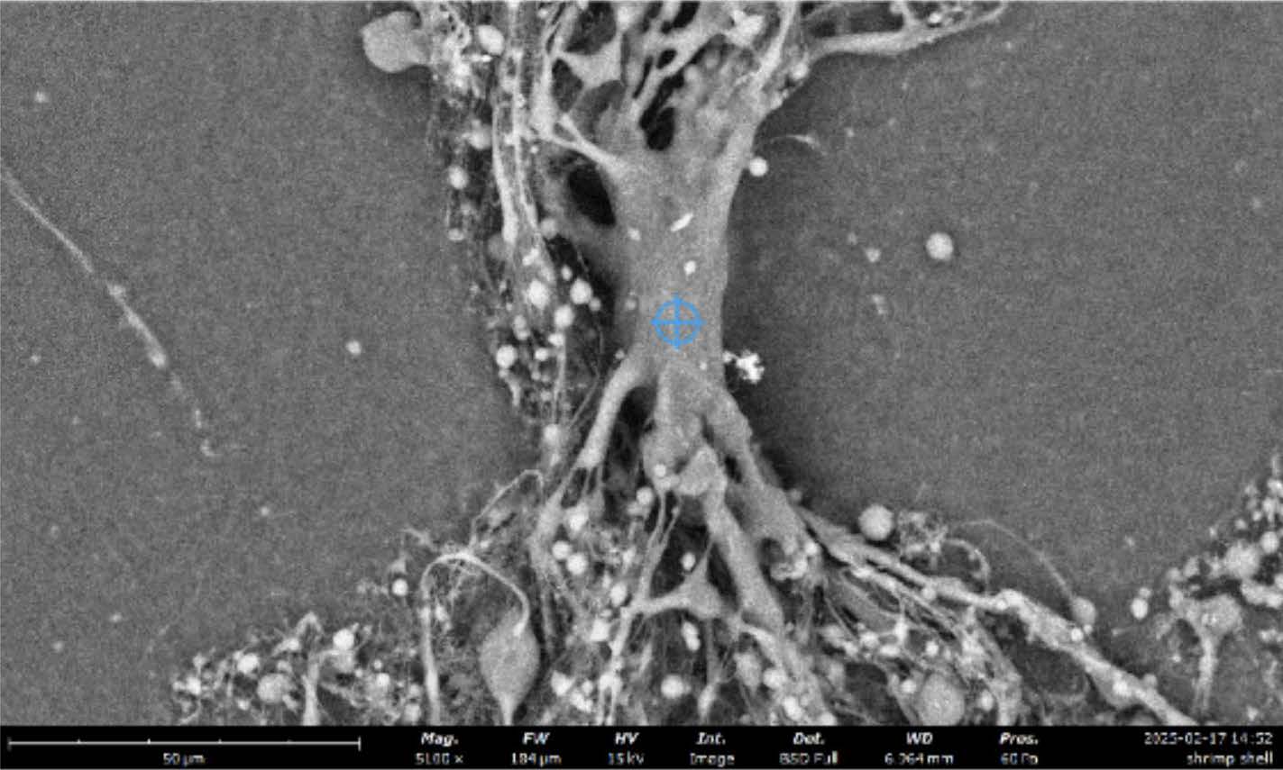
| Figure 1. Scanning electron microscope (SEM) image of electrospun chitosan fibers (Klossner et al., 2008), magnified 5100×. |
|
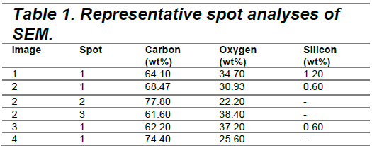
Elemental composition shows predominantly carbon and oxygen
Elemental analysis via SEM-EDS (Energy Dispersive X-ray Spectroscopy) was conducted on fibers from multiple SEM images. All samples showed carbon and oxygen as the primary elements, consistent with the expected composition of chitosan and PEO. Small amounts of silicon were also detected in some spots, likely due to environmental contamination or substrate interference. SEM-EDS elemental analysis confirmed that the nanofibers primarily contained carbon and oxygen, consistent with chitosan-PEO composition, with minor silicon traces likely due to contamination or substrate interference.(Table 1). These minuscule amounts were sporadic, likely due to contamination in the lab.Table 1 – Representative spot analyses of SEM
|
The carbon-to-oxygen ratios aligned with expectations for organic polymers, validating the incorporation of both chitosan and PEO. The lack of unexpected elements confirms the chemical purity of the fibers aside from trace silicon. The carbon-to-oxygen ratios confirmed successful incorporation of chitosan and PEO, and the absence of unexpected elements indicated high chemical purity of the nanofibers, aside from trace silicon.
Observed trends and fiber quality
The presence of higher carbon content in certain spots (up to 77.8 wt%) may reflect regions with increased PEO content, which contains proportionally more carbon than chitosan. Conversely, areas with higher oxygen content may be enriched with chitosan or indicate localized moisture retention. The consistent absence of nitrogen in SEM-EDS data, despite being a component of chitosan’s amine groups, is likely due to the low detection sensitivity for light elements in EDS at the magnifications and voltages used.
The successful electrospinning and nanoscale morphology of chitosan-PEO fibers provide a promising foundation for the development of biodegradable, high-strength fabrics. These results represent an early step toward the goal of integrating chitin-based fibers into impact-resistant materials like bulletproof vests
SUBLIMATION OF CHITIN GEL
Freeze thawing:
We conducted freeze-thaw cycles in order to transform our chitin powder and formic acid solution into a solid for further testing. The freeze-thaw cycling protocol effectively transformed the chitin-formic acid solution into a semi-rigid gel. Air-dried sheets exhibited a translucent ivory appearance with visible lamellar structures under oblique lighting. This reproducibility stemmed from uniform moisture expulsion during the 48-hour compression phase, with some observed delamination and warping due to folding of the oven paper. These defects resulted in increased difficulty with combining layers in later steps.
Mechanical testing:
Standard Tensile Testing
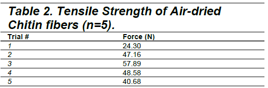
The Logger Pro system revealed significant performance variations, with recorded forces spanning 24.30 N to 57.89 N across five trials. The trials produced peak forces of 24.30-57.89 N, yielding a mean tensile strength of 43.72 ± 12.01 MPa.
|
Impact Resistance
Impact resistance testing with a 50 g weight drop generated 0.40 ± 0.10 cm gelatin indentations. High-speed video analysis identified two-phase energy absorption: initial sheet flexure absorbed kinetic energy within 5 ms, followed by residual energy transfer into gelatin deformation. No permanent damage occurred to the sheet. More trials are needed to confirm the resistant nature of the sheets.
Scanning electron microscope (SEM)
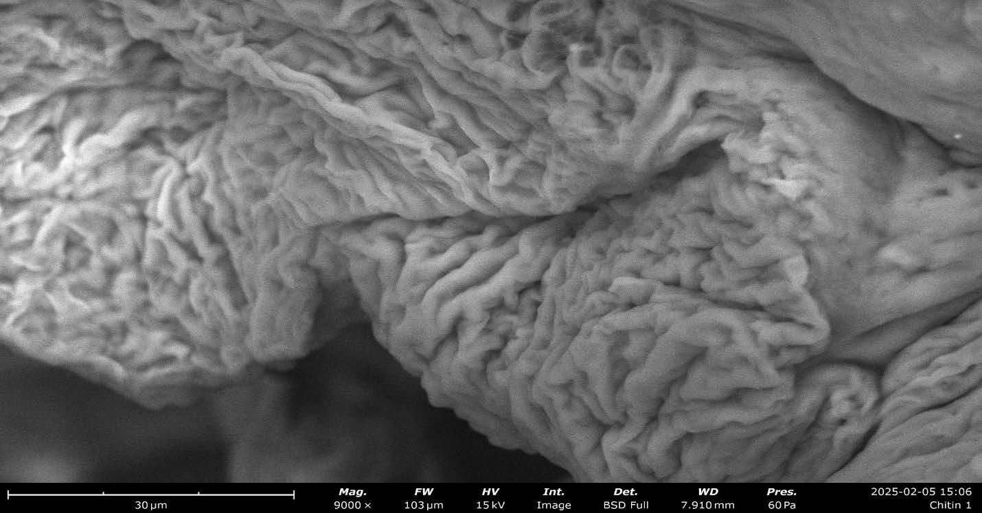
Scanning electron microscopy (SEM) reveals a layered and fibrous microstructure in the dried chitin sheet derived from the turbid gel we created (Figure 2). At 9000x magnification, SEM images show a network of interconnected fibers with varying degrees of alignment and porosity on the surface. Elemental analysis identifies carbon (C), oxygen (O), and nitrogen (N) and confirms that they exist in proportions consistent with the elemental composition of chitin typically found in shrimp shells, thereby supporting the material’s identity as chitin.
| Figure 2. Scanning electron microscopy (SEM) image of an air-dried chitin sheet at 9000× magnification. |
|
When compared to the reference SEM images of chitosan fibers ( Huang et. al, 2022) the shrimp shell-derived chitin exhibits notable differences. At higher concentrations, the referenced chitosan fibers show a more compact and layered arrangement with increasing concentration (Figure 3), whereas our sample derived from shrimp shells (Figure 2) appears to have a rougher surface texture and a less dense packing of fibers. Moreover, the shrimp shell-derived chitin seems to possess a greater degree of surface irregularity compared to the smoother, more uniform texture observed in the reference image, which are all smooth. These distinctions in surface texture and fiber arrangement may be attributed to differences in the source material, processing techniques, and potential variations in the degree of deacetylation between chitin and chitosan.
| Figure 3. SEM Micrographs showing the Entire fiber Width at Low Magnification (inset, second column), the Surface at Intermediate Magnification (second column), and the Relative Chitin Fibril Alignment at High Magnification (third column) from Huang et. al 2022. |
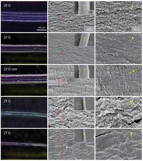
|
Discussions
This study demonstrates the feasibility of electrospinning chitosan and polyethylene oxide (PEO) into nanofibers that exhibit consistent morphology and chemical composition, marking a significant foundational step towards the development of impact-resistant, biodegradable materials. Previous studies have explored the properties of electrospun chitosan and PEO fibers, noting their potential for applications in protective gear due to their nanofiber structure, which typically features high surface area-to-volume ratios and interconnected porous networks—traits that are also utilized in contemporary soft body armor technologies (Benzait & Trabzon, 2018; Koosha & Mirzadeh, 2015).
While our results do not yet confirm the mechanical strength or ballistic properties necessary for protective applications, they validate critical aspects of the production process that are essential for future scaling and functional testing. The fibers produced in our experiments were continuous and nanoscale, with diameters aligning with expected results from electrospinning, signifying that our methods are well established within the field (Nitti et al., 2018; Liverani et al., 2018). The analysis through SEM combined with EDS corroborates the chemical purity of the fibers, revealing a consistent presence of carbon and oxygen typical of organic polymer backbones, alongside only trace contamination from silicon. This compositional integrity is vital for upcoming performance evaluations, particularly regarding the strength and elasticity of the material, which are crucial metrics for any potential armor solutions (Liverani et al., 2018).
Despite these promising findings, several challenges persist before chitosan-based nanofibers can be deemed suitable for ballistic protection. Notably, our investigation did not assess mechanical properties such as tensile strength, flexibility, or impact resistance, all of which are essential for materials intended to replicate or replace established options like Kevlar (Su et al., 2016). Furthermore, the random orientation of the nanofibers limits their capacity to effectively resist directional forces, which is critical for current armor composites. Hence, future studies must delve into techniques for aligning fibers either during or post-electrospinning or consider weaving them into composite fabrics to optimize performance (Su et al., 2021).
Another limitation highlighted in our study pertains to the reliance on PEO addition, which facilitates the electrospinning process. While effective at enhancing the process, PEO may adversely affect the final mechanical properties or the biodegradability of the resultant composite material. Investigating alternative cross-linkers or post-processing treatments could improve fiber strength while still aligning with sustainability goals (Santiago-Castillo et al., 2022). Ultimately, this project lays a robust groundwork for producing nanofibrous mats using chitosan, a biopolymer known for its structural and antimicrobial advantages, thus establishing a platform for further advancements like stress testing, weaving, and impact simulations. Continued refinement efforts could make chitosan-based nanofibers viable as a sustainable, multifunctional alternative to synthetic polymers in body armor and other protective applications (Zhang et al., 2015).
Next Steps
To build on our current findings, our next steps will focus on improving the structural integrity and usability of our chitosan-based fibers. First, we plan to optimize the electrospinning process by experimenting with different chitosan-to-PEO ratios, as chitosan requires an electrospinning agent, such as PEO, for successful electrospinning. We can also experiment with varying solvent concentrations and spinning parameters to enhance fiber consistency and mechanical strength. Additionally, we aim to begin preliminary testing on weaving the electrospun nanofibers into layered fabric composites. These composites will be used in future experiments simulating real-world impact to evaluate their potential as a biodegradable alternative to Kevlar in protective gear such as bulletproof vests.
In addition to improving fiber strength and exploring fabric weaving techniques, we plan to functionalize our chitosan-based nanofibers by incorporating antimicrobial agents or bioactive nanoparticles during the electrospinning process. Given chitosan’s inherent antimicrobial properties, enhancing this feature could enable the development of multi-functional materials for applications in both protective wear and biomedical settings, such as wound dressings or infection-resistant vests.
In the future, we plan to synthetically place the gene that codes for chitin production in bacteria to express chitin. Bacterial biosynthesis is preferable to extraction from natural sources because microbial production can yield chitin samples with fewer impurities. In addition, bacterial biosynthesis offers an environmentally sustainable alternative to extraction which often involves shellfish farming which generates significant waste and can harm marine ecosystems. Microbial biosynthesis allows for the usage of the entire microbial biomass, reducing waste that would otherwise be discarded into the environment (Chakravarty, J et al.,2018).
Author Contributions
CT was primarily responsible for the electrospinning experiments, including preparation, electrospinning, collection, and analysis.MS, MC, and IL worked on initial cloning steps for synthetic production of chitin. MC also helped with electrospinning.
FW and AL were responsible for all the methods and procedures that fall under sublimation of chitin gel, including preparation, set up, measurements, and analysis. FW, MC, AL, MS, CT, IL worked on the manuscript and editing. Acknowledgements
We would like to extend our heartfelt gratitude to Dr. Hsu for his guidance and unwavering support throughout this project, as well as his invaluable advice. We also wish to thank Mr. Dezieck for imparting his expertise in materials science and chemistry, which greatly contributed to the success of our experiments. Special thanks to Mr. Tsao for assisting with the ordering process and ensuring that we had all necessary materials. Our TA, Isabelle, provided continuous support and aid, and we truly appreciate her dedication. Lastly, we would like to express our gratitude to Ms. Read and Mr. Frankenberg for their ongoing support of the scientific research program. Their contributions were essential in making this study possible.
References
Abu-Danso, E., Peräniemi, S., Leiviskä, T., Kim, T., Tripathi, K. M., & Bhatnagar, A. (2020). Synthesis of chitosan-based activated carbon–silver nanocomposite and its application for water treatment: Removal of humic acid, silver ions, and 4-chlorophenol. International Journal of Biological Macromolecules, 150, 1091–1100. https://doi.org/10.1016/j.ijbiomac.2020.02.111
Aldana, A. A., Abraham, G. A., & Alvarez-Lorenzo, C. (2015). Smart polymers for biomedical applications. In A. A. Aldana & G. A. Abraham (Eds.), Smart polymers and their applications (pp. 237–270). Woodhead Publishing. https://doi.org/10.1016/B978-0-08-100069-9.00008-7
Anitha, A., Sowmya, S., Kumar, P. T. S., Deepthi, S., Chennazhi, K. P., Ehrlich, H., Tsurkan, M., & Jayakumar, R. (2014). Chitin and chitosan in selected biomedical applications. Progress in Polymer Science, 39(9), 1644–1667. https://doi.org/10.1016/j.progpolymsci.2014.07.001
Aranaz, I., Harris, R., & Heras, A. (2010). Chitosan amphiphilic derivatives. Chemistry and Chemical Technology, 4(1), 35–40.
Azevedo, S., Costa, A. M. S., Andersen, A., Choi, I. S., Birkedal, H., & Mano, J. F. (2019). Bioinspired ultrafast self-gelling hydrogels using molecular recognition for 3D cell culture. Advanced Functional Materials, 29(14), 1806443. https://doi.org/10.1002/adfm.201806443
Benhabiles, M. S., Salah, R., Lounici, H., Drouiche, N., Goosen, M. F. A., & Mameri, N. (2012). Antibacterial activity of chitin, chitosan and its oligomers prepared from shrimp shell waste. Food Hydrocolloids, 29(1), 48–56. https://doi.org/10.1016/j.foodhyd.2012.02.013
Bhattarai, N., Gunn, J., & Zhang, M. (2010). Chitosan-based hydrogels for controlled, localized drug delivery. Advanced Drug Delivery Reviews, 62(1), 83–99. https://doi.org/10.1016/j.addr.2009.07.019
Boeris, V., Nerli, B., & Romanini, D. (2009). Synthesis and characterization of N-acylated chitosan as potential matrix for hydrophobic drug delivery. Carbohydrate Polymers, 77(3), 572–578. https://doi.org/10.1016/j.carbpol.2009.02.015
Calvo, P., Remuñán-López, C., Vila-Jato, J. L., & Alonso, M. J. (1997). Novel hydrophilic chitosan–polyethylene oxide nanoparticles as protein carriers. Journal of Applied Polymer Science, 63(1), 125–132. https://doi.org/10.1002/(SICI)1097-4628(19970103)63:1<125::AID-APP14>3.0.CO;2-E
Chandy, T., & Sharma, C. P. (1990). Chitosan—as a biomaterial. Biomaterials, Artificial Cells and Artificial Organs, 18(1), 1–24. https://doi.org/10.3109/10731199009117674
Chen et al. (2022). Investigation on Residual Strength and Failure Mechanism of the Ceramic/UHMWPE Armors after Ballistic Tests. Materials. https://doi.org/10.3390/ma15030901
Chen, L., Zhu, C., Fan, D., Liu, B., Ma, X., Duan, Z., … & Zhou, Y. (2011). A human‐like collagen/chitosan electrospun nanofibrous scaffold from aqueous solution: electrospun mechanism and biocompatibility. Journal of Biomedical Materials Research Part A, 99A(3), 395–409. https://doi.org/10.1002/jbm.a.33202
Cui, J., Yu, Z., & Lau, D. (2016). Effect of acetyl group on mechanical properties of chitin/chitosan nanocrystal: a molecular dynamics study. International Journal of Molecular Sciences, 17(1), 61. https://doi.org/10.3390/ijms17010061
Cunniff. (1996). A Semiempirical Model for the Ballistic Impact Performance of Textile-Based Personnel Armor. Textile Research Journal. https://doi.org/10.1177/004051759606600107
Decker et al. (2007). Stab resistance of shear thickening fluid (STF)-treated fabrics. Composites Science and Technology. https://doi.org/10.1016/j.compscitech.2006.08.007
Desai, K., Kit, K., Li, J., & Zivanovic, S. (2008). Morphological and surface properties of electrospun chitosan nanofibers. Biomacromolecules, 9(3), 1000–1006. https://doi.org/10.1021/bm701017z
Ding, B., Cai, J., Huang, J., Zhang, L., Chen, Y., Shi, X., … & Kuga, S. (2012). Facile preparation of robust and biocompatible chitin aerogels. Journal of Materials Chemistry, 22(12), 5801. https://doi.org/10.1039/c2jm16032c
Du et al. (2015). The use of ramie fibers as reinforcements in composites. https://doi.org/10.1533/9781782421276.1.104
Duan, B., Chang, C., & Zhang, L. (2013). Structure and properties of films fabricated from chitin solution by coagulating with heating. Journal of Applied Polymer Science, 131(4). https://doi.org/10.1002/app.39538
Fernandez, J., & Ingber, D. (2013). Bioinspired chitinous material solutions for environmental sustainability and medicine. Advanced Functional Materials, 23(36), 4454–4466. https://doi.org/10.1002/adfm.201300053
Gadgey, K. K., & Bahekar, A. (2017). Studies on extraction methods of chitin from crab shell and investigation of its mechanical properties. International Journal of Mechanical Engineering and Technology, 8(2), 220–231.
Guleria et al. (2020). Fabrication of Kevlar®-reinforced ultra-high molecular weight polyethylene composite through microwave-assisted compression molding for body armor applications. Journal of Reinforced Plastics and Composites. https://doi.org/10.1177/0731684420959449
Huang, J., Zhong, Y., Zhang, X., Xu, H., Zhu, C., & Cai, J. (2021). Continuous pilot‐scale wet‐spinning of biocompatible chitin/chitosan multifilaments from an aqueous koh/urea solution. Macromolecular Rapid Communications, 42(16). https://doi.org/10.1002/marc.202100252
Huang, W., Montroni, D., Wang, T., Murata, S., Arakaki, A., Nemoto, M., & Kisailus, D. (2022). Direct Ink Write Printing of Chitin-Based Gel Fibers with Customizable Fibril Alignment, Porosity, and Mechanical Properties for Biomedical Applications. Journal of Functional Biomaterials, 13(2), 83. https://doi.org/10.3390/jfb13020083
Ibitoye, E., Lokman, I., Noor, M., Goh, Y., Zuki, A., & Jimoh, A. (2018). Extraction and physicochemical characterization of chitin and chitosan isolated from house cricket. Biomedical Materials, 13(2), 025009. https://doi.org/10.1088/1748-605x/aa9dde
Jin, H., Fridrikh, S., Rutledge, G., & Kaplan, D. (2002). Electrospinning Bombyx mori silk with poly(ethylene oxide). Biomacromolecules, 3(6), 1233–1239. https://doi.org/10.1021/bm025581u
Khodadadi et al. (2019). Ballistic performance of Kevlar fabric impregnated with nanosilica/PEG shear thickening fluid. Composites Part B Engineering. https://doi.org/10.1016/j.compositesb.2018.12.121
Klossner, R., Queen, H., Coughlin, A., & Krause, W. (2008). Correlation of chitosan’s rheological properties and its ability to electrospin. Biomacromolecules, 9(10), 2947–2953. https://doi.org/10.1021/bm800738u
Klumdoung, P., & Pankaew, P. (2017). The development of electrospinning apparatus to fabricate nanofiber for future material applications. Applied Mechanics and Materials, 866, 244–247. https://doi.org/10.4028/www.scientific.net/amm.866.244
Koosha, M., & Mirzadeh, H. (2015). Electrospinning, mechanical properties, and cell behavior study of chitosan/pvananofibers. Journal of Biomedical Materials Research Part A, 103(9), 3081–3093. https://doi.org/10.1002/jbm.a.35443
Lertsutthiwong, P., Rojsitthisak, P., & Nimmannit, U. (2009). Preparation of chitosan nanoparticles using water-in-oil emulsion method. Journal of Metals, Materials and Minerals, 19(1), 1–5.
Muzzarelli, R. A. A., El Mehtedi, M., & Bottegoni, C. (2012). Developments of chitin and chitosan in tissue engineering, wound healing, and orthopedic surgery. Biomedical Materials, 7(4), 045009. https://doi.org/10.1088/1748-6041/7/4/045009
Negi, Y., Mohabeer, C., & Agnihotri, P. K. (2019). Synthesis and characterization of bulletproof vests made from aramid and UHMWPE. Materials Today: Proceedings, 18, 2330–2336. https://doi.org/10.1016/j.matpr.2019.07.189
Ogunsona, E. O., Misra, M., & Mohanty, A. K. (2017). Insight into the performance of polylactide and durable biobased polymer blends: A review. Journal of Applied Polymer Science, 134(45). https://doi.org/10.1002/app.45419
Park, Y., Lee, D. Y., Park, D., Choi, Y., Lim, J., & Youn, J. R. (2016). High-strength chitosan fibers reinforced with graphene oxide for wearable electronics. RSC Advances, 6(100), 98075–98083. https://doi.org/10.1039/C6RA17925A
Park, B. D., & Lee, S. M. (2004). Hot-pressing behavior and bonding mechanism in the bonding of wood using high temperature and pressure. Journal of Adhesion Science and Technology, e202513
Chung, Y. C., Chen, C. Y., & Liaw, L. Y. (2003). Antibacterial activity of chitosan in water. Bioresource Technology, 88(2), 179–184. https://doi.org/10.1016/S0960-8524(02)00279-X
Dash, M., Chiellini, F., Ottenbrite, R. M., & Chiellini, E. (2011). Chitosan—A versatile semi-synthetic polymer in biomedical applications. Progress in Polymer Science, 36(8), 981–1014. https://doi.org/10.1016/j.progpolymsci.2011.02.001
Dutta, P. K., Dutta, J., & Tripathi, V. S. (2004). Chitin and chitosan: Chemistry, properties and applications. Journal of Scientific and Industrial Research, 63(1), 20–31.
Elieh-Ali-Komi, D., & Hamblin, M. R. (2016). Chitin and chitosan: Production and application of versatile biomedical nanomaterials. International Journal of Advanced Research, 4(3), 411–427.
Fang, J., Tung, Y.-T., & Lin, Y.-C. (2020). Antimicrobial activity of chitosan-based matrices against oral pathogens. Materials, 13(22), 5306. https://doi.org/10.3390/ma13225306
Goy, R. C., Morais, S. T. B., & Assis, O. B. G. (2009). Evaluation of the antimicrobial activity of chitosan and its quaternized derivative on E. coli and S. aureus growth. Revista Brasileira de Farmacognosia, 19, 777–782. https://doi.org/10.1590/S0102-695X2009000500022
Hadwiger, L. A. (2013). Multiple effects of chitosan on plant systems: Solid science or hype. Plant Science, 208, 42–49. https://doi.org/10.1016/j.plantsci.2013.03.012
Helander, I. M., Nurmiaho-Lassila, E. L., Ahvenainen, R., Rhoades, J., & Roller, S. (2001). Chitosan disrupts the barrier properties of the outer membrane of Gram-negative bacteria. International Journal of Food Microbiology, 71(2–3), 235–244. https://doi.org/10.1016/S0168-1605(01)00609-7
Il’ina, A. V., & Varlamov, V. P. (2005). Antimicrobial activity of chitosan. Applied Biochemistry and Microbiology, 41, 286–289. https://doi.org/10.1007/s10438-005-0050-2
Islam, S., Bhuiyan, M. A. R., & Islam, M. N. (2017). Chitin and chitosan: Structure, properties and applications in biomedical engineering. Journal of Polymers and the Environment, 25, 854–866. https://doi.org/10.1007/s10924-016-0865-5
Jayakumar, R., Prabaharan, M., Kumar, P. S., Nair, S. V., & Tamura, H. (2011). Biomaterials based on chitin and chitosan in wound dressing applications. Biotechnology Advances, 29(3), 322–337. https://doi.org/10.1016/j.biotechadv.2011.01.005
Kim, I. Y., Seo, S. J., Moon, H. S., Yoo, M. K., Park, I. Y., Kim, B. C., & Cho, C. S. (2008). Chitosan and its derivatives for tissue engineering applications. Biotechnology Advances, 26(1), 1–21. https://doi.org/10.1016/j.biotechadv.2007.07.009
Kumar, M. N. V. R. (2000). A review of chitin and chitosan applications. Reactive and Functional Polymers, 46(1), 1–27. https://doi.org/10.1016/S1381-5148(00)00038-9
Kumirska, J., Weinhold, M. X., Czerwicka, M., Kaczyński, Z., & Stepnowski, P. (2011). Influence of the chemical structure and physicochemical properties of chitosan on its antibacterial activity. Progress on Chemistry and Application of Chitin and Its Derivatives, 16, 5–20.
Kurita, K. (2006). Chitin and chitosan: Functional biopolymers from marine crustaceans. Marine Biotechnology, 8, 203–226. https://doi.org/10.1007/s10126-005-0097-5
Lertsutthiwong, P., How, N. C., Chandrkrachang, S., & Stevens, W. F. (2009). Effect of chemical treatment on the properties of chitosan nanoparticles. Journal of Metals, Materials and Minerals, 19(1), 1–5.
Muzzarelli, R. A. A., El Mehtedi, M., & Mattioli-Belmonte, M. (2012). Emerging biomedical applications of nano-chitins and nano-chitosans obtained via advanced eco-friendly technologies from marine resources. Marine Drugs, 12(11), 5468–5502. https://doi.org/10.3390/md12115468
Negi, Y. S., Choudhary, V., & Samad, A. (2019). Fundamentals and frontiers of functional biocomposites. In Functionalized engineering materials and their applications (pp. 289–309). Apple Academic Press.
Ogunsona, E. O., Misra, M., & Mohanty, A. K. (2017). Advances in the fabrication and application of bio-based polymer blends and composites. In M. Jawaid, M. S. Salit, & M. Tahir (Eds.), Bio-based polymers and nanocomposites (pp. 1–24). Springer. https://doi.org/10.1007/978-3-319-41189-3_1
Park, J. H., Kim, M., Kang, S., & Lee, M. S. (2016). Flexible and transparent chitosan-based resistive switching memory devices for organic electronics. ACS Applied Materials & Interfaces, 8(44), 30575–30582. https://doi.org/10.1021/acsami.6b09127
Park, S., & Lee, P. W. (2004). Effect of pressing temperature on the properties of wood-based panels. Journal of the Korean Wood Science and Technology, 32(6), 17–25.
Qi, G., & Yu, D. (2018). Preparation and characterization of ultrafine chitin nanofibers by electrospinning. Carbohydrate Polymers, 180, 223–229. https://doi.org/10.1016/j.carbpol.2017.10.013
Rinaudo, M. (2006). Chitin and chitosan: Properties and applications. Progress in Polymer Science, 31(7), 603–632. https://doi.org/10.1016/j.progpolymsci.2006.06.001
Shahbazi, M. A., Shavandi, A., Maleki, H., Nie, L., Ebrahimi, A., Hamishehkar, H., & Milojevic, M. (2015). Chitosan-based nanocomposites for biomedical applications. In Nanobiomaterials in Soft Tissue Engineering (pp. 43–76). Elsevier. https://doi.org/10.1016/B978-0-323-42865-6.00003-6
Shen, J., Shi, M., Yan, B., Ma, H., Li, N., Hu, Y., & Ye, M. (2011). Covalently immobilized chitosan on graphene oxide for improved mechanical and antibacterial properties. Carbohydrate Polymers, 84(1), 310–316. https://doi.org/10.1016/j.carbpol.2010.11.029
Shukla, S. R., & Cheryan, M. (2001). Performance of ultrafiltration membranes for treatment of whey protein-based effluents. Journal of Membrane Science, 193(1), 9–19. https://doi.org/10.1016/S0376-7388(01)00506-3
Smith, L. A., Ma, P. X., & Laurencin, C. T. (2013). Electrospinning: A powerful technique for fabricating nanofibrous scaffolds for regenerative medicine. Nanotechnology in Tissue Engineering and Regenerative Medicine, 73–94. https://doi.org/10.1016/B978-0-12-398520-0.00006-0
Tan, H., Ma, R., Lin, C., Liu, Z., & Tang, T. (2013). Quaternized chitosan as an antimicrobial agent: Antimicrobial activity, mechanism of action and biomedical applications in orthopedics. International Journal of Molecular Sciences, 14(1), 1854–1869. https://doi.org/10.3390/ijms14011854
Van der Lubbe, W. H. J., & De Haan, J. L. (2006). High-performance fibers for ballistic protection. In J. W. S. Hearle (Ed.), High-performance fibers (pp. 295–308). Woodhead Publishing.
Wang, H., Song, S., & Li, J. (2016). Electrospinning of polysaccharides: A review. Carbohydrate Polymers, 148, 324–333. https://doi.org/10.1016/j.carbpol.2016.04.073
Wang, Y., Wang, L., Li, T., & Ma, X. (2020). Recent advances in chitosan-based electrospun nanofibers for biomedical applications. Carbohydrate Polymers, 241, 116388. https://doi.org/10.1016/j.carbpol.2020.116388
Yan, L., & Chouw, N. (2013). Natural fibre-reinforced polymer composites: A review. Materials and Design, 49, 69–79. https://doi.org/10.1016/j.matdes.2013.01.025
Yadav, M., Goswami, P., Paritosh, K., Kumar, M., & Pareek, N. (2019). Seafood waste: A source for preparation of commercially employable chitin/chitosan materials. Bioresources and Bioprocessing, 6(1), 1–13. https://doi.org/10.1186/s40643-019-0277-y
Yang, Y., Wei, Y., Wang, S., & Wang, C. (2016). Electrospinning of chitosan/polyethylene oxide blend nanofibers for wound dressing applications. Carbohydrate Polymers, 148, 176–182. https://doi.org/10.1016/j.carbpol.2016.04.061
Zeng, J., Xu, X., Chen, X., Liang, Q., Bian, X., Yang, L., & Jing, X. (2003). Biodegradable electrospun fibers for drug delivery. Journal of Controlled Release, 92(3), 227–231. https://doi.org/10.1016/S0168-3659(03)00303-6