Annie Chang, Ryan Chen, Harriet Chi, Cate Huang, Angelina S. Kuo, Lindsey Liaw, Jocelyn Weng, Sophie Yang, John Young, Iris Yu, Taipei American School, Department of Scientific Research, Taipei, Taipei, Taiwan
Reviewed on 3 May 2025; Accepted on 9 June 2025; Published on 27 October 2025
With help from the 2025 BioTreks Production Team.
Silk fibroin is a promising biomaterial for bone grafts that could revolutionize the healing of bone injuries and defects. Our research aims to develop a bioengineered composite material for bone grafts by genetically engineering Escherichia coli (E. coli) to produce the fibroin heavy chain (Fib-H), a key silk fibroin protein, as a sustainable alternative to natural silk. We designed a gene sequence containing sixteen repeated motifs of alternating hydrophobic and hydrophilic residues (GAGAS). We introduced this recombinant gene into E. coli for protein expression and production, and we are in the process of obtaining preliminary results. Simultaneously, we developed an initial method to solubilize silk fibroin from silkworm cocoons, which showed successful polymerization of methacrylated silk fibroin crosslinked by ultraviolet (UV) exposure; the resulting methacrylated silk fibroin will be evaluated for its compatibility with 3D printing techniques. Future work includes refining Fib-H expression, testing the mechanical properties of the 3D-printed scaffolds, and evaluating their biocompatibility. By integrating synthetic biology and materials engineering, we aim to develop a sustainable and effective silk-based bone graft to enhance bone regeneration.
Keywords: Silk fibroin, bone tissue, and photocrosslinking
Authors are listed in alphabetical order. Jonathan Hsu mentored the group. Please direct all correspondence to hsujo@tas.edu.tw.


Background
Bone graft surgery is a critical medical procedure that utilizes bone tissue transplantation to repair or reconstruct compromised areas in the body, particularly to treat bone defects, fractures, or injuries that exhibit poor healing potential. Bone grafts can be categorized chiefly into two types: autografts, which are harvested from the patient’s body, and allografts, which are sourced from deceased donors. Autografts are often regarded as the gold standard due to their optimal integration and healing properties; however, they require an additional surgical site, leading to increased pain and prolonged recovery periods (Baldwin et al., 2019). Moreover, the availability of sufficient donor bone can be a restricting factor, calling for multiple surgical interventions(Baldwin et al., 2019). While allografts eliminate the requirement for a second surgical site, they can carry significant drawbacks, including the risks of immune rejection, infection, and limited availability. These limitations emphasize the urgent need for alternative solutions to enhance bone regeneration (Betz et al., 2002; Baldwin et al., 2019).
In this context, our research explores the potential of silk fibroin as a compelling alternative material, distinguished by its superior material strength and biocompatibility (Xiang-Long et al., 2019). Silk fibroin is a fibrous protein secreted by the silkworm Bombyx mori, which has garnered attention in the bone tissue engineering field for its advantageous properties. A key aspect of this project revolves around a “scaffold,” which we define as a three-dimensional structural framework designed to support cell attachment and promote the growth of new bone tissue. Rather than seeking to create a hard material that indiscriminately replaces bone, we aim to develop a scaffold specifically engineered for bone tissue to facilitate regenerative processes (Cheng et al., 2016). Thus, the term “bone graft” refers to this scaffold, which is intended to foster an environment conducive to osteogenesis while gradually assimilating into the body as new bone is formed.
In our study, we concentrate on utilizing silk fibroin, particularly Fibroin heavy chain Fib-H, which serves as the main structural component responsible for the mechanical strength and durability of the silk. Fib-H consists of repetitive amino acid sequences, particularly glycine, and alanine, forming a highly crystalline configuration that imparts remarkable tensile strength, flexibility, and resilience (Leng-Duei et al., 2015). In this study, we aim to employ recombinant DNA technology to produce silk fibroin in E. coli, thereby circumventing the ethical and resource-intensive challenges associated with traditional silk farming practices.
Following the expression and purification of the Fib-H protein, the objective is to convert it into a photopolymerizable format by generating methacrylated silk fibroin (Sil-MA). This conversion involves introducing methacrylate groups to the silk fibroin, allowing the material to crosslink when exposed to UV light (Xi-He et al., 2024). Crosslinking is a critical step, as it solidifies the material, facilitating the formation of stable and biocompatible scaffolds for bone graft applications(Koh et al., 2015). The Sil-MA modification results in covalently bonded structures in the presence of UV light and a photoinitiator, whereby free radicals generated during this reaction lead to the polymerization of the scaffold material (He et al., 2024).
This approach seeks to ascertain the feasibility of 3D printing with silk fibroin and aims to provide innovative, sustainable, and effective scaffolds that enhance tissue integration and bone regeneration (Rockwood et al., 2011; Zafar et al., 2015; Marcos-Campos et al., 2012). By clarifying our objectives and delineating the capabilities of hybrid materials, we strive to address the limitations associated with traditional bone grafts. Ultimately, our research aims to create a dual-function scaffold, that offers temporary structural support for bone regeneration while biodegrading in sync with the body’s natural healing processes (Elsevier, n.d.).
This paper will present detailed insights into the development of engineered scaffolds and the anticipated outcomes relating to their application in bone tissue engineering.
Materials and Methods
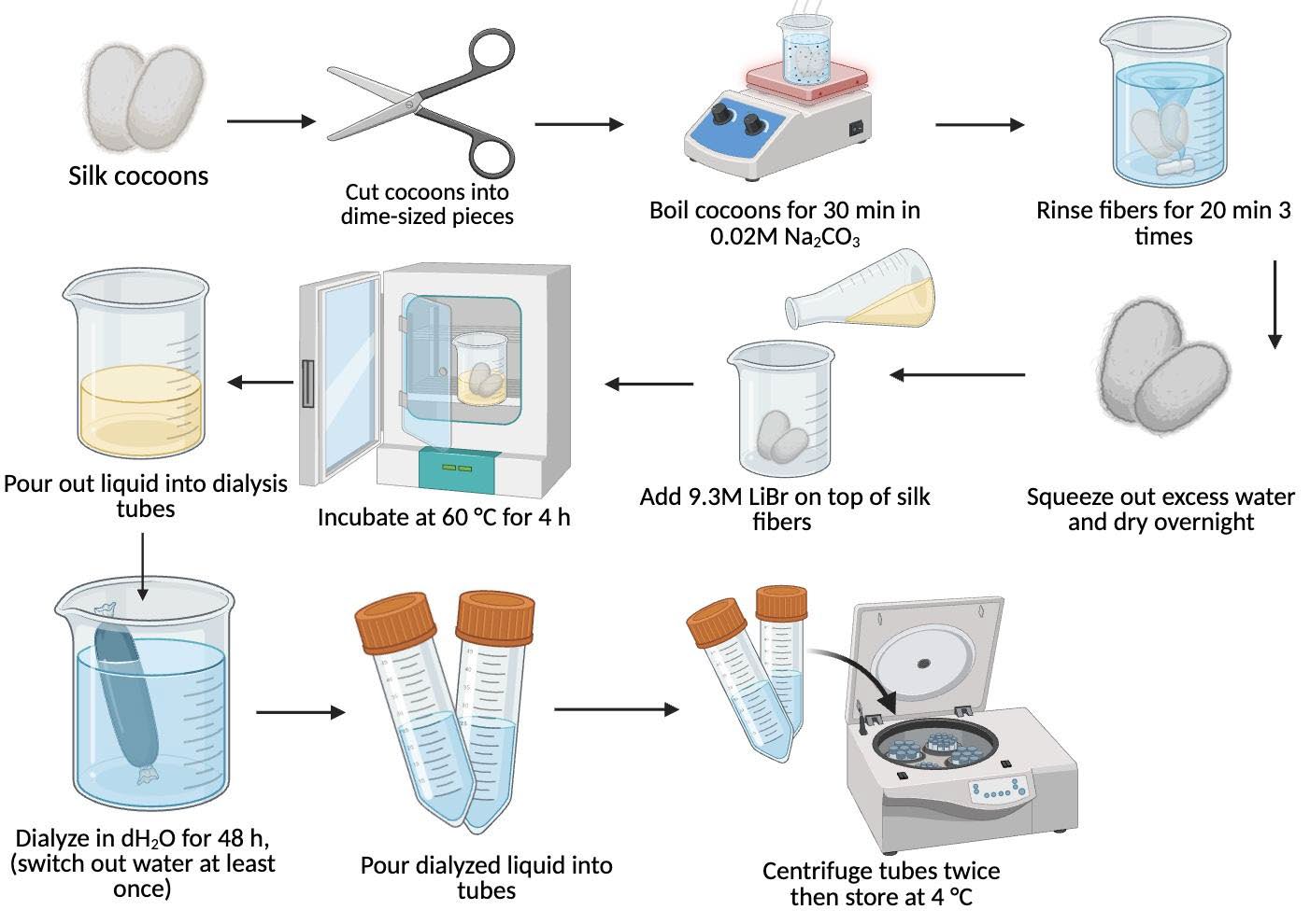
Degumming of silk cocoons. Raw Bombyx mori cocoons were cut into dime-sized pieces to maximize surface area. A total of 5 g of this cut cocoon material was combined with 4.24 g of 0.02 M sodium carbonate (Na2CO3) in 2 L of water, which was pre-heated on a hot plate under aluminum foil protection (Figure 1). The Na2CO3 was gradually added to ensure complete incorporation while stirring continuously and making sure it did not boil over. Once the solution reached a boil, the cocoon pieces were introduced and allowed to boil for 30 minutes (Figure 1). Following degumming, the resulting wet cocoon fibers were removed and subsequently washed in distilled water three times—each wash lasting 20 minutes—to effectively remove residual sericin (Figure 1). The washed fibers were then returned to a beaker containing 1 L of distilled water and stirred for an additional 20 minutes to ensure further extraction of sericin, as reported in similar methodologies employing water degumming (Figure 1) (Rockwood et al., 2011; Atay et al., 2025; Echeverri et al., 2019).
| Figure 1. General overview of the degumming and dialysis process. |
|
Cloning:
Gene Design and Synthesis
The Fibroin heavy chain (Fib-H) is a critical structural protein of silk fibroin, known for its mechanical properties. The Fib-H gene consists of both repetitive and non-repetitive regions. For this study, a gene containing 16 GAGAS amino acid sequence repeats was engineered and chosen based on prior work demonstrating their role in silk protein function. The designed sequence incorporated essential elements for expression in E. coli, including a T7 promoter, a ribosome binding site (RBS), a Cytiva Protein Select Tag for enhanced protein production, a 6× histidine tag for purification, and both N-terminal and C-terminal sequences. The gene was synthesized by Genomics BioSci & Tech Co., Ltd. (Taipei, Taiwan).
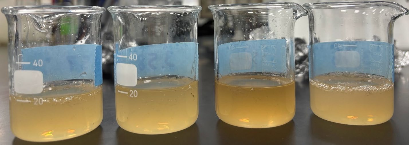
| Figure 2. The result of dissolved silk cocoons in 0.02 M sodium carbonate (Na2CO3) and 2 L of water. |
|
PCR Amplification and Subcloning
To isolate the gene of interest, we designed forward (5’-AAAACCATGGATGG TTAAAATTGTGAGCCG-3’) and reverse (5’-TTTTCTCGAGTTAGTGATGGTGATGATG-3’) primers that specifically amplified the Fib-H gene region, excluding the RBS. The gene was amplified by PCR using a high-fidelity polymerase, followed by analysis on an1% agarose gel to confirm the gene size. The resulting PCR product was then subcloned into the pET-14b expression vector for subsequent protein expression in E. coli.
Agarose Gel Electrophoresis
We used gel that was made with 1% agarose (Bioman Scientific) and 1x TAE (Geneaid). We added 0.005% SeeingSafe (Seeing Bioscience). We ran the gels at 120V for 10-20 minutes.
Transformation and Expression Testing
The synthesized plasmid was first transformed into E. coli DH5ɑ cells for amplification. Following confirmation of plasmid integrity, the plasmid was transferred into E. coli BL21(DE3) cells for expression purposes. Protein expression was assessed using IPTG induction, where overnight cultures of E. coli BL21 containing the Fib-H plasmid were diluted into fresh LB medium and incubated for 3–4 hours to maintain cells in the log phase. Afterward, 100 µM IPTG was added to one culture to induce protein expression, while the other served as an uninduced control. The cultures were then incubated for 4 hours at 30°C.
Following induction, cells were collected by centrifugation at 12,000 rpm for 1 minute. The resulting cell pellets were resuspended in 50 µL of 2× Laemmli sample buffer containing beta-mercaptoethanol and then heated at 95°C for 5 minutes. We loaded 15 µL of the heated samples into an SDS-PAGE gel, which was run at 120 V for 45 minutes. Gels were stained with Coomassie Brilliant Blue for 1 hour, then destained in a solution of 10% acetic acid and 40% methanol until the background was cleared.
Protein Purification
In the event of successful protein expression, we purified the Fib-H protein using Cytiva Protein Select Resin following the standard protocol outlined by Cytiva (n.d.). This purification method would allow for the isolation of the recombinant protein from E. coli lysates for further structural and functional analyses.
Dissolving of degummed silk cocoons
After completion of the degumming process, the purified silk fibroin was divided equally into two 10 mL beakers. In each beaker, a solution was prepared by adding 1 g of 9.3 M lithium bromide (LiBr) per (grams of silk fibroin × 4), and the mixtures were incubated at 60 °C overnight to ensure complete dissolution of the fibroin protein.
Methacrylation and Dialysis
Glycidyl methacrylate (GMA) was added dropwise to the dissolved silk fibroin at a 97% concentration while stirring at 300 rpm. The resulting solutions were transferred into dialysis bags and immersed in 2 L of distilled water to remove LiBr, unreacted GMA, and other byproducts. The water was dialyzed for 4 days to ensure the LiBr was removed. This followed protocols similar to those described in established silk fibroin purification studies (Rockwood et al., 2011; Atay et al., 2025; Sah & Pramanik, 2010).
Freeze-drying and Photopolymerization
After dialysis, the solution was frozen for 24 hours, followed by vacuum freeze-drying for 48 hours to eliminate excess moisture. The freeze-dried product, referred to as Sil-MA, was then mechanically broken into smaller pieces to increase surface area, facilitating future dissolution. For subsequent application in bio-3D printing, Lithium Phenyl-2,4,6-trimethylbenzoylphosphinate (LAP) was added to the mixture as a photoinitiator, enabling rapid crosslinking of the fibroin upon exposure to UV light (Hong et al., 2020). After applying a 365 nm UV light, the crosslinking reaction yielded a solid structure suitable for fabricating biocompatible scaffolds in digital light processing (DLP) printing applications (Hong et al., 2020).
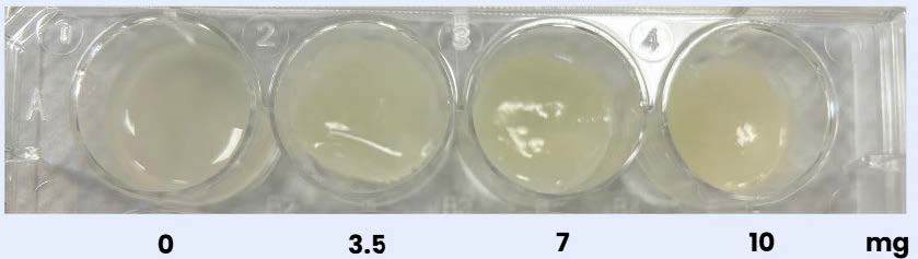
During the step of adding LAP, different concentrations of LAP were tested in the Sil-MA, including 0mg/ml, 3.5mg/ml, 7mg/ml, and 10.5mg/ml in separate test tubes. The solutions were then exposed to UV light for 10 minutes, and the differences in structure were observed.
Results
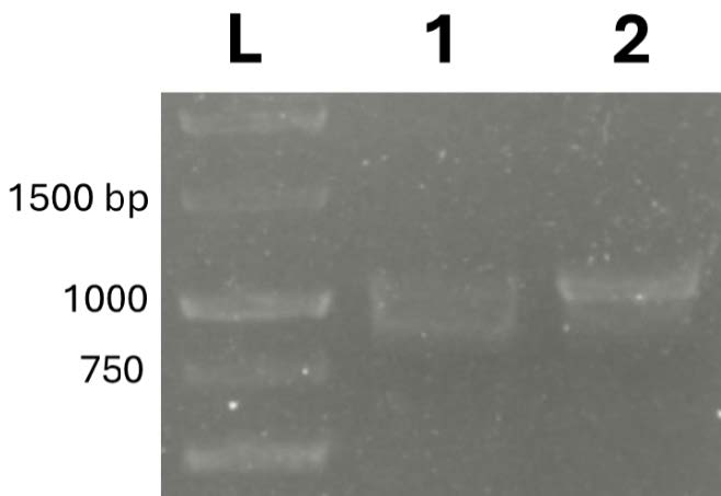
We purified the Fib-H protein by cloning a fragment of the Fib-H gene into an expression vector. We successfully amplified the Fib-H gene, as confirmed by a single band at the expected size of ~1056 bp (Figure 3). Using varying template concentrations, we obtained consistent results, which validated our primer design. We then subcloned the resulting PCR product into the pET-14b vector for downstream protein expression.
| Figure 3. Gel electrophoresis of PCR-amplified Fib-H gene. |
|
PCR Amplification and Gene Validation
Initial Expression Attempts
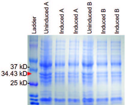
We initially tested protein expression by transforming the Fib-H plasmid into E. coli BL21(DE3) cells, followed by IPTG induction. However, SDS-PAGE analysis of the induced samples revealed consistent banding in the induced and uninduced samples at 34.43 kDa, indicating an error with protein expression. This lack of differential expression may have been due to several factors, such as leaky expression before induction, incorrect plasmid construction, or issues with codon optimization for E. coli.
| Figure 4. Preliminary SDS-PAGE of Fib-H protein expression in E. coli BL21(DE3). |
|
In response to this issue, we proceeded to subclone the PCR-amplified Fib-H gene into the pET-14b expression vector, which is known to offer a stronger T7 promoter system and better overall expression efficiency in E. coli BL21 cells. The new plasmid construct was confirmed, and we are currently conducting IPTG induction tests with the optimized plasmid for improved protein production.
| Figure 5. Photopolymerization of Sil-MA with different LAP concentrations. |
|
Polymerization of silk fibroin using 365 nm UV light produced soft, gel-like layers. While results remain qualitative, preliminary trials indicate that increasing silk fibroin concentration increases rigidity and strength. In contrast, varying LAP concentration and extending UV exposure time did not yield noticeable differences in gel texture. However, there were slight differences in coloration, with prolonged exposure resulting in yellowish hues. Prior to polymerization, we concentrated the silk fibroin via centrifugation and attempted to quantify its concentration by spectrophotometry, but it was too opaque. We encountered difficulties due to the opaqueness of the solution, thus limiting accurate measurement. In addition,we discovered that incubation at 60 degrees celsius after photopolymerization slightly increased the product’s hardness.
Discussions
Our project revolves around developing silk bone grafts that leverage the properties of silk fibroin to support bone regeneration in medical contexts. Several benefits and challenges are inherent in the pursuit of this innovative approach.
From a benefits standpoint, silk fibroin offers a biocompatible and biodegradable platform for bone grafting applications (Koh et al., 2015). Its excellent mechanical properties can be enhanced through techniques such as double-crosslinking, which involves crosslinking the silk fibroin with two distinct agents to improve the rigidity and stability of the scaffold. This enhanced mechanical strength is crucial for load-bearing applications, potentially making silk-based grafts more suitable replacements for conventional materials currently used in orthopedic procedures (Baldwin et al., 2019).
However, challenges remain in the process of optimizing the properties of the material for clinical application. For instance, the efficiency of dissolving Sil-MA poses a significant barrier; ensuring complete and effective solvation is critical for scaling up the production of silk scaffolds (He et al., 2024). There is also the ongoing necessity to validate the biological performance of these materials through rigorous testing for biocompatibility and their capacity to promote cellular activities aligned with bone regeneration.
Future improvements could focus on refining the double-crosslinking process (Koh et al., 2015) and exploring additional pairing agents that would further enhance the mechanical properties of the scaffold. Additionally, interdisciplinary collaboration may reveal novel methodologies for incorporating other bioactive materials, such as growth factors (Chen et al., 2020), which could significantly improve cellular integration and tissue regeneration (Barlian et al., 2020; Guo et al., 2021).
Moving forward, we will focus on developing accurate, quantitative methods to measure silk fibroin concentration and assess the mechanical properties of the final product. The opacity of the silk solution limited spectrophotometric accuracy, so we plan to explore alternative techniques, such as gravimetric analysis or low-wavelength absorbance methods. We also aim to investigate the effects of thermal post-processing and UV exposure parameters on material performance more systematically. By standardizing these measurements, we can better optimize silk-based materials for targeted applications (García-Fernández et al., 2024).
Cloning:
The successful amplification of the Fib-H gene using custom-designed primers suggests the accuracy of our gene synthesis and the effectiveness of the primer design. The observed band at 1056 bp supports that the gene was correctly amplified without nonspecific products, which is essential for downstream cloning accuracy.
The problems with protein expression in the initial IPTG induction test highlight a common challenge in synthetic biology: even well-designed genes may not express optimally without the properexpression context. Our data suggest a leaky T7 promoter system due to both the induced and uninduced protein samples (Zhang et al., 2021). This challenge underscores the complexity of expressing repetitive or synthetic sequences in E. coli, where codon usage, mRNA stability, or structural issues may reduce expression (Rockwood et al., 2011).
Subcloning the gene into the pET-14b vector was a critical next step to address these limitations. This vector offers a high-copy backbone, optimized T7 promoter system, and enhanced translation signals—all of which can improve protein yield. Although expression testing of the new construct is still in progress, this troubleshooting reflects a rational and iterative approach to design optimization, which is central to synthetic biology workflows.
In a broader context, these early findings validate our design of the Fib-H construct and provide a clear path toward functional expression and purification. Once protein expression is achieved, the construct has strong potential to support further analysis of synthetic silk properties, with applications in biomaterials, tissue engineering, and structural biopolymers.
Next Steps
As our next steps, we will clone the gene encoding Fib-H into a suitable expression vector under the control of a tightly regulated promoter to minimize leaky expression. Following transformation into E. coli, we will optimize induction parameters such as temperature, inducer concentration, and post-induction time to enhance protein yield and solubility. The expressed protein will then be purified and characterized using SDS-PAGE and spectroscopy techniques. Finally, we will evaluate the mechanical and structural properties of the purified Fib-H to assess its similarity to native silk fibroin through rheological testing and microscopy.
In our future research endeavors, we aim to rigorously evaluate the biocompatibility and tensile strength of silk bone grafts, with a concentrated focus on their applicability within real-world medical environments. Our assessment will investigate the integration of silk fibroin with surrounding biological tissues and its efficacy in supporting bone regeneration. This comprehensive evaluation will encompass in vitro cell culture experiments, in which we will study cell behavior and proliferation in response to silk fibroin scaffolds, alongside mechanical testing to ascertain the material’s functional and durability attributes. Previous studies have demonstrated that silk fibroin exhibits favorable mechanical properties and biocompatibility, supporting long-term cell growth and maintaining functional morphology in cultured cells (Chen et al., 2020; Ni et al., 2018).
A significant aspect of our research is the exploration of double-crosslinking, a technique that may enhance the rigidity and stability of silk fibroin scaffolds. Double-crosslinking refers to of employing two distinct crosslinking agents or methods to create a more densely interconnected network of polymer chains within the biomaterial. This approach can yield a scaffold with superior mechanical properties by improving its structural integrity, reducing biodegradability, and enhancing overall stability. Specifically, double-crosslinking may lead to an increased elastic modulus and tensile strength, making the silk fibroin more suitable for load-bearing applications, such as in orthopedic implants or bone grafts (Rnjak‐Kovacina et al., 2015; Guo et al., 2021).
In tandem with this, we will investigate various methodologies for transforming silk fibroin into filament-like structures tailored for 3D printing applications. Developing such structures is anticipated to facilitate improved fabrication processes while optimizing the physical properties of the grafts (Nashchekina et al., 2024; Mu et al., 2022). Furthermore, we are dedicating efforts to enhancing the dissolution process of Sil-MA (Silk Methacrylate), aiming to ensure efficient solvation in solution without the onset of complications that could impede scalability. The capability to effectively dissolve large quantities of Sil-MA will be pivotal for successfully scalling our manufacturing processes (Jose et al., 2015).
Furthermore, it is essential to ascertain whether Sil-MA can achieve a proper hardening phase under ultraviolet (UV) light exposure. This characteristic is particularly vital for its intended application in digital light processing (DLP) 3D printers, as it produces high-quality, biocompatible scaffolds crafted with precise control over both shape and structural characteristics.
In addition to these efforts, we are contemplating the incorporation of collagen solutions and hydroxyapatite nanoparticles into our silk-based scaffolds. Collagen, a principal constituent of bone tissue, is anticipated to enhance the biological compatibility of the scaffold, thereby facilitating improved cell attachment, proliferation, and function. On the other hand, hydroxyapatite nanoparticles, which emulate an inorganic component of natural bone, are projected to augment mineralization processes and bolster the mechanical strength of the silk fibroin matrix. The synergistic combination of silk fibroin, collagen, and hydroxyapatite nanoparticles may culminate in a composite biomaterial endowed with exceptional properties, offering a more effective solution for facilitating bone regeneration (Barlian et al., 2020; Hu et al., 2020). Studies have demonstrated that silk fibroin composites reinforced with hydroxyapatite exhibit improved mechanical properties and support enhanced biological functionality (Mu et al., 2019).
In summary, our future research will encompass a thorough exploration of double-cross linking techniques, optimization of Sil-MA dissolution, and the strategic integration of collagen and hydroxyapatite, all aimed at advancing the development of innovative silk-based scaffolds for bone grafting applications.
Author Contributions
HC, CH, AK, LL, and IY contributed to the degumming and dissolving processes central to this project. JW and SY led the cloning efforts, including gene design and selection, transformation, expression, and protein purification. RC and JY focused on developing methods to render silk fibroin suitable for 3D printing through photopolymerization and freeze-drying techniques. AC helped with editing the paper.
Acknowledgements
Thank you to Dr. Hsu for his help and support throughout our research, Ms. Amanda Ferrante and Dr. William Chen for sharing their knowledge and guidance, Mr. Tsao for ordering lab materials, and Dr. Becky Reed for making this project possible.
References
Atay, I., Asad, E., Yağci, M. B., Sürme, S., Kavaklı, İ. H., Yılgör, E., … & Yılgör, İ. (2025). Simple and green process for silk fibroin production by water degumming. ACS Omega, 10(1), 272-280. https://doi.org/10.1021/acsomega.4c05531.
Baldwin, P., Li, D. J., Auston, D. A., Mir, H. S., Yoon, R. S., & Koval, K. J. (2019). Autograft, Allograft, and Bone Graft Substitutes: Clinical Evidence and Indications for Use in the Setting of Orthopaedic Trauma Surgery. Journal of orthopaedic trauma, 33(4), 203–213. https://doi.org/10.1097/BOT.0000000000001420.
Barlian, A., Judawisastra, H., Ridwan, A., Wahyuni, A. R., & Lingga, M. E. (2020). Chondrogenic differentiation of wharton’s jelly mesenchymal stem cells on silk spidroin-fibroin mix scaffold supplemented with l-ascorbic acid and platelet rich plasma. Scientific Reports, 10(1). https://doi.org/10.1038/s41598-020-76466-8
Betz R. R. (2002). Limitations of autograft and allograft: new synthetic solutions. Orthopedics, 25(5 Suppl), s561–s570. https://doi.org/10.3928/0147-7447-20020502-04.
Chen, W., Xu, Y., Li, H., Dai, Y., Zhou, G., Zhou, Z., … & Liu, H. (2020). Tanshinone iia delivery silk fibroin scaffolds significantly enhance articular cartilage defect repairing via promoting cartilage regeneration. ACS Applied Materials &Amp; Interfaces, 12(19), 21470-21480. https://doi.org/10.1021/acsami.0c03822.
Cheng, M., Wahafu, T., Jiang, G., Liu, W., Qiao, Y., Peng, X., … & Liu, X. (2016). A novel open-porous magnesium scaffold with controllable microstructures and properties for bone regeneration. Scientific Reports, 6(1). https://doi.org/10.1038/srep24134.Cytiva. (n.d.). Instructions for use: Cytiva Protein Select Resin. Cytiva. Retrieved from https://manualzz.com/doc/77567136/cytiva-instructions-for-use-instructions-for-use
Echeverri, E., Grajales, D., Gutiérrez-Restrepo, S., & Orozco, C. P. O. (2019). Effective sericin-fibroin separation from bombyx mori silkworms fibers and low-cost salt removal from fibroin solution. Revista Facultad De Ingeniería Universidad De Antioquia, (94), 97-101. https://doi.org/10.17533/udea.redin.20190731.
Elsevier. (n.d.). Silk fibroin. In ScienceDirect Topics. https://www.sciencedirect.com/topics/medicine-and-dentistry/silk-fibroin
García-Fernández, L., Sánchez-Porras, D., Pérez-Rigueiro, J., & González-Nieto, D. (2024). Comparative analysis of silk fibroin membranes across cross-linking methods: Physical and structural characterization for ophthalmic applications. ACS Omega, 9(35), 31456–31468. https://doi.org/10.1021/acsomega.4c02204.
Guo, X., Nan, L., Lu, S., Zhang, F., & Zuo, B. (2021). Preparation and biocompatibility characterization of silk fibroin 3d scaffolds. ACS Applied Bio Materials, 4(2), 1369-1380. https://doi.org/10.1021/acsabm.0c01239.
Hong, H., Lee, O. J., Lee, Y. J., Lee, J. S., Ajiteru, O., Lee, H., … & Park, C. H. (2020). Cytocompatibility of modified silk fibroin with glycidyl methacrylate for tissue engineering and biomedical applications. Biomolecules, 11(1), 35. https://doi.org/10.3390/biom11010035.
Hu, K., Hu, M., Xiao, Y., Cui, Y., Jia, Y., Yang, G., … & Cui, F. (2020). Preparation recombination human‐like collagen/fibroin scaffold and promoting the cell compatibility with osteoblasts. Journal of Biomedical Materials Research Part A, 109(3), 346-353. https://doi.org/10.1002/jbm.a.37027.
Jose, R. R., Brown, J. E., Polido, K. E., Omenetto, F. G., & Kaplan, D. L. (2015). Polyol-silk bioink formulations as two-part room-temperature curable materials for 3d printing. ACS Biomaterials Science &Amp; Engineering, 1(9), 780-788. https://doi.org/10.1021/acsbiomaterials.5b00160.
Koh, L.-D., Cheng, Y., Teng, C.-P., Khin, Y.-W., Loh, X.-J., Tee, S.-Y., Low, M., Ye, E., Yu, H.-D., Zhang, Y.-W., & Han, M.-Y. (2015). Structures, mechanical properties and applications of silk fibroin materials. Progress in Polymer Science, 46, 86–110. https://doi.org/10.1016/j.progpolymsci.2015.02.001
Marcos-Campos, I., Marolt, D., Petridis, P., Bhumiratana, S., Schmidt, D. F., & Vunjak‐Novakovic, G. (2012). Bone scaffold architecture modulates the development of mineralized bone matrix by human embryonic stem cells. Biomaterials, 33(33), 8329-8342. https://doi.org/10.1016/j.biomaterials.2012.08.013.
Mu, X., González‐Obeso, C., Xia, Z., Sahoo, J. K., Li, G., Cebe, P., … & Kaplan, D. L. (2022). 3d printing of monolithic proteinaceous cantilevers using regenerated silk fibroin. Molecules, 27(7), 2148. https://doi.org/10.3390/molecules27072148.
Mu, X., Wang, Y., Guo, C., Li, Y., Ling, S., Huang, W., … & Kaplan, D. L. (2019). 3d printing of silk protein structures by aqueous solvent‐directed molecular assembly. Macromolecular Bioscience, 20(1). https://doi.org/10.1002/mabi.201900191.
Nashchekina, Y. A., Militsina, A., Elokhovskiy, V., Иванькова, Е. М., Нащекин, А. В., Kamalov, A., … & Yudin, V. E. (2024). Precisely printable silk fibroin/carboxymethyl cellulose/alginate bioink for 3d printing. Polymers, 16(8), 1027. https://doi.org/10.3390/polym16081027.
Ni, Y., Jiang, Y., Wang, K., Shao, Z., Chen, X., Sun, S., … & Li, W. (2018). Chondrocytes cultured in silk-based biomaterials maintain function and cell morphology. The International Journal of Artificial Organs, 42(1), 31-41. https://doi.org/10.1177/0391398818806156.
Rnjak‐Kovacina, J., DesRochers, T. M., Burke, K. A., & Kaplan, D. L. (2015). The effect of sterilization on silk fibroin biomaterial properties. Macromolecular Bioscience, 15(6), 861-874. https://doi.org/10.1002/mabi.201500013.
Rockwood, D. N., Preda, R. C., Yücel, T., Wang, X., Lovett, M. L., & Kaplan, D. L. (2011). Materials fabrication from Bombyx mori silk fibroin. Nature protocols, 6(10), 1612–1631. https://doi.org/10.1038/nprot.2011.379.
Sah, M. K. and Pramanik, K. (2010). Regenerated silk fibroin from b. mori silk cocoon for tissue engineering applications. International Journal of Environmental Science and Development, 404-408. https://doi.org/10.7763/ijesd.2010.v1.78.
Xiang-Long Lin, Li-Lan Gao, Rui-xin Li, Wei Cheng, Chun-Qiu Zhang, Xi-zheng Zhang,
Mechanical property and biocompatibility of silk fibroin–collagen type II composite membrane, Materials Science and Engineering: C, Volume 105, 2019, 110018, ISSN 0928-4931, https://doi.org/10.1016/j.msec.2019.110018.
Xi He, RuiDeng Wang, Fang Zhou, Haifeng Liu, Recent advances in photo-crosslinkable methacrylated silk (Sil-MA)-based scaffolds for regenerative medicine: A review, International Journal of Biological Macromolecules, Volume 256, Part 1, 2024, 128031, ISSN 0141-8130, https://doi.org/10.1016/j.ijbiomac.2023.128031.
Zafar, M. S., Belton, D. J., Hanby, B., Kaplan, D. L., & Perry, C. C. (2015). Functional material features of Bombyx mori silk light versus heavy chain proteins. Biomacromolecules, 16(2), 606–614. https://doi.org/10.1021/bm501667j.
Zhang, X., Wang, Y., Wang, Y., & Zhang, Y. (2021). Regulating the T7 RNA polymerase expression in E. coli BL21 (DE3) to improve recombinant protein production. Microbial Cell Factories, 20(1), 1–14. https://doi.org/10.1186/s12934-021-01680-6