
Tejas Reddy Kasaram and Rruchir Rupkumar, Dublin High School, Dublin, California, United States
Reviewed on 9 May 2026; Accepted on 22 June 2026; Published on 26 October 2026
With help from the 2026 BioTreks Production Team.
Rheumatoid arthritis is a chronic autoimmune disease affecting over 18 million people worldwide, marked by the immune system’s loss of tolerance to citrullinated self‑proteins. A key driver of this breakdown is the overactivation of Peptidylarginine deiminase enzymes, which become a nuisance by excessively converting arginine residues to citrulline in structural proteins such as vimentin, fibrinogen, and collagen. This abnormal citrullination alters protein shape and charge, creating neoantigens that the immune system misidentifies as foreign. Autoreactive B cells recognizing these neoantigens escape tolerance, expand within synovial tissue, and produce anti‑citrullinated protein antibodies (ACPAs) that sustain inflammation and joint destruction. Current therapies like methotrexate and tumor necrosis factor-alpha inhibitors suppress symptoms but fail to eliminate these autoreactive B cells. This study introduces a chimeric autoantibody receptor T‑cell engineered with a citrullinated vimentin peptide to selectively target ACPA‑producing B cells.
Keywords: CAR-T cell therapy, CAAR-T cells, rheumatoid arthritis, ACPA, citrullinated proteins, autoimmune disease, CRISPR/Cas9
Authors are listed in alphabetical order. Preethi Rupkumar mentored the group. Please direct all correspondence to mailto:prerup28@gmail.com.
Background
Rheumatoid arthritis (RA) is a chronic autoimmune disorder affecting approximately 1% of the global population. There is no one definitive aggressor for this disorder, but rather a culmination of environmental and genetic factors, chiefly composed of smoking (Malmström et al., 2017), presence of the human leukocyte antigen-DR beta 1 specific epitope (HLA-DRB1 SE) genetic marker, and declining sex hormones such as estrogen and progesterone linked with a disproportionately higher impact on women. The disease is driven by a molecular cascade where an intercellular influx of calcium ions leads to Peptidylarginine deiminase (PAD) enzymes activating, converting arginine residues in proteins into citrulline, triggering B-cells to produce anti-citrullinated protein antibodies (ACPAs) that recognize and attack synovial joint tissue. ACPAs bind to citrullinated targets within inflamed synovium and activate complement pathways and Fc receptors on macrophages, perpetuating chronic inflammation through cytokine release and neutrophil extracellular trap (NET) formation (Figure 1). This autoimmune attack results in progressive cartilage degradation, bone erosion, and, in severe cases, joint destruction requiring surgical intervention.
| Figure 1- The figure titled “Rheumatoid Arthritis Pathogenesis” summarizes how genetic, environmental, and immune factors interact to cause rheumatoid arthritis. It shows that certain genes (like HLA-DRB1) and triggers such as smoking or infections lead to PAD enzyme activation, which modifies proteins (citrullination) and prompts T-cell and B-cell activation. These immune cells produce autoantibodies (ACPAs) that form complexes in the joint, driving inflammation through cytokines like TNF-α and interleukin-6 (IL-6). Over time, this chronic inflammation causes cartilage degradation and bone erosion, completing a cycle of joint damage and further immune activation. | 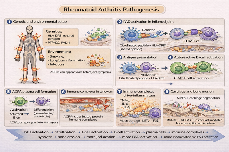 |
|---|
Existing therapeutic approaches, including disease-modifying antirheumatic drugs (DMARDs) such as methotrexate and biologic tumor necrosis factor-alpha (TNF-α) inhibitors, focus on broad immunosuppression to manage inflammation. While these treatments can slow disease progression and alleviate symptoms, they fail to address the root cause: the autoreactive B-cell clones producing ACPAs. Consequently, patients face lifelong medication dependency, increased susceptibility to infections due to systemic immune suppression, elevated risks of thrombotic events, and a frustrating trial-and-error approach to finding effective treatments due to weak predictive biomarkers.
Chimeric antigen receptor (CAR) T-cell therapy represents a paradigm shift toward precision medicine in autoimmune disease treatment. Originally developed for hematologic malignancies, mainly blood cancer, CAR-T therapy has recently demonstrated remarkable success in autoimmune diseases, with CD19-targeted CAR-T cells achieving drug-free remission in patients with systemic lupus erythematosus (SLE) and other refractory autoimmune conditions (Yu et al., 2026). A 2025 meta-analysis of 47 patients receiving CAR-T therapy for refractory autoimmune diseases found that all patients achieved an immunosuppression-free state at last follow-up, with acceptable safety profiles despite 79 cases of manageable cytokine release syndrome. In myasthenia gravis clinical trials, 57% of patients achieved minimal symptom expression by month 6, maintaining remission through month 12 after six short weekly infusions. Notably, one lupus patient has remained in drug-free remission for 4 years after a single CAR-T treatment, demonstrating the potential for durable immune reset. (Yu et al., 2026)
Building on this success, a variation of CAR T-cells, chimeric autoantibody receptor (CAAR) T-cells offer an even more refined approach by targeting only disease-specific B-cells rather than all CD19-positive B-cells. By engineering T-cells to express receptors that use disease-specific autoantigens as "bait," CAAR-T cells can selectively bind to and eliminate autoreactive B-cells displaying the corresponding autoantibody on their surface. Recent studies have demonstrated this concept's feasibility in multiple sclerosis (Reincke et al., 2023) (targeting NMDA receptor-specific B-cells) and Graves' disease (eliminating thyroid-stimulating hormone receptor autoantibody-producing B-cells) (Cheever et al., 2025).
The objective of this study was to design a conceptual CitB-CAAR T-cell construct that targets ACPA-producing B-cells in RA using citrullinated vimentin as molecular bait, incorporating second-generation costimulatory domains for enhanced persistence, safety mechanisms to prevent off-target toxicity, and resistance to T-cell exhaustion. This proof-of-concept design demonstrates how targeted cellular immunotherapy can address the immunological root cause of RA while preserving healthy immune function.
Systems level
The CitB-CAAR T-cell therapeutic system operates through a multi-stage process that begins with patient leukapheresis to collect autologous T-cells, followed by ex vivo genetic engineering to introduce the CAAR construct, expansion in bioreactors, and reinfusion into the patient. Upon infusion, these engineered T-cells circulate through the bloodstream and migrate to lymphoid tissues and inflamed synovial joints where ACPA-producing B-cells reside.
The therapeutic mechanism relies on molecular recognition between the citrullinated vimentin "bait" displayed on the CAAR and the ACPA autoantibodies expressed as B-cell receptors on pathogenic B-cells. This binding forms an immunological synapse that triggers intracellular signaling cascades, leading to CAAR-T cell activation, proliferation, and cytotoxic function. Activated CAAR-T cells release perforin and granzyme B, which induce apoptosis in the target B-cells within 30 min of engagement.
A critical safety feature is the synNotch AND gate circuit, which requires dual recognition of both citrullinated antigen and CD19 before full T-cell activation occurs. This Boolean logic gate functions as an "IF-THEN" circuit: the synNotch receptor first recognizes citrullinated antigen and undergoes cleavage to release a transcriptional activation domain, which then induces expression of the CD19-targeting CAR. Only when both antigens are detected does the CAAR-T cell execute its cytotoxic program, minimizing off-target destruction of healthy B cells that express CD19 but lack ACPA receptors, which would cause a severely weakened immune system, leading to increased risks of infections.
Following successful elimination of autoreactive B-cells, the immune system undergoes reconstitution over 6-12 months, with healthy B-cell populations repopulating from bone marrow precursors. Memory CAAR-T cells persist in circulation to provide long-term surveillance against any emerging autoreactive B-cell clones, potentially enabling durable drug-free remission.
Device level
Extracellular antigen recognition domain
The antigen-binding domain consists of citrullinated vimentin peptide (citVim60-71), a 12-amino acid sequence identified as a dominant ACPA epitope in seropositive RA patients. Citrullinated vimentin was selected over other potential ACPA targets because it is one of the most prevalent and disease-specific autoantigens in RA, recognized by ACPAs in approximately 70% of seropositive patients. By displaying this citrullinated peptide on the CAAR surface, the engineered T-cells act as "decoys" that capture B-cells displaying ACPA-containing B-cell receptors, enabling selective targeting of pathogenic B-cells while sparing healthy immune cells.
Spacer and transmembrane domains
The spacer region utilizes a 12-amino acid IgG4 hinge domain to optimize the immunological synapse distance at approximately 15 nm. This distance is critical for effective T-cell receptor engagement with target B-cells, as it positions the antigen-binding domain at the optimal distance for receptor-ligand interaction while maintaining flexibility for conformational changes during activation. The transmembrane domain incorporates CD8α for stable membrane anchoring and receptor clustering, which facilitates efficient downstream signaling by concentrating signaling molecules at the site of antigen engagement.
Intracellular signaling architecture
The CAAR construct employs a second-generation design with multiple costimulatory domains to enhance T-cell function, persistence, and antitumor activity. The intracellular signaling architecture comprises three domains with distinct but complementary functions: Inducible T-cell COStimulator (ICOS) for rapid T-cell proliferation upon antigen engagement, 4-1BB (CD137) for establishing long-term memory T-cell populations capable of multi-year persistence, and CD3ζ containing three immunoreceptor tyrosine-based activation motifs (ITAMs) that trigger cytotoxic granule release.
ICOS provides powerful proliferative signals that enable rapid CAAR-T cell expansion upon encountering target antigens. The 4-1BB costimulatory domain activates a distinct molecular program that supports memory T-cell development and long-term persistence, critical for maintaining durable remission. Preclinical and clinical data confirm that 4-1BB-based CARs direct differentiation of T cells into memory phenotypes with enhanced survival capacity compared to CD28-based designs. The CD3ζ domain provides the primary activation signal through its ITAM motifs, which recruit kinases that initiate the signaling cascade leading to cytotoxic function.
Parts level
The complete CitB-CAAR construct is assembled through a modular design that can be integrated into T-cells using lentiviral or retroviral vectors. Gene expression is driven by the murine stem cell virus (MSCV) long terminal repeat promoter, selected for its resistance to epigenetic silencing and ability to maintain high-level transgene expression in primary T-cells over extended periods. The MSCV promoter ensures stable, long-term expression of the CAAR construct, critical for maintaining therapeutic efficacy over months to years.
The genetic construct is organized in the following 5' to 3' configuration:
MSCV Promoter: Drives constitutive, high-level expression resistant to silencing
Signal Peptide: Directs the nascent protein to the endoplasmic reticulum for proper membrane trafficking
citVim60-71 Antigen Domain: 12-amino acid citrullinated vimentin peptide for ACPA recognition
IgG4 Hinge: 12-amino acid spacer optimizing immunological synapse distance (15 nm)
CD8α Transmembrane Domain: Provides stable membrane anchoring and receptor clustering
ICOS Costimulatory Domain: Signals for rapid T-cell proliferation
4-1BB Costimulatory Domain: Promotes memory formation and long-term persistence
CD3ζ Signaling Domain: Three ITAMs trigger cytotoxic function via perforin/granzyme B release
The synNotch and gate are expressed from a separate cassette to provide the dual-antigen safety mechanism. Additionally, an interleukin-10 (IL-10) expression cassette under constitutive promoter control enables anti-inflammatory cytokine secretion. CRISPR-Cas9 components targeting PD-1 and TIGIT genes can be delivered during T-cell engineering either through electroporation of ribonucleoprotein complexes or via additional viral vectors (Figure 2).
| Figure 2- This figure illustrates how CitB‑CAAR T cells are engineered to target and eliminate autoreactive B cells responsible for rheumatoid arthritis (RA). The CitB‑CAAR T cell displays citrullinated vimentin as “bait,” connected by an IgG4 hinge that allows it to bind specifically to ACPA B‑cell receptors on autoreactive B cells. Once bound, the T cell forms an immunological synapse and kills the B cell using perforin and granzyme. It also includes design features like PD‑1/TIGIT knockout to prevent exhaustion and IL‑10 secretion to reduce inflammation ensuring sustained activity and lower immune toxicity. In short, this approach selectively removes harmful B cells while calming inflammation in RA. | 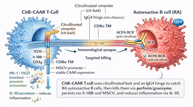 |
|---|
Safety
To enhance safety and prevent off-target toxicity, the design incorporates a synNotch AND gate circuit requiring dual recognition of both citrullinated antigen and CD19 surface marker before full T-cell activation. The synNotch receptor recognizes the citrullinated antigen via its extracellular recognition domain and undergoes cleavage to release a transcriptional activation domain that induces CAR expression. This two-step process ensures that CAAR-T cells only target cells expressing both markers, dramatically reducing the risk of on-target off-tumor toxicity. Additionally, synNotch CAR circuits can inhibit CAR-mediated tonic signaling, thereby promoting the maintenance of long-lived memory and a non-exhausted phenotype.
The construct design also includes constitutive IL-10 secretion to dampen local inflammation and prevent cytokine release syndrome (CRS), a potentially serious complication observed in CAR-T therapy. To maintain sustained cytotoxic function and prevent T-cell exhaustion, CRISPR-Cas9-mediated knockout of PD-1 and TIGIT checkpoint receptors is incorporated. These checkpoint molecules normally suppress T-cell activity to prevent autoimmunity, but their removal in the therapeutic context ensures that CAAR-T cells maintain their killing capacity even in the immunosuppressive tumor microenvironment of inflamed synovial tissue. This design would be built with a Biosafety Level 2 (BSL-2) biosafety framework, which is appropriate for moderate-risk biological materials and requires restricted access, personal protective equipment (PPE), and biosafety cabinet use for aerosol-generating procedures.
Discussions
The CitB-CAAR T-cell design represents a fundamental shift from symptom management to targeting the immunological root cause of rheumatoid arthritis. By selectively destroying ACPA-producing B-cells while preserving healthy immune populations, this approach addresses a critical limitation of current therapies that rely on broad immunosuppression. Recent clinical trial successes with CD19 CAR-T therapy in lupus and other autoimmune diseases validate this recognize-and-eliminate strategy, achieving drug-free remission where traditional immunosuppression has failed for decades.
The incorporation of second-generation costimulatory domains (ICOS and 4-1BB) is expected to provide both rapid therapeutic response and long-term persistence. While CD28-based CARs promote rapid expansion, 4-1BB-based designs have demonstrated superior memory formation and durability in clinical applications. The combination of ICOS for immediate proliferative response and 4-1BB for sustained memory development may provide optimal therapeutic kinetics.
The synNotch AND gate safety mechanism addresses a critical concern in CAAR-T therapy: preventing off-target destruction of healthy B-cells. By requiring recognition of both citrullinated antigen and CD19, the system ensures that only ACPA-displaying B-cells are targeted. This dual-antigen requirement provides substantially greater specificity than CD19-targeting alone, which eliminates all B-cells regardless of their antigenic specificity. Furthermore, synNotch circuits have been shown to inhibit tonic signaling and promote maintenance of non-exhausted T-cell phenotypes, potentially enhancing long-term efficacy.
CRISPR-mediated knockout of PD-1 and TIGIT checkpoint receptors aims to prevent T-cell exhaustion in the chronic inflammatory environment of RA synovial tissue. These checkpoint molecules normally dampen T-cell responses, and their expression on CAR-T cells can limit therapeutic efficacy in solid tumors and chronic inflammatory conditions. Removing these negative regulators should maintain sustained cytotoxic function even in immunosuppressive microenvironments.
A key advantage of the CAAR-T approach is its modularity and adaptability to other autoimmune diseases. By simply swapping the citrullinated vimentin "bait" for other disease-specific autoantigens, the same engineering framework can be applied to treat multiple sclerosis (using myelin oligodendrocyte glycoprotein or aquaporin-4), type 1 diabetes (using insulin or GAD65), and other antibody-mediated autoimmune conditions. This platform technology could transform the treatment paradigm across multiple autoimmune diseases.
However, several challenges must be acknowledged. The safety profile of CAAR-T therapy in RA remains to be established through clinical trials, with particular attention to CRS, which occurred in the majority of patients in one meta-analysis but was manageable. The immunosuppressive effects of lymphodepleting chemotherapy required before CAR-T infusion carry risks of infection. Additionally, the long-term consequences of eliminating ACPA-producing B-cells and maintaining engineered T-cells for years require careful monitoring. Manufacturing complexity and cost remain significant barriers to widespread clinical implementation.
Next steps
Clinical translation of the CitB-CAAR T-cell design requires systematic validation through several discrete research activities. The first priority is lentiviral vector cloning to assemble the complete CAAR construct, including the citVim60-71 antigen domain, IgG4 hinge, CD8α transmembrane domain, and ICOS-4-1BB-CD3ζ signaling cassette. This genetic construct must be validated through sequence verification and functional testing in mammalian cell expression systems.
Following successful vector construction, the CAAR construct should be introduced into primary human T-cells isolated from healthy donor peripheral blood mononuclear cells (PBMCs). Lentiviral transduction efficiency can be assessed by flow cytometry to quantify the percentage of T-cells expressing the CAAR construct on their surface. Transduced T-cells should then be expanded in vitro using anti-CD3/CD28 activation beads and interleukin-2 (IL-2) supplementation to generate sufficient cell numbers for functional assays.
In vitro cytotoxicity testing represents a critical validation step to demonstrate that CitB-CAAR T-cells can selectively kill ACPA-producing B-cells. This can be modeled by engineering target B-cell lines to express ACPA-like B-cell receptors specific for citrullinated vimentin, then co-culturing them with CitB-CAAR T-cells at various effector:target ratios. Cell death can be quantified using flow cytometry-based assays measuring apoptosis markers or chromium-51 release assays. Specificity should be confirmed by demonstrating that CitB-CAAR T-cells do not kill control B-cells lacking ACPA receptors or expressing non-citrullinated vimentin.
The synNotch AND gate safety mechanism requires independent validation to confirm that dual antigen recognition is necessary for full T-cell activation. This can be tested using target cells expressing only CD19, only citrullinated antigen, or both markers, measuring CAAR-T cell activation through CD69 upregulation, cytokine secretion (interferon-gamma (IFN-γ), TNF-α), and cytotoxic function. Successful validation would show minimal activation against single-positive targets and robust activation only against double-positive targets.
Subsequent research stages include preclinical validation in animal models of RA, such as collagen-induced arthritis in mice or humanized mouse models bearing human synovial tissue. These studies would assess in vivo trafficking, persistence, efficacy in reducing joint inflammation, and safety profiles. If preclinical results demonstrate acceptable safety and efficacy, Phase I clinical trials could be initiated in patients with refractory RA who have failed multiple lines of conventional therapy.
All of these research activities can be adapted to the capabilities of a high school synthetic biology laboratory with appropriate biosafety containment and mentor supervision. The molecular cloning, vector construction, and initial cell culture work provide hands-on experience with the foundational techniques underlying modern cell and gene therapy development.
Author contributions
RR conceived the CitB-CAAR T-cell design, researched and selected costimulatory domains and safety mechanisms, analyzed clinical trial data from related CAR-T therapies, predicted therapeutic outcomes based on published literature, and took the lead in writing the manuscript. TRK conducted the background research on rheumatoid arthritis, how it works on a molecular scale, identifying causes, identifying root molecular cause, and identifying CAR-T methodology as effective for this disease. In addition, TRK also analyzed clinical trial data, and contributed ideas for CAAR-T cell design.
Acknowledgements
The authors would like to thank William Chen for their guidance and support throughout this project. Special thanks to the Bio Builder synthetic biology program for providing the opportunity to explore cutting-edge therapeutic design. Gratitude is also extended to the researchers whose published work on CAR-T and CAAR-T cell therapies provided the scientific foundation for this conceptual design.
References
Cheever, A., Lindsay, H. G., Kang, C. C., Hansen, M., Demars, K., O'Neill, K. L., & Weber, K. S. (2025). Chimeric autoantibody receptor T cells specifically eliminate Graves' Disease autoreactive B cells. Frontiers in immunology, 16, 1562662. https://doi.org/10.3389/fimmu.2025.1562662
Malmström, V., Catrina, A. I., & Klareskog, L. (2017). The immunopathogenesis of seropositive rheumatoid arthritis: from triggering to targeting. Nature reviews. Immunology, 17(1), 60–75. https://doi.org/10.1038/nri.2016.124
Reincke, S. M., von Wardenburg, N., Homeyer, M. A., Kornau, H. C., Spagni, G., Li, L. Y., Kreye, J., Sánchez-Sendín, E., Blumenau, S., Stappert, D., Radbruch, H., Hauser, A. E., Künkele, A., Edes, I., Schmitz, D., & Prüss, H. (2023). Chimeric autoantibody receptor T cells deplete NMDA receptor-specific B cells. Cell, 186(23), 5084–5097.e18. https://doi.org/10.1016/j.cell.2023.10.001
Yu, B., Xu, J., & Cui, Y. (2026). From technological iteration to clinical breakthrough: advances of CAR-T cell therapy in autoimmune diseases. Annals of medicine, 58(1), 2627057. https://doi.org/10.1080/07853890.2026.2627057