
Raghav Bajaj, Arush Maridu, Shaun Philip Mathew, Meera Paramagurusamy, Sarvesh Sathiyamoorthi, Ishani Sharma, Mukul Vishwakumar, and Vedanth Vivekanand, South Forsyth High School, Cumming, Georgia, United States of America
Reviewed on 9 May 2026; Accepted on 22 June 2026; Published on 26 October 2026
With help from the 2026 BioTreks Production Team.
In today’s world, constant use of digital devices has made blue light exposure unavoidable. Prolonged exposure leads to increased oxidative stress due to excessive reactive oxygen species (ROS), contributing to cellular damage in retinal tissue. Excessive ROS disrupts mitochondrial function, causes tissue damage, and can lead to cell death. Current approaches to managing oxidative stress caused by blue-light exposure primarily focus on reducing blue-light exposure rather than addressing the underlying causes of oxidative stress. Our project aims to both detect and mitigate the excess levels of oxidative stress induced by blue light. The first plasmid construct functions as a ROS biosensor by using a ROS-responsive biosensor to activate green fluorescent protein (GFP) expression under high oxidative stress conditions, allowing elevated ROS levels to be visually detected. The second plasmid construct expresses superoxide dismutase (SOD2), an antioxidant enzyme, to reduce ROS. Together, these constructs form a system that both detects and minimizes oxidative stress, allowing the cell to continue normal functions while reducing the negative effects of excess ROS. The plasmids are constructed and validated in Escherichia coli (E. coli), an efficient cellular chassis for transformation, then fed to C. elegans, transparent nematodes that enable organism-level ROS reporting. This blue light–regulated oxidative stress sensing system links an external stimulus to a genetic response, enabling precise monitoring of cellular health. This innovation assists in countering the harmful effects of blue light and provides a foundation for developing technologies that can both sense and therapeutically respond to cellular stress in real time, with potential applications in ocular health, disease monitoring, and antioxidant therapies.
Keywords: Synthetic biology, oxidative stress, reactive oxygen species (ROS), blue light, antioxidant
Authors are listed in alphabetical order. Peter Horanyi mentored the group. Please direct all correspondence to mailto:f41100@forsyth.k12.ga.us.
Background
In recent decades, the proliferation of digital devices such as smartphones, computers, and televisions has increased dramatically. These devices emit blue light from LED-based screens, resulting in prolonged exposure for many individuals. While blue light plays an important role in regulating circadian rhythms and alertness, excessive exposure has raised concerns regarding its potential effects on ocular health, particularly in retinal cells (Cougnard-Gregoire et al., 2023).
The retina is a highly metabolically active tissue that relies heavily on mitochondrial function to meet its high energy demands. Retinal cells, especially photoreceptors and retinal pigment epithelial (RPE) cells, contain large numbers of mitochondria and are therefore particularly vulnerable to oxidative stress. Studies have shown that high levels of blue light exposure increase the production of reactive oxygen species (ROS) within retinal cells (Godley et al., 2005). Additionally, ROS can damage cellular components such as proteins, lipids, and DNA, ultimately impairing mitochondrial function and contributing to cell death (Dröge, 2002).
Under normal conditions, cells maintain a balance between ROS production and antioxidant enzymes such as superoxide dismutase (SOD), which help prevent oxidative damage. However, excessive blue light exposure can overwhelm these systems, leading to oxidative stress and mitochondrial dysfunction (Beatty et al., 2000). Some studies suggest that chronic oxidative damage in retinal tissue may be associated with retinal degeneration and visual impairment, including age-related macular degeneration, although the relationship between blue light exposure and these conditions remains under investigation.
Although preventative measures such as limiting screen time and using blue light–filtering glasses exist, many individuals do not adhere to recommended exposure limits.
Analysis of the Data Collected:
To test this hypothesis, our team conducted an anonymous survey to assess compliance with screen-time restrictions among adolescents and young adults who regularly use digital devices. Participants were found through school networks and outside clubs, resulting in a wide variety of responses. We found that, among 72 participants, 90% reported bypassing screen-time restrictions. Additionally, social media and entertainment accounted for approximately 80% of these bypass instances. These findings suggest that behavioral interventions alone may be insufficient to fully mitigate cellular damage associated with chronic exposure (Singh et al., 2021) (Figures 1 and 2).
| Figure 1: Distribution of screen time use by purpose | 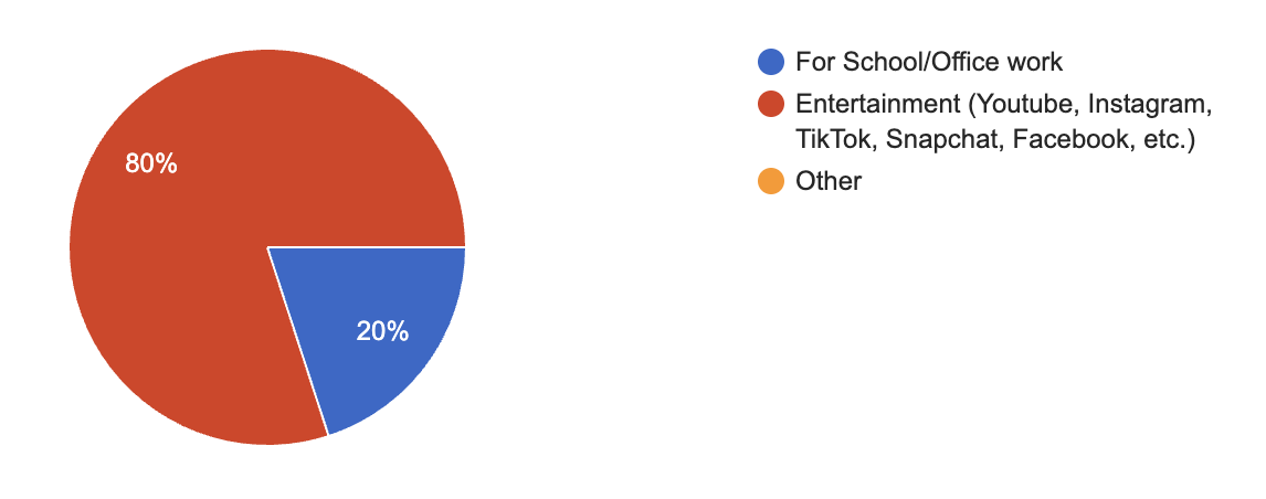 |
|---|
| Figure 2: Average screen time (# of hours) of students & adults | 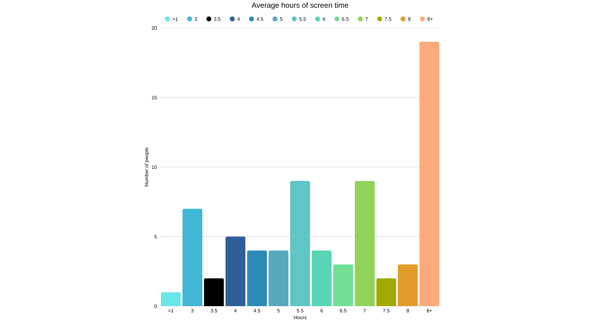 |
|---|
To address this real-world problem, synthetic biology offers an innovative and promising approach to target oxidative stress at the cellular level instead of relying on external prevention. In this project, we propose the design of a plasmid that both detects elevated ROS levels and responds by enhancing the activity of the SOD antioxidant within retinal cells, thereby reducing ROS accumulation and protecting mitochondrial integrity. This approach enhances intrinsic cellular resilience, providing a potentially more robust and long-term solution to blue light-induced oxidative damage.
Systems level
To address oxidative stress caused by prolonged blue-light exposure, our system was designed to detect blue light as an environmental stimulus and trigger a protective cellular response. Blue light in the 400-500 nm range serves as the system input, where it is absorbed by intracellular photosensitizers, particularly flavins (Hunter et al., 2012). Additionally, digital devices also emit light in the same range. When these flavins absorb the blue light, they become “excited” and start transferring energy to nearby oxygen molecules, generating reactive oxygen species (ROS). While antioxidant systems normally regulate ROS levels, prolonged or intense blue light exposure increases ROS production beyond this capacity, leading to oxidative damage and cellular degeneration (Kuse et al., 2014).
| Figure 3: Genetic circuit design showing two expression constructs assembled in E. coli and later introduced into C. elegans to test gene expression. | 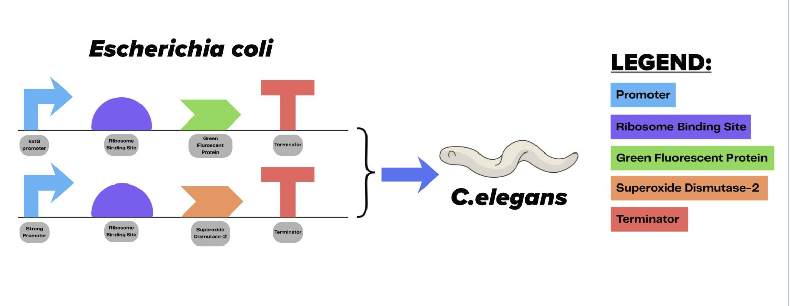 |
|---|
Because direct experimentation in retinal cells is not available to our team, this system models the underlying mechanism of blue-light induced oxidative stress rather than the retinal tissue itself. The key process driving this damage is not unique to retinal cells, but instead depends on photochemical reactions involving intracellular photosensitizers. Both bacteria and simple multicellular organisms contain endogenous flavins that absorb blue light and undergo the same excitation and energy-transfer reactions observed in retinal cells. As a result, these organisms are able to generate ROS through the same energy-transfer reactions observed in retinal cells. This allows blue-light-induced oxidative stress to be accurately reproduced in experimentally accessible systems while maintaining the core biological mechanism (De Magalhães Filho et al., 2018).
To capture and respond to oxidative stress at the cellular level, E. coli was selected as a sensing and response platform due to its well-characterized genetics, rapid growth, ease of genetic engineering, and extensive use in synthetic biology research. ROS generated inside the bacterial cytoplasm activates the transcription factor OxyR, which undergoes a conformational change upon oxidation and binds to DNA sequences upstream of antioxidant genes, including the katG promoter (Zheng et al., 1998; Storz et al., 1990). By placing engineered genes under control of this promoter, oxidative stress is converted into a measurable output.
The system uses two plasmids in which one plasmid reports oxidative stress and the second drives an antioxidant response. When intracellular ROS levels rise and activate OxyR, the katG promoter on the reporter plasmid induces fluorescence through the green fluorescent protein (GFP) gene, providing a visible readout of oxidative stress (Storz et al., 1990; Zheng et al., 1998). The second plasmid functions as the response component by expressing superoxide dismutase 2 (SOD2), an enzyme that reduces superoxide radicals and mitigates oxidative damage (McCord & Fridovich, 1969; Imlay, 2008) (Figure 3).
To confirm that activation of the system is driven by oxidative stress rather than light exposure alone, hydrogen peroxide is used as a positive control. Hydrogen peroxide directly oxidizes OxyR and activates the same promoter, allowing validation that both the reporter and response outputs are ROS-dependent (Storz et al., 1990; Zheng et al., 1998). Comparable responses under hydrogen peroxide treatment and blue light exposure support the conclusion that blue light activates the system through ROS generation (De Magalhães Filho et al., 2018).
To examine whether this stress-responsive behavior can function within a living organism, C. elegans is used as a model organism. C. elegans is mainly used to study organism responses, including oxidative stress pathways, since it is easy to culture and its structure allows for observation of fluorescent markers in vivo. The Bristol N2 strain allows visualization of fluorescent signals and has well-characterized oxidative stress pathways (Brenner, 1974). Since C. elegans naturally consume E. coli, engineered bacteria can be delivered through feeding, enabling evaluation of ROS detection and antioxidant response within a living organism (De Magalhaes Filho et al., 2018) (Figure 4).
| Figure 4: Overview of our system-level design. Both plasmids will be tested and amplified in E. coli before transforming them into C. elegans | 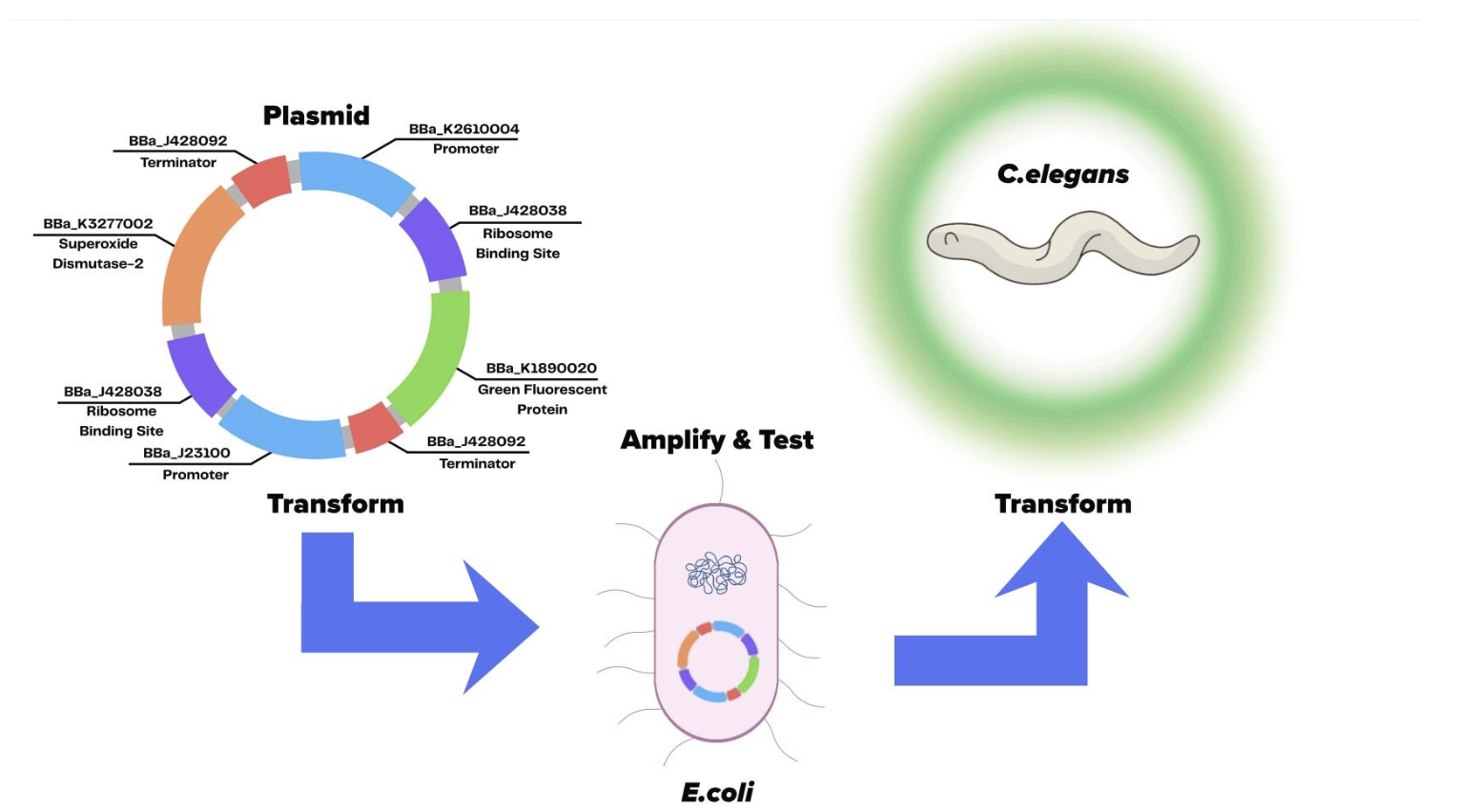 |
|---|
Together, this system establishes a continuous, light-responsive pathway in which blue light induces ROS through flavin excitation, ROS activates a genetic stress sensor, and this signal triggers both detection and targeted antioxidant defense. By engineering a system that responds directly to oxidative stress rather than blocking light exposure directly, the approach provides a biologically stable framework for studying and mitigating blue-light-induced cellular damage.
Device level
The device consists of an engineered system built within E. coli using the pSB1C3 plasmid backbone. This high-copy BioBrick vector was selected for its stability, compatibility with standard parts, and its ability to maintain controlled expression in oxidative-stress-responsive conditions. The plasmids carry chloramphenicol resistance and function reliably in E. coli DH5α, serving as the main host organism for device construction and verification (Škrlj et al., 2009). These markers would be removed if adapted for therapeutic applications.
Two plasmids will be used, each serving a distinct role within the system. The first plasmid acts as a sensing and reporting module, utilizing the ROS-responsive katG promoter to drive gene expression in response to increased ROS levels (BBa_K1890020). This module produces a measurable fluorescence signal (GFP), allowing visual and quantitative detection of oxidative stress in cells (Belkin et al., 1996).
The second plasmid functions as a response module, containing a SOD2 gene that produces an antioxidant response. Upon activation by the same ROS-sensitive promoter, the module produces enzymes that neutralize harmful ROS, thereby reducing oxidative damage. Together, these functional components form a system that enables E. coli to report and lessen damage in oxidative conditions (Imlay, 2013).
These two plasmids will be constructed using Golden Gate Assembly, which utilizes Type IIS restriction enzymes and DNA ligase in an iterative thermocycling reaction. This allows multiple DNA fragments with overhangs to be optimally joined in a fixed order, ensuring the seamless assembly and functionality of the standard plasmid parts (Bird et al., 2022) (Figure 5).
| Figure 5: Plasmid assembly strategy using Golden Gate cloning to build two expression plasmids before transformation into E. coli. | 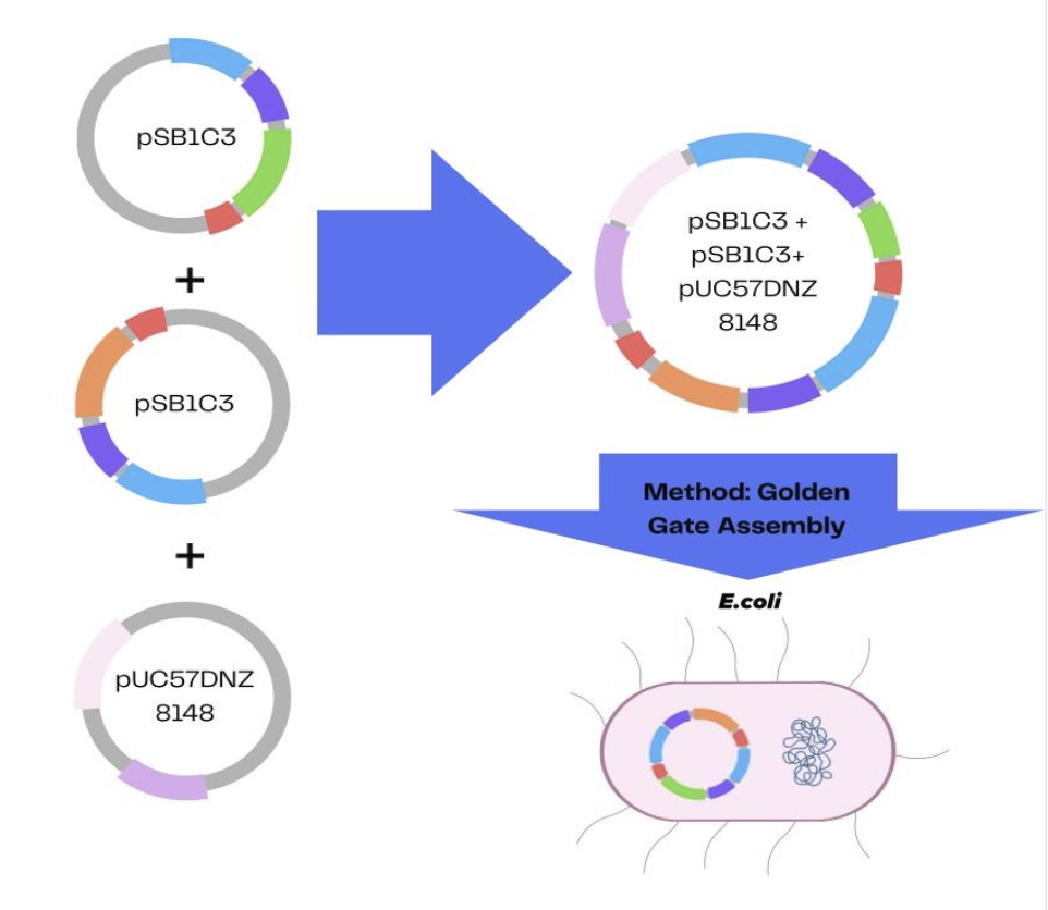 |
|---|
Together, these two engineered plasmids form an integrated device in which E. coli DH5α acts as the cellular chassis. Plasmid DNA is typically introduced into DH5α cells using chemical transformation or electroporation, and successful transformant cells are selected through the antibiotic resistance markers carried on the plasmid (Kostylev et al., 2015).
To extend functionality beyond a single-cell system, C. elegans is used as the model organism. Engineered E. coli are introduced as a food source for C. elegans, allowing the bacteria to function within the worm’s intestinal environment (Rutter et al., 2019). As the worms ingest the bacteria, the reporting system can function in the intestinal environment and therefore enable the assessment of oxidative stress detection and removal on an organismal level.
During plasmid construction, antibiotic resistance markers identify the cells that have taken up the plasmids. For both the ROS-responsive reporter plasmid and the antioxidant response plasmid, chloramphenicol resistance is also included as a selectable marker in E. coli. Chloramphenicol resistance is used as a selection marker because only bacteria carrying the plasmid express chloramphenicol acetyltransferase (CAT), which inactivates the antibiotic, allowing them to survive and grow, while non-transformed cells are inhibited and die.
Not only do these markers support genetic maintenance, but they also enhance the device’s safety by enabling researchers to control and verify its presence before application of the bacteria (Jang & Magnuson, 2013). After verification, the markers can be removed to prevent needless spreading of antibiotic resistance.
Parts level
The first plasmid functions as a ROS-responsive reporter module. The plasmid begins with the oxidative stress-inducible katG promoter (BBa_K2610004), which is activated by the transcription factor OxyR under elevated oxidative stress, particularly in the presence of hydrogen peroxide. Downstream of the promoter is a strong ribosome-binding site (BBa_J428038) that promotes efficient translation initiation. This is followed by the GFP reporter gene (BBa_K1890020), which produces a fluorescent signal when high ROS levels are induced. The construct ends with a double transcription terminator (BBa_J428092, derived from BBa_B0015) to ensure reliable termination. Together, these components form an operon that converts oxidative stress into a measurable fluorescence output, allowing for the necessary energy expenditure of the plasmids to be viable, which would not have been done in a singular plasmid.
The second plasmid serves as the antioxidant response module by reducing the ROS levels within the cell. This construct includes a strong promoter (BBa_J23100) that allows for the expression of the antioxidant gene when activated. Similar to the first plasmid, the second plasmid also contains the same ribosome binding site (BBa_J428038), a sequence that base-pairs with rRNA of the 30S subunit to drive translation initiation of the coding sequence. The primary gene of this plasmid encodes a superoxide dismutase enzyme (BBa_K3277002), which breaks down superoxide molecules into less harmful molecules. SOD functions as an antioxidant that decreases intracellular ROS that accumulate due to excess blue-light stress, mitigating potential retinal damage. As in the reporter plasmid, a double transcription terminator (BBa_J428092) is included to ensure controlled gene expression. Both plasmids are designed so that gene expression is linked to the oxidative stress conditions, allowing the system to function only when ROS levels are elevated.
Safety
The safety and ethical foundation of this project begins with using low-risk model organisms. Both the N2 Bristol strain of C. elegans, and non-pathogenic E. coli are classified as Biosafety Level 1 (BSL-1), meaning they are not known to cause disease in healthy adult humans (Centers for Disease Control and Prevention [CDC], 2020). All procedures follow standard laboratory safety protocols, including the use of personal protective equipment (PPE), proper disposal of biological waste (Stiernagle, 2006).
Environmental containment is further strengthened by biological safeguards. The engineered E. coli strains would likely be outcompeted by wild type counterparts in a natural environment, serving as an innate safeguard against the environmental spread of synthetic traits (Kuldell et al., 2015). Additionally, antibiotic resistance markers are used only during selection.
Beyond physical containment, using light to control genes raises important questions about accidental activation and ethics. As our genetic "switch" (the katG promoter) responds to oxidative stress, it could be turned on accidentally by environmental factors like heat or normal room lighting instead of our specific blue-light source. ROS are not merely harmful byproducts, but also essential signaling molecules that play important roles in regulating normal cellular processes (Dröge, 2002). If our system accidentally overrides these signals, it might stop the body from removing unhealthy cells. Care is taken to ensure that the system does not interfere with essential biological processes by tuning the sensitivity and threshold of the synthetic circuit so it responds only to pathologically elevated ROS levels. Furthermore, while using light to treat disease is a breakthrough, the power to control living systems must be managed carefully to prevent misuse. By targeting only elevated ROS levels and focusing on neutralizing excessive stress, the design is ensured not to interfere with the natural signals the cell needs to stay healthy (Ighodaro & Akinloye, 2018).
This project also draws insight from Lai et al. (2022), whose work on reducing presbyopia through oxidative stress reduction with flavonoid-producing gut bacteria is closely related to our project. This study proposed engineering Bifidobacterium longum to produce the antioxidant quercetin for the treatment of presbyopia, an oxidative stress-related condition. While this approach explores systemic delivery of antioxidants through the gut, it introduces additional challenges such as large plasmid size, transformation difficulty, and uncertainty in developing compounds to ocular tissues. In contrast, our system focuses on localized sensing and response within a targeted area, to help reduce complexity and improve effectiveness.
Discussions
While forming our proposal, we evaluated various challenges related to the development and implementation of our research.
Challenge 1: Effects of reducing intracellular ROS levels
One major challenge is the risk of excessive antioxidant activity at the cellular level. The engineered plasmid is designed to express Superoxide Dismutase 2 (SOD2), an enzyme that catalyzes the dismutation of superoxide radicals (a type of ROS) into hydrogen peroxide and oxygen, where there is an excess concentration of ROS. However, overproduction of antioxidants may disrupt cellular balance. Many studies and experiments, including this proposal, have mainly “focused on oxidative stress (OS), characterized by an overproduction of reactive oxygen (ROS)…and its detrimental effects on inflammation and disease pathogenesis” (Bellanti et al., 2025). However, it is also important to consider the opposite condition: reactive stress (RS). RS is characterized by the overabundance of reducing agents, leading to the cell having an overall reduced state (Bellanti et al., 2025). To mitigate this, the plasmid will be programmed to pause the production of antioxidants when an adequate amount is reached. In the plasmid design, the katG promoter will be controlled by a redox-sensitive transcription factor, oxyR. The katG promoter will be repressed once ROS levels decrease, allowing transcription of the downstream gene sequence and reducing SOD2 translation and expression. Then, the antioxidant release will slowly come to a stop.
Challenge 2: Signal transmission utilizing ROS
In terms of communication between cells, ROS is an important component of cell signaling. H2O2 (Hydrogen peroxide) is a common ROS. It is “…an essential redox signaling molecule…” (Averill-Bates, 2024) and is linked to many cellular processes, including activation or repression of transcription factors, programmed cell death, and phosphorylation cascades (Averill-Bates, 2024). In other words, the proposed technology has the potential for off-target effects and may disturb other cellular processes by trying to reduce the amount of ROS. However, it is important to note that the project aims to reduce the concentration of ROS to a healthier amount, not completely eliminate it, which would compromise the health of the cell. The plasmid will also be engineered to specifically target excess ROS. This approach allows the cell to maintain normal function while reducing the harmful effects of excess ROS.
Challenge 3: Implications of the final product on society
Although reducing the amount of ROS in retinal cells has many benefits, it also raises concerns about underlying risks, such as biosafety. Biosafety risks are defined as “biorisks that arise from human actions that are not deliberate or intentional” (Zeng et al., 2022). The proposal aims to genetically engineer a plasmid that produces antioxidants in environments where there is a significantly higher concentration of ROS. The solution will reduce such risks by strongly adhering to lab protocol, and monitoring all model organisms by keeping them contained safely. In addition to this, many scientists in the field discern that development of synthetic biology is still in its infancy, such that “the use of synthetic biology to reconstruct and synthesize life forms from scratch is currently limited…” (Li et al., 2021). To address this, the project proposes to utilize C. elegans, more commonly known as roundworms, as the model organism. C. elegans are well studied organisms, and are fairly simplistic organisms, containing only 959 somatic cells. By working with organisms that are well studied and characterized, the project minimizes the opportunities for unpredictable behaviors, thereby significantly reducing biosafety risks.
Main Challenge: Transformation Efficiency of the Plasmid
Transformation efficiency can be described as a quantitative measure of how successful the uptake of foreign DNA was in the host cell. Our plasmid faces the risk of having a low transformation rate, as it involves multiple plasmid parts. This introduces many points where the uptake could go wrong. This proposal will use the Golden Gate assembly method to increase the efficiency rate of transformation. Golden Gate assembly is an assembly method that utilizes Type II restriction endonucleases, allowing for parts to have overhangs for ligation at will (Biró et al., 2024). This means that multiple parts can be attached and assembled in the plasmid and have seamless joining. This challenge can also be addressed by exploring the optimal conditions for producing competent cells. These specific factors can include the ionic species the strain has been prepared in, plasmid concentration, or ice bath time (Liu et al., 2014).
Next steps
Blue light functions as both an environmental stressor and a controllable contact point, which scientists to link these engineered systems with authentic oxidative-stress signals. Multiple studies establish that blue light with wavelengths between 400 and 500 nanometers causes oxidative stress in retinal cells because it increases ROS levels. Together with mitochondrial destruction and the initiation of inflammatory pathways, this results in retinal degeneration and age-related macular disease. (Yeh et al., 2025) The use of blue light as a control mechanism enables built systems to connect with authentic biological stress signals. The biosensing processes that need exact time measurements and low harmful effects make light the perfect solution because it lets scientists create chemical compounds for biological testing, which need both features.
Our designed E. coli biosensor employs the cell’s natural oxidative-stress control systems to enhance both accuracy and operational capacity. The OxyR and SoxRS regulons function as established systems that respond to hydrogen peroxide and superoxide, yet serve as base elements in synthetic biology because they function as ROS-responsive promoters (Imlay, 2013). The system produces a measurable output when ROS levels exceed normal physiological thresholds, as the fluorescent reporter is regulated by ROS-responsive promoters. This approach is far more practical for educational labs than chemical PFN probes, even though it is a validated method for quantifying ROS in mammalian oxidative-stress models. (The use of E. coli DH5α leads to improved safety, which the system scales through its genetic components and its capacity to track fluorescence.
The biosensor’s actual performance shows its usefulness in real-world applications that extend beyond just the lab. The low-cost genetically encoded ROS detector can assist in three areas: antioxidant screening, early-stage research on oxidative-stress-related diseases, and the detection of stress-related microRNAs through integrated toehold switches. The system grows its versatility through the integration of programmable RNA, which includes toehold switches. Research also demonstrates that microRNAs (miRNAs) function as vital regulatory components that control ROS-mediated pathways in cancer and melanoma (Kozar et al., 2021) because oxidative stress alters genetic and epigenetic processes. (Leung & Sharp, 2010) The development of a toehold switch that detects stress-related miRNA would help the biosensor to measure both chemical ROS and genetic miRNA indicators of oxidative stress.
This system requires multiple steps that need to be completed before it can function outside the lab. Researchers need to improve the genetic constructs through methods such as adjusting promoter strength with decreasing background fluorescence and checking the dose response through testing actual concentrations of blue light exposure. The implementation of biosafety containment strategies, which include kill switches and cell-free expression systems, will protect against any potential biosafety risks that exist outside the lab. This biosensor system also requires development into portable formats like adhesive patches or eye drops to enable the population to operate this technology without needing special equipment; however, translating an engineered bacterial system into these environments comes with its own biosafety and regulatory challenges. Engineered E. coli strains face a low survival rate in ocular environments due to tear-film turnover, antimicrobial peptides, and osmotic stress, which limits the engineered strain’s ability to act as therapeutic or diagnostic agents without extra stabilization strategies. Any deployment outside the lab would also need containment mechanisms – such as kill switches or cell-free expression systems – to prevent any unintended colonization or environmental release. Furthermore, topical use of a genetically modified organism would fall under FDA/NIH regulation and would require plenty of safety testing, toxicity assessments, and evaluation of immune responses before the system can be applied in the real-world. This development process requires these specific steps to change the system from a theoretical demonstration into an operational research instrument that lets scientists investigate oxidative stress while using light-based biotechnology.
Author contributions
SS developed the core concepts of the project, drafted the abstract, refined the overall paper, and ensured the introduction clearly articulated the rationale for selecting blue light-induced oxidative stress as the study subject. IS conducted an extensive literature review, compiling the information regarding the exposure to blue light and created the survey to establish the project’s scientific foundation. AM designed the systems-level approach for the blue light-responsive ROS response pathway, defined its operational logic, and assisted in comparing alternative genetic circuits. MV was responsible for planning the plasmid constructs and cloning strategies which included checking part compatibility and coming up with a validation strategy. VV searched and evaluated genetic parts from the registry to help with sensing and antioxidant expression. Additionally, VV compared promoter strengths and regulatory elements. RB looked into biosafety issues, ethical concerns, and the risks that might result from the deployment of the system. MP pointed out the project limitations and discussed the experimental constraints and other broader challenges of the work component. Additionally, MP helped design all the figures. Lastly, SM gave an overview of the project output and talked about the subsequent uses, as well as the long-term directions, in the closing.
Acknowledgements
This project was completed through our participation in the BioBuilderClub, founded by our teacher, Mrs. Pamala Seeley. We sincerely thank the BioBuilderClub for providing us with the opportunity to engage in authentic scientific research.
We are especially grateful to Mrs. Pamala Seeley and Dr. Peter Horanyi for their unwavering support, guidance, and encouragement throughout every stage of the project. Their expertise, insightful feedback, and dedication were instrumental in shaping our research and deepening our understanding of the subject.
References
Averill-Bates, D. (2024). Reactive oxygen species and cell signaling. Review. Biochimica et Biophysica Acta (BBA) – Molecular Cell Research, 1871(2). https://doi.org/10.1016/j.bbamcr.2023.119573
Beatty, S., Koh, H.-H., Phil, M., Henson, D., & Boulton, M. (2000). The role of oxidative stress in the pathogenesis of age-related macular degeneration. Survey of Ophthalmology, 45(2), 115–134. https://doi.org/10.1016/s0039-6257(00)00140-5
Belkin, S., Smulski, D. R., Vollmer, A. C., Van Dyk, T. K., & LaRossa, R. A. (1996). Oxidative stress detection with Escherichia coli harboring a katG’::lux fusion. Applied and Environmental Microbiology, 62(7), 2252–2256. https://doi.org/10.1128/aem.62.7.2252-2256.1996
Bird, J. E., Marles-Wright, J., & Giachino, A. (2022). A user’s guide to Golden Gate cloning methods and standards. ACS Synthetic Biology, 11(11), 3551–3563. https://doi.org/10.1021/acssynbio.2c00355
Brenner, S. (1974). The genetics of Caenorhabditis elegans. Genetics, 77(1), 71–94. https://doi.org/10.1093/genetics/77.1.71
Centers for Disease Control and Prevention (CDC). (2020). Biosafety in microbiological and biomedical laboratories (BMBL) (6th ed.). U.S. Department of Health and Human Services. https://www.cdc.gov/labs/bmbl/index.html
Cougnard-Gregoire, A., Merle, B. M. J., Aslam, T., Seddon, J. M., Aknin, I., Klaver, C. C. W., Garhöfer, G., Layana, A. G., Minnella, A. M., Silva, R., & Delcourt, C. (2023). Blue Light Exposure: Ocular Hazards and Prevention-A Narrative Review. Ophthalmology and therapy, 12(2), 755–788. https://doi.org/10.1007/s40123-023-00675-3
De Magalhaes Filho, C. D., Henriquez, B., Seah, N. E., Evans, R. M., Lapierre, L. R., & Dillin, A. (2018). Visible light reduces C. elegans longevity. Nature Communications, 9(1). https://doi.org/10.1038/s41467-018-02934-5
Dröge, W. (2002). Free radicals in the physiological control of cell function. Physiological Reviews, 82(1), 47–95. https://doi.org/10.1152/physrev.00018.2001
Godley, B. F., Shamsi, F. A., Liang, F.-Q., Jarrett, S. G., Davies, S., & Boulton, M. (2005). Blue light induces mitochondrial DNA damage and free radical production in epithelial cells. Journal of Biological Chemistry, 280(22), 21061–21066. https://doi.org/10.1074/jbc.m502194200
Hunter, J. J., Morgan, J. I. W., Merigan, W. H., Sliney, D. H., Sparrow, J. R., & Williams, D. R. (2012). The susceptibility of the retina to photochemical damage from visible light. Progress in Retinal and Eye Research, 31(1), 28–42. https://doi.org/10.1016/j.preteyeres.2011.11.001
iGEM Registry. iGEM Registry. (n.d.). https://registry.igem.org/
Ighodaro, O. M., & Akinloye, O. A. (2018). First line defence antioxidants-superoxide dismutase (SOD), catalase (CAT) and glutathione peroxidase (GPX): Their fundamental role in the entire antioxidant defence grid. Alexandria Journal of Medicine, 54(4), 287–293. https://doi.org/10.1016/j.ajme.2017.09.001
Imlay, J. A. (2013). The molecular mechanisms and physiological consequences of oxidative stress: Lessons from a model bacterium. Nature Reviews Microbiology, 11(7), 443–454. https://doi.org/10.1038/nrmicro3032
Imlay, J. A. (2015). Diagnosing oxidative stress in bacteria: Not as easy as you might think. Current Opinion in Microbiology, 24, 124–131. https://doi.org/10.1016/j.mib.2015.01.004
Ito, H., Ito, Y., Kataoka, K., Ueno, S., Takeuchi, J., Nakano, Y., Fujita, A., Horiguchi, E., Kaneko, H., Iwase, T., & Terasaki, H. (2020). Association between retinal layer thickness and perfusion status in extramacular areas in diabetic retinopathy. American Journal of Ophthalmology, 215, 25–36. https://doi.org/10.1016/j.ajo.2020.03.019
Jang, C.-W., & Magnuson, T. (2013). A novel selection marker for efficient DNA cloning and recombineering in E. coli. PLoS ONE, 8(2). https://doi.org/10.1371/journal.pone.0057075
Kostylev, M., Otwell, A. E., Richardson, R. E., & Suzuki, Y. (2015). Cloning should be simple: Escherichia coli DH5α-mediated assembly of multiple DNA fragments with short end homologies. PLoS ONE, 10(9), e0137466. https://doi.org/10.1371/journal.pone.0137466
Kozar, I., Philippidou, D., Margue, C., Gay, L. A., Renne, R., & Kreis, S. (2021). Cross‑linking ligation and sequencing of hybrids (qCLASH) reveals an unpredicted miRNA targetome in melanoma cells. Cancers, 13(5), 1096. https://doi.org/10.3390/cancers13051096
Kuldell, N., Bernstein, R., Ingram, K., & Hart, K. M. (2015). BioBuilder: Synthetic biology in the lab. O’Reilly Media, Inc.
Kuse, Y., Ogawa, K., Tsuruma, K., Shimazawa, M., & Hara, H. (2014). Damage of photoreceptor-derived cells in culture induced by light emitting diode-derived blue light. Scientific Reports, 4, 5223. https://doi.org/10.1038/srep05223
Lai, B., Li, D., Meng, C., & Weaver, P. (2022). Decreasing the incidence of presbyopia through oxidative stress reduction with flavonoid-producing gut bacteria. BioTreks. https://biotreks.org/e202209/
Leung, A. K., & Sharp, P. A. (2010). MicroRNA functions in stress responses. Molecular Cell, 40(2), 205–215. https://doi.org/10.1016/j.molcel.2010.09.027
Li, J., Zhao, H., Zheng, L., & An, W. (2021). Advances in synthetic biology and biosafety governance. Frontiers in Bioengineering and Biotechnology, 9, 598087. https://doi.org/10.3389/fbioe.2021.598087
McCord, J. M., & Fridovich, I. (1969). Superoxide dismutase: An enzymic function for erythrocuprein (hemocuprein). Journal of Biological Chemistry, 244(22), 6049–6055. https://doi.org/10.1016/s0021-9258(18)63504-5
Rutter, J. W., Ozdemir, T., Galimov, E. R., Quintaneiro, L. M., Rosa, L., Thomas, G. M., Cabreiro, F., & Barnes, C. P. (2019). Detecting changes in the Caenorhabditis elegans intestinal environment using an engineered bacterial biosensor. ACS Synthetic Biology, 8(12), 2620–2628. https://doi.org/10.1021/acssynbio.9b00166
Škrlj, N., Erčulj, N., & Dolinar, M. (2009). A versatile bacterial expression vector based on the synthetic biology plasmid pSB1. Protein Expression and Purification, 64(2), 198–204. https://doi.org/10.1016/j.pep.2008.10.019
Stiernagle, T. (2006). Maintenance of C. elegans. WormBook. http://www.wormbook.org/chapters/www_strainmaintain/strainmaintain.html
Storz, G., Tartaglia, L. A., & Ames, B. N. (1990). Transcriptional regulator of oxidative stress-inducible genes: Direct activation by oxidation. Science, 248(4952), 189–194. https://doi.org/10.1126/science.2183352
Yeh, W.-J., Yan, C., & Wu, C.-H. (2025). Photoprotective effects of phytochemicals on blue light-induced retinal damage: Current evidence and future perspectives. Nutrients, 17(2), 331. https://doi.org/10.3390/nu17020331
Zeng, X., Jiang, H., Yang, G., Ou, Y., Lu, S., Jiang, J., Lei, R., & Su, L. (2022). Regulation and management of the biosecurity for synthetic biology. Synthetic and Systems Biotechnology, 7(2), 784–790. https://doi.org/10.1016/j.synbio.2022.03.005
Zheng, M., Åslund, F., & Storz, G. (1998). Activation of the OxyR transcription factor by reversible disulfide bond formation. Science, 279(5357), 1718–1722. https://doi.org/10.1126/science.279.5357.1718