
Maya Vaden and Jungin Yoon, Western Reserve Academy, Hudson, Ohio, United States
Reviewed on 9 May 2026; Accepted on 22 June 2026; Published on 26 October 2026
With help from the 2026 BioTreks Production Team.
Heavy metal use in cosmetics poses detrimental health risks. In the United States, 41% of consumers aged 30 to 59 use makeup daily, and over 90% of cosmetics contain one or more of the following toxic elements: mercury, lead, or cadmium. Heavy metals are often used in cosmetics to boost their function. For example, mercury in whitening creams inhibits melanin production, a substance responsible for skin pigmentation. When cosmetics are directly applied to the skin, metal ions enter the body through hair follicles and sweat pores, then reach the bloodstream, allowing them to bind to proteins, disrupt their function by accumulating in various organs, causing health issues such as dizziness, insomnia, and neurological damage. However, current testing requires specialized laboratories, trained personnel, and complex procedures. Our project aims to develop an in vitro test solution that detects heavy metals in cosmetics. The presence of metal ions will bind to metal-responsive proteins, triggering the corresponding promoter to initiate the translation of fluorescent protein genes, revealing different colors. The team started by designing the lead detection system, creating the linear DNA construct and integrating it with a cell-free TX-TL kit. They then combined the system with different concentrations of lead for preliminary testing. While the first trial showed no fluorescence, the second trial showed fluorescence in the positive control, proving the system works properly. As development continues in producing the red fluorescence, this project will not only empower consumers to test their products at home but also enable cosmetic manufacturers to evaluate the composition of raw materials.
Keywords: Heavy metals, fluorescence, cosmetic, biosensor, in vitro, cell-free, lead, TX-TL kit
Authors are listed in alphabetical order. Hiroko Kaczmarek mentored the group. Please direct all correspondence to mailto:marmersteinv@wra.net.





Background
The global cosmetics industry has experienced a recent expansion, with market valuations projected to reach $417.24 billion by 2030 ("Cosmetics Market," 2023). This growth is reinforced by high-frequency consumer habits, as it is estimated that in the United States, 41% of consumers aged 30 to 59 use makeup daily, and 25% use it several times a week (Djordjevic, 2026). However, this rapidly growing industry involving heavy metal components is responsible for exposing humans to heavy metals that can harm our health. These toxic elements are often introduced as pigments or contaminants during the manufacturing process. Due to cosmetic formulations frequently utilizing oils, surfactants, and penetration enhancers to mimic skin properties, they inadvertently facilitate dermal absorption of heavy metals (Okereke et al., 2015).
| Figure 1: Penetration of heavy metals through human skin. Heavy metal-containing cosmetics come into direct contact with the surface of the skin, facilitating the transdermal absorption as it disrupts normal protein function. Created in BioRender. Yoon, J. (2026) https://BioRender.com/lqipkax | 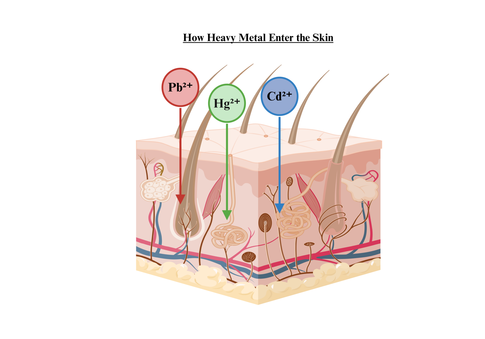 |
|---|
As cosmetics are directly applied to the skin, the metal ions can diffuse through the intercellular lipid pathways of the stratum corneum, particularly when in small, soluble, and lipophilic complexes such as mercury-thiol complexes (Tchounwou et al., 2012). Hair follicles and sweat glands bypass the stratum corneum, allowing nanoparticles or metal ions suspended in creams to accumulate within these appendages. Additionally, surfactants, solvents such as alcohols, and penetration enhancers in cosmetic formulations disrupt the lipid matrix of the stratum corneum, facilitating deeper penetration. Green, blue, and red arrows in Figure 1 indicate the pathways through which metal ions may enter systemic circulation (Okereke et al., 2015).
Table 1: Comparison of mercury, lead, and cadmium. The table summarizes the typical uses of each metal, the maximum permissible limits set by the FDA for cosmetic products, and the associated health risks resulting from exposure or intoxication (FDA's Testing, n.d.).
| Heavy Metal | Usage | FDA Limit (ppm) | Side Effects |
|---|---|---|---|
| Mercury | Whitens skin by inhibiting melanin | 1 | Dizziness, insomnia, neurological damage |
| Lead | Color additive in lip products and eyeliners | 20 | Miscarriage, irregular menstrual cycles, hormonal changes, diminished fertility |
| Cadmium | Expands color tones and creams | No limit (EU: 20 ppm) | Proteinuria, carcinogenicity, genetic disorders |
Heavy metals are used in order to enhance the performance of the products, but unfortunately, many consumers are unaware of the risks of inadvertent heavy metal consumption through cosmetics. Mercury is the most frequent additive in skin whitening products (SLPs) as it suppresses the production of melanin, a substance that's responsible for skin pigmentation. As seen in Table 1, the FDA-recommended concentration is under one part per million (ppm), but many cosmetic manufacturers use above 1000 ppm of mercury to maximize their whitening effect (FDA's Testing, n.d.; Sun et al., 2017). Side effects of mercury intoxication include dizziness, insomnia, and neurological damage (Fernandes Azevedo et al., 2012). Mercury is particularly hazardous due to its affinity for thiol (-SH) groups in proteins that make up body tissues, specifically targeting cysteine residues (Bjørklund et al., 2019). This interaction distorts the tertiary structure of proteins, resulting in a loss of biological function (Kang et al., 2024). Lead acts as a color additive in most lip products, eyeliners, and mascara, enabling cosmetics to feature certain colors (Raza-Naqvi et al., 2022). Continuous absorption of lead featured cosmetics is detrimental to the human body, as blood lead concentrations of 3.5 µg/dL or higher lead to effects such as miscarriage, irregular menstrual cycles, changes in hormones, and diminished fertility in both genders (Lead Poisoning, 2024). Similarly to mercury, lead (Pb²⁺) toxicity arises from the ability to mimic and interfere with essential divalent ions like calcium (Ca²⁺) (Virgolini & Aschner, 2021). By targeting hydroxyl (-OH) groups and disrupting cellular signaling, lead exposure causes systemic neurotoxicity and hormonal imbalances.
Within the cosmetics industry, cadmium is eminently used in the form of cadmium sulfide, a yellow pigment that creates specific shades. The application of cadmium sulfide is widespread across numerous product manufacturing due to its versatility in producing more intense, longer-lasting hues and its expansion of color tones in eyeliners, lip gloss, and beauty creams (Omenka & Adeyi, 2016). Prolonged exposure of 0.005 mg/kg/day for more than two weeks (Cadmium, n.d.) may lead to kidney disorders, proteinuria (high levels of protein in urine), potential carcinogenic exposure, and genetic disorders (Raza-Naqvi et al., 2022). Despite these well-documented hazards, the FDA has not yet established a set limit in cadmium overuse in cosmetic manufacturing, exposing consumers to unmitigated health risks (Table 1). These findings underscore the need not only for more rigorous regulatory enforcement but also for the advancement of accessible, consumer-oriented testing methodologies that enable individuals to evaluate product safety independently.
Current testing methods for identifying heavy metal contaminants in cosmetics present challenges, largely due to technological complexity. For instance, gold-standard detection methods such as Inductively Coupled Plasma Mass Spectrometry (ICP-MS) and Atomic Absorption Spectroscopy (AAS) are inaccessible to the general public. These techniques require expensive instrumentation, professional laboratories, and complex sample preparation (Arshad et al., 2020). Existing biosensors also lack the capacity for recognizing multiple heavy metals at once. Since heavy metals often coexist in a single product to enhance pigment intensity, and also given how consumers often use multiple cosmetic products simultaneously, a sensor limited to a single output provides an incomplete assessment of risk (Fernandes Azevedo et al., 2012).
| Figure 2: In vitro genetic circuit. The left side illustrates the transcriptional machinery extracted from cells for use in a cell-free system. Specific genetic components involved in metal ion detection are displayed within the big tube. Smaller tubes with green, red, and blue indicate possible outcomes following the genetic pathway. Created in BioRender. Yoon, J. (2026) https://BioRender.com/8qs91s1 | 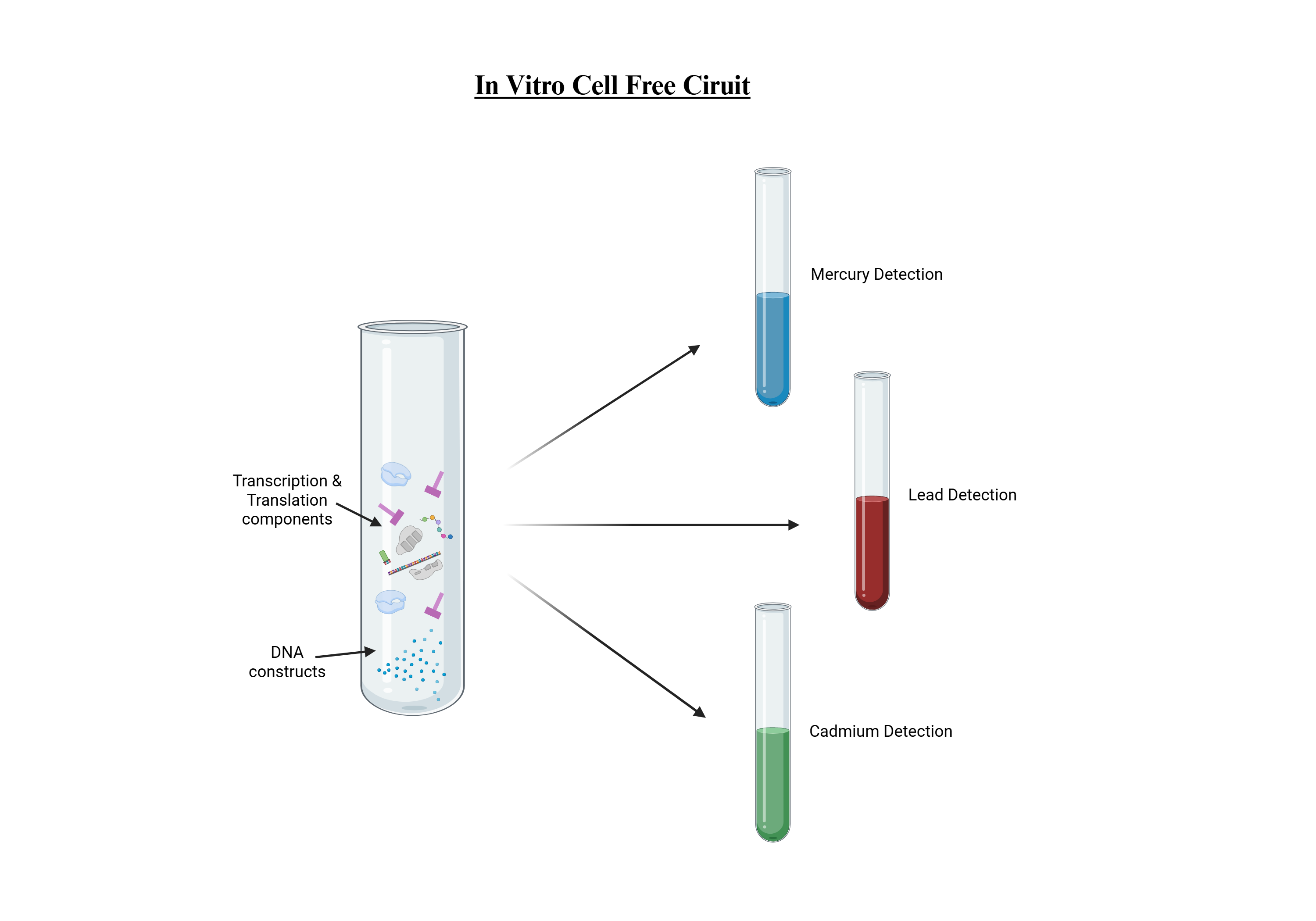 |
|---|
Therefore, our project aims to develop a three-in-one in vitro test solution that detects the presence of mercury, lead, and cadmium through a cell-free TX-TL (Transcription-Translation) system. By using a cell-free platform, the biosensor ensures enhanced user safety by eliminating the need for live organisms or genetically modified organisms within a household setting. Furthermore, the in vitro machinery will remain functional in toxic environments that would otherwise be deadly to living host cells (Wu et al., 2008). Our system will be constituted of a total of three distinct detection pathways that feature linear DNA constructs and transcriptional machinery for each heavy metal as illustrated in Figure 2.
The presence of each heavy metal will trigger a distinct color change, allowing users to easily identify the substance. Each circuit features a metal-responsive promoter linked to a fluorescent reporter protein that ultimately produces a visible signal when heavy metals are present. For the mercury pathway, in the presence of the mercury ion, the MerR transcriptional regulator will be activated to initiate the pMerT promoter, and code for GFP, a green fluorescent protein. The lead pathway utilizes the PbrR transcriptional regulator, which drives the pPbrA promoter to code for the expression of mCherry, a type of red fluorescent protein. Similarly, the cadmium pathway utilizes the CadR transcriptional regulator and the pCadA promoter to drive expression of CFP, a cyan fluorescent protein. These proteins will let the users check which metals are present, depending on which color or mix of colors they see. Given the fact that the kit does not require any maintenance to sustain circuit viability, it allows for use in both household and industrial settings. Therefore, our project will empower consumers to evaluate the risks of their own products, as well as manufacturers to evaluate the contents of their raw materials.
Materials and methods
In this study, we integrated the customized linear DNAs and TX-TL kits to express the mCherry red fluorescence for the lead detection system.
Linear DNA design and synthesis
The genetic design of the heavy metal biosensing circuits was engineered using Benchling molecular biology software. To facilitate rapid prototyping and avoid the metabolic constraints associated with host-cell transformation, the project utilized customized linear DNA. Each circuit was designed with a T7 promoter (derived from the iGEM Registry of Standard Biological Parts), which was placed upstream of the regulatory sequences to drive robust transcription within the cell-free environment and also the self-expression of the regulatory promoter. For the lead-sensing circuit, the system was split into a two-part assembly: a regulatory gBlock expressing the PbrR transcription factor and a reporter gBlock containing the pPbrA promoter fused to the mCherry red fluorescent protein sequence.
The T7 deGFP control linear DNA sequence was brought from the myTXTL plasmid sequences website in linear FASTA format, and the pbrR sequence was obtained from the iGEM Registry of Standard Biological Parts (BBa_K1701001). All sequences were imported into Benchling with topology set to linear. DNA elements were annotated using Benchling’s auto-annotation function; however, the DNA elements needed to be added manually due to the imperfection of the auto-annotation function, which includes the T7 promoter, UTR1, and the T7 terminator, using the positional information from the GenBank format. The regulatory gBlock was created by duplicating the Linear_T7 deGFP file, removing the deGFP sequence with the stop codon (5’-TAA-3’) removed. The reporter gBlock was created by replacing the sequence ranging from the T7 promoter to the T7 terminator with the 936 bp pPbrA promoter and mCherry sequence. The finalized linear DNA designs for both regulatory and reporter DNA are shown in Figure 3.
| Figure 3. (A) The overview of the T7 deGFP plasmid. (B) Restriction site maps for linear DNA T7 pbrR (left) and linear DNA pPbrA promoter mCherry (right). Both Linear DNA constructs were created through Benchling. | 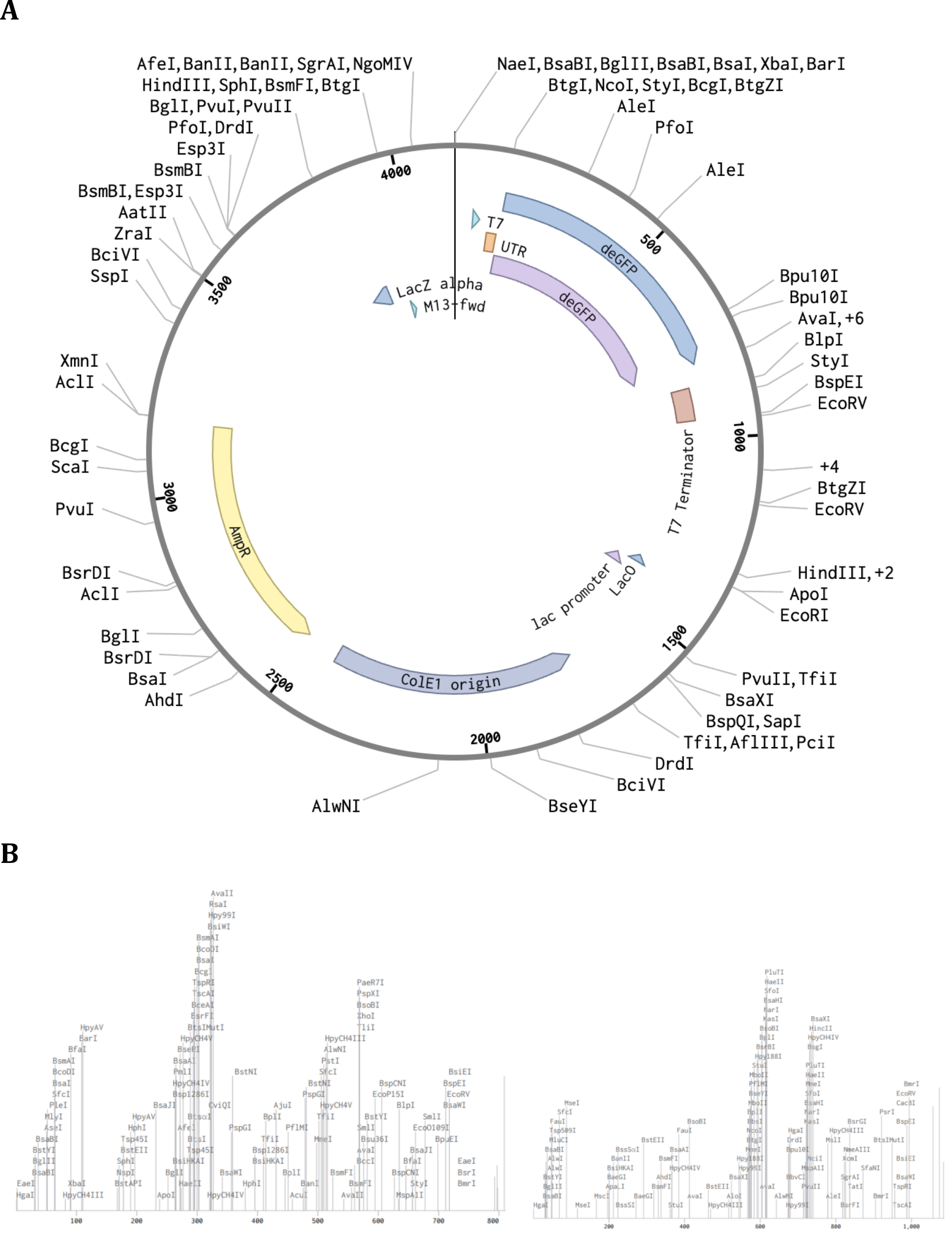 |
|---|
Lead dilution
Creating a lead calibration curve was essential in order to test out different contamination levels. To quantify lead contamination at different concentrations, we created a calibration curve using a 1000 ppm lead reference standard solution from Fisher Scientific. Six sterile 25 mL tubes were labeled according to the following target concentrations, as the FDA sets a limit of 10 ppm for lead in cosmetics and 20 ppm for lead in color additives: 0 ppm (Control), 1 ppm, 5 ppm, 10 ppm, 100 ppm, and lastly 1000 ppm (Lead in Cosmetics, 2022). Following the calculated amount of deionized water for each concentration demonstrated in Table 2, the deionized water was first distributed into each tube using a micropipette, followed by the set volume of 1000 ppm stock solution to create the targeted concentration. Each tube was shortly centrifuged and vortexed to ensure the solutions were fully mixed. All prepared concentrations of solutions were stored at 4°C before usage and integration.
Table 2: Lead dilution concentration chart. Each row demonstrates the target concentration of lead, followed by the needed volume of the 1000 ppm standard lead solution in µL, and the volume of di water in mL.
| Concentration (ppm) | Volume of 1000 ppm Stock (µL) | Volume of di Water (mL) | Calculation () |
|---|---|---|---|
| 1 ppm | 10 µL | 9.990 mL | |
| 5 ppm | 50 µL | 9.950 mL | |
| 10 ppm | 100 µL | 9.900 mL | |
| 100 ppm (high) | 1000 µL | 9.000 mL | |
| 1000 ppm | 10.000 mL | 0.000 mL |
gBlock resuspension
IDT gBlocks came accompanied by a specifications sheet that states the molecular weight and the specific mass of each fragment. Each tube was reconstituted in DNase/RNase-free water to reach the target concentration for later TX-TL kit integration, rather than the manufacturer’s recommendations. Calculations were involved in order to determine the appropriate solvent volume for each gBlock. The number of moles in each tube was determined by dividing the supplied mass () by the molecular weight listed on the specification sheet. The required solvent was then calculated by dividing the number of moles by ().
The pPbrA promoter mCherry gBlock has a molecular weight of and a supplied mass of 1000 ng (1490 fmol); therefore, 15.52 µL of DNase/RNase-free water was added to reach the target concentration. For the T7 promoter-driven pbrR gBlock with a molecular weight of with a supplied mass of 1000 ng (1998 fmol), the amount of 20.81 µL of DNase/RNase-free water was added. After the DNase/RNase water was added to the respective tubes, the gBlocks were briefly vortexed and centrifuged, then incubated at 50°C for 20 minutes.
TX-TL kit integration
Experimental assays were concluded using the myTXTL Pro Master Mix system (Daciel Arbor Biosciences), which provides the necessary transcriptional and translational machinery in vitro. Although the manual provided maps out a 12 µL reaction volume and the ratio of the components as demonstrated in Table 3, each reaction was scaled up to 13 µL for anticipated reaction volume loss during pipetting. Each reaction was assembled on ice to preserve enzymatic activity and combined in the order recommended by the manual provided by Arbor Biosciences.
| Component | 12 µL reaction | 50 µL reaction | µL reaction |
|---|---|---|---|
| Pro Master Mix | 9.0 µL | 37.5 µL | µL |
| Pro Helper Plasmid | 0.5 µL | 2.0 µL | µL |
| Template DNA | 2.5 µL | 10.5 µL | µL |
| Total Volume | 12.0 µL | 50.0 µL | µL |
Table 3: Chart indicating the amount of components needed to assemble the myTXTL reactions. Each reaction consisted of Pro Master Mix, Pro Helper Plasmid, and a 96 nM Template DNA in order to create the reactions. Component volumes are set for two example reaction sizes (12 µL and 50 µL), while a reaction of size V μL could be created by multiplying the desired V μL by the indicated ratios per component. In this case.
Each reaction consisted of 9.0 µL myTXTL Pro Master Mix, 0.5 µL Pro Helper Plasmid (pTXTL-P70a-T7RNAP), 1.66 µL of linear T7-driven pbrR DNA (96 nM stock), 0.83 µL of linear pPbrA-driven mCherry DNA (96 nM stock), and 1 µL of lead(II) solution at the desired concentration created via the lead dilution lab (Table 4). The ratio between linear T7-driven pbrR DNA and linear pPbrA-driven mCherry DNA was adjusted to 2:1 for optimized mCherry expression.
To ensure the reliability of the results, a positive control was prepared by utilizing 2.5 µL of T7-deGFP plasmid instead of the linear DNA templates (Table 4). A negative control reaction was similarly prepared, as it consisted of nuclease-free water in place of lead and DNA templates (Table 4). After assembling both the positive control and the negative control reactions, all reactions were briefly vortexed and centrifuged for 5 seconds prior to incubation.
| Component | Positive control (µL) | Negative control (µL) |
|---|---|---|
| myTXTL Pro Master Mix | 9.0 | 9.0 |
| Pro Helper Plasmid | 0.5 | 0.5 |
| T7-deGFP plasmid (96 nM) | 2.5 | — |
| DI water | 1 | 3.5 |
| Total | 13.0 | 13.0 |
Table 4: Positive and negative control reaction setup for myTXTL reactions. The positive control contains 9.0 µL of myTXTL Pro Master Mix, 0.5 µL of Pro Helper Plasmid, 2.5 µL of T7-deGFP plasmid, and 1 µL of DI water to create a total of 13.0 µL of reaction. The negative control contains 9.0 µL of myTXTL Pro Master Mix, 0.5 µL of Pro Helper Plasmid, and 3.5 µL of DI water to create a total of 13.0 µL of reaction.
Incubation was performed at 27°C for 16 hours to allow for full protein maturation. The reactions were placed on ice to terminate continued expression. Fluorescence was monitored and documented using the P51™ Molecular Fluorescence Viewer. This portable blue-light visualization platform allowed for the detection of mCherry expression without the need for high-cost laboratory plate readers or fluorescence microscopy.
Laboratory safety
Given that lead is a potent neurotoxin, all laboratory activities were conducted under a strict chemical safety framework. Working on lead does not apply in the BSL (Biosafety Level) categorization; instead of following BSL levels, all work should be accompanied by complementary PPE, aerosol control, and Hygiene Parallels. All dilution and TX-TL assembly was carried out in a ventilated fume hood while wearing appropriate PPE, including double nitrile gloves, lab coats, and safety goggles. To prevent cross-contamination and environmental exposure, all pipette tips that were used and were in contact with the 1000 ppm standard lead solutions were disposed of in a sealed, puncture-resistant waste container in accordance with local environmental health and safety (EHS) regulations.
Results
The results of this study were found through two distinct experimental trials focused on optimizing the ratio of the genetic components to ensure effective lead detection. In the first trial, the cell-free reactions were assembled using a 1:2 molar ratio of the regulator construct, linear T7_pbrR DNA, to the reporter construct, linear pPbrA-mCherry DNA. Despite the presence of lead dilutions, this configuration failed to yield a detectable fluorescent signal as demonstrated in Figure 4. The positive control for this trial exhibited no activity (Figure 4), suggesting that the lower concentration of the T7-driven regulator may have been insufficient to activate transcription-translation or that the lead concentration settled to the bottom, making it unable to interact with the genetic constructs suspended in the TX-TL extract.
| Figure 4: The first trial conducted with a ratio of 1:2 of the regulator and the reporter genes. Samples from left to right: Positive control, Negative control, 1 ppm, 5 ppm, 10 ppm, 100 ppm, 1000 ppm. The TX-TL solutions are mixed with different concentrations of Lead and placed under the P51™ Molecular Fluorescence Viewer. No signs of green fluorescence in the positive control and the different concentrations. | 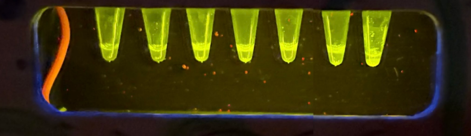 |
|---|
A second trial was conducted with an adjusted molar ratio of 2:1 between the regulator (T7-driven pbrR DNA) and the reporter (pPbrA-driven mCherry DNA) linear DNA genes, respectively. By doubling the concentration of the T7_pbrR DNA relative to the pPbrA-mCherry reporter, the system was optimized to ensure an adequate supply of the PbrR transcription factor, which is capable of responding to the lead. In this trial, both the positive and negative controls functioned as intended as seen in Figure 5. The positive control showed green fluorescence, validating the enzymatic viability of the TX-TL master mix, while the negative control remained dark, proving that the pPbrA promoter works in the absence of lead (Figure 5). However, despite the successful validation of the controls and the optimized DNA ratio, the experimental tubes containing lead standards did not produce a visible mCherry fluorescent output.
| Figure 5: The second trial conducted with a ratio of 2:1 of the regulator and the reporter genes. Samples from left to right: Positive control, Negative control, 1 ppm, 5 ppm, 10 ppm, 100 ppm, 1000 ppm. The TX-TL solutions are mixed with different concentrations of Lead and placed under the P51™ Molecular Fluorescence Viewer. Green fluorescence is observed in the positive control, but no fluorescence was observed in the different concentrations. | 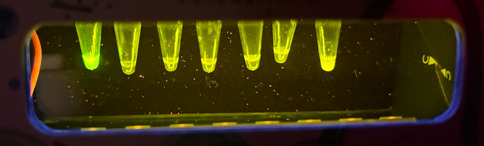 |
|---|
Overall, the visualization of the GFP in the positive control confirms that the TX-TL system is functioning correctly. Similarly, the absence of fluorescence in the negative control, which does not contain any DNA components, proves that the linear DNA components (reporter genes and fluorescent proteins) were the parts responsible for the expression of fluorescence.
Discussions
The objective of the project was to create an in vitro cell-free detection system for heavy metals: mercury, lead, and cadmium by integrating the synthesized linear DNA and TX-TL kit to produce a portable kit that produces fluorescence that indicates heavy metal contamination. By using mCherry as a fluorescent reporter, the system was designed to provide a clear, visual indication of metal presence. While the lead-sensing circuit was the primary focus of this phase, the lack of observed fluorescence across the varying lead concentrations (1 ppm to 1000 ppm) necessitates an evaluation of the biochemical and physical properties in the cell-free TX-TL environment.
Despite all the successful integration of the TX-TL kit and the necessary components, we weren’t able to observe the mCherry RFP reactions across the tubes with the different concentrations of lead. This absence of fluorescence suggests multiple potential causes related to plasmid ratio imbalance. In Trial 1, the 1:2 regulator-to-reporter ratio may have resulted in a dilution of the PbrR transcription factor. If the concentration of the regulator DNA is too low relative to the reporter, the system cannot produce enough PbrR protein to saturate the available pPbrA promoter sites, even in the presence of lead. Additionally, in Trial 2, while the 2:1 ratio was intended to favor regulator production, it is possible that the total DNA concentration reached a saturation point (Nagaraj et al., 2017).
The lead-responsive regulatory elements may have failed to function due to improper protein folding (Shin & Noireaux, 2012). Because linear DNA templates are susceptible to exonuclease degradation in certain extracts, the effective lifespan of the pbrR gene may have been too short to establish a functional concentration of protein (Sun et al., 2013).
An observation after incubation was the formation of white pellets at the base of the reaction tubes with different standard lead concentration solutions. We hypothesize that these pellets may be due to the heaviness of the lead as it reacted with the ions and components of the TX-TL system, such as proteins or nucleic acids. This sequestration of lead ions effectively reduced their bioavailability (Fowler, 1998). This physical separation, combined with the potential denaturation of essential enzymes by the heavy metal ions, likely inhibited the protein synthesis process entirely.
Next steps
While the TX-TL kit provides the necessary transcriptional and translational mechanisms, further optimization is needed. The 2:1 ratio of T7-driven PbrR DNA to pPbrA-mCherry DNA made significantly more progress compared to the 1:2 ratio, as the 2:1 ratio solution showed green fluorescence in the positive control. This progress suggests that increasing the dosage of the T7-driven regulator is a critical factor in overcoming the metabolic threshold of the in vitro environment; however, this ratio must continue to be tested and changed in order to identify the optimal conditions that yield the signaling of mCherry fluorescence. If the ratio adjustment enables the expression of mCherry fluorescence, we hope to quantify the fluorescence intensity using ImageJ for a more precise system performance.
Additionally, the physical behavior of the lead standard within the reaction remains a problem. To address the potential separation of lead ions, further experiments will utilize a shaking incubator to provide continuous agitation overnight. Another experiment consideration our team hopes to conduct in the future is to try redoing the lead dilution and TX-TL integrating with a different standard lead solution from a different company. By keeping the reaction in a state of constant suspension, we aim to prevent the lead ions from settling due to their high density.
Once the lead-sensing circuit is confirmed, we will expand to other heavy metals, cadmium and mercury as well. The linear DNAs will be designed using similar T7 promoters to drive the expression of metal-specific regulators. By integrating these pathways into a single assay, the final part of the project will provide a comprehensive, portable tool for the rapid assessment of heavy metal contamination in commercial cosmetics.
Author contributions
JY and MV conducted the early research process. JY and MV contributed to the writing and proofreading of the whole paper. JY and MV worked in the lab. JY and MV created the video. JY designed the images and graphics for this project.
Acknowledgements
We would like to extend our gratitude to the Western Reserve Academy Biobuilder Program for providing us with the opportunity and the resources that enabled us to conduct our research. We are also very grateful to Ms. Marmerstein for her support and guidance throughout our project. Her advice on sources, lab experiments, how to safely execute our hypothesis, and her feedback on all steps were crucial; we extend our most sincere thanks. We also deeply thank and appreciate our mentor, Dr. Hiroko Kaczmarek, for her unwavering, unconditional support, guidance, and feedback for every step of our project, from the early research process to even after. She guided us through difficulties and experimental errors, even helping us in areas beyond our scope of work. Without their support, the project would not have been possible. Finally, we would like to express our gratitude to the BioTreks Organization for providing the platform and community to share our research with our peers.
References
Bjørklund, G., Crisponi, G., Nurchi, V. M., Cappai, R., Buha Djordjevic, A., & Aaseth, J. (2019). A review on coordination properties of thiol-containing chelating agents towards mercury, cadmium, and lead. Molecules, 24(18), 3247. https://doi.org/10.3390/molecules24183247
Cadmium [Fact sheet]. (n.d.). World Health Organization. Retrieved May 1, 2026, from https://archive.epa.gov/epawaste/hazard/wastemin/web/pdf/cadmium.pdf
Cosmetics Market to hit USD 417.24 billion by 2030 | With a CAGR of 4.2%. (2023). Yahoo! Finance. https://finance.yahoo.com/news/cosmetics-market-hit-usd-417-083800842.html
Djordjevic, M. (2026, February 25). 15 fascinating beauty industry statistics you should know. SaveMyCent. Retrieved April 29, 2026, from https://savemycent.com/beauty-industry-statistics/
FDA's testing of cosmetics for arsenic, cadmium, chromium, cobalt, lead, mercury, and nickel content. (n.d.). U.S. Food & Drug Administration. Retrieved April 29, 2026, from https://www.fda.gov/cosmetics/potential-contaminants-cosmetics/fdas-testing-cosmetics-arsenic-cadmium-chromium-cobalt-lead-mercury-and-nickel-content
Fernandes Azevedo, B., Barros Furieri, L., Peçanha, F. M., Wiggers, G. A., Frizera Vassallo, P., Ronacher Simões, M., Fiorim, J., Rossi de Batista, P., Fioresi, M., Rossoni, L., Stefanon, I., Alonso, M. J., Salaices, M., & Valentim Vassallo, D. (2012). Toxic effects of mercury on the cardiovascular and central nervous systems. Journal of Biomedicine and Biotechnology, 2012, 1-11. https://doi.org/10.1155/2012/949048
Fowler, B. A. (1998). Roles of lead-binding proteins in mediating lead bioavailability. Environmental Health Perspectives, 106(suppl 6), 1585-1587. https://doi.org/10.1289/ehp.98106s61585
Kang, B., Wang, J., Guo, S., & Yang, L. (2024). Mercury-induced toxicity: Mechanisms, molecular pathways, and gene regulation. Science of the Total Environment, 943, 173577. https://doi.org/10.1016/j.scitotenv.2024.173577
Lead in Cosmetics. (2022, February 25). U.S. Food & Drug Administration. Retrieved May 1, 2026, from https://www.fda.gov/cosmetics/potential-contaminants-cosmetics/lead-cosmetics
Lead poisoning [Fact sheet]. (2024, September 27). World Health Organization. Retrieved May 1, 2026, from https://www.who.int/news-room/fact-sheets/detail/lead-poisoning-and-health
Nagaraj, V. H., Greene, J. M., Sengupta, A. M., & Sontag, E. D. (2017). Translation inhibition and resource balance in the tx-tl cell-free gene expression system. Synthetic Biology, 2(1). https://doi.org/10.1093/synbio/ysx005
Okereke, J. N., Udebuani, A. C., Ezeji, E. U., Obasi, K. O., & Nnoli, M. C. (2015). Possible health implications associated with cosmetics: A review. Science Journal of Public Health, 3(5), 58-63. https://doi.org/10.11648/j.sjph.s.2015030501.21
Omenka, S. S., & Adeyi, A. A. (2016). Heavy metal content of selected personal care products (PCPs) available in Ibadan, Nigeria and their toxic effects. Toxicology Reports, 3(14), 628-635. https://doi.org/10.1016/j.toxrep.2016.07.006
Raza-Naqvi, S. A., Idrees, F., Sherazi, T. A., Anjum-Shahzad, S., Ul-Hassan, S., & Ashraf, N. (2022). Toxicology of heavy metals used in cosmetics. Journal of the Chilean Chemical Society, 67(3), 5615-5622. https://doi.org/10.4067/s0717-97072022000305615
Shin, J., & Noireaux, V. (2012). An E. coli cell-free expression toolbox: Application to synthetic gene circuits and artificial cells. ACS Synthetic Biology, 1(1), 29-41. https://doi.org/10.1021/sb200016s
Sun, G.-F., Hu, W.-T., Yuan, Z.-H., Zhang, B.-A., & Lu, H. (2017). Characteristics of mercury intoxication induced by skin-lightening products. Chinese Medical Journal, 130(24), 3003-3004. https://doi.org/10.4103/0366-6999.220312
Sun, Z. Z., Hayes, C. A., Shin, J., Caschera, F., Murray, R. M., & Noireaux, V. (2013). Protocols for implementing an Escherichia coli based tx-tl cell-free expression system for synthetic biology. Journal of Visualized Experiments, (79). https://doi.org/10.3791/50762
Tchounwou, P. B., Yedjou, C. G., Patlolla, A. K., & Sutton, D. J. (2012). Heavy metal toxicity and the environment. Experientia Supplementum, 133-164. https://doi.org/10.1007/978-3-7643-8340-4_6
Virgolini, M. B., & Aschner, M. (2021). Molecular mechanisms of lead neurotoxicity. Advances in Neurotoxicology, 159-213. https://doi.org/10.1016/bs.ant.2020.11.002
Wu, Y., Zhou, Y., Song, J., Hu, X., Ding, Y., & Zhang, Z. (2008). Using green and red fluorescent proteins to teach protein expression, purification, and crystallization. Biochemistry and Molecular Biology Education, 36(1), 43-54. https://doi.org/10.1002/bmb.117