
Alida Karsten, Alex Lee, Lydia Lockwood, and Delaney Pittinger, Western Reserve Academy, Hudson, Ohio, United States
Reviewed on 9 May 2026; Accepted on 22 June 2026; Published on 26 October 2026
With help from the 2026 BioTreks Production Team.
Atopic dermatitis, or eczema, is a chronic inflammatory disease that impacts 204 million globally. It is characterized by dryness, itching, and irritation of the skin caused by reduced moisture retention and increased susceptibility to irritants, commonly Staphylococcus aureus. While some find success in treating the disease with topical corticosteroids, those treatments generally come with side effects, such as skin thinning, acne, and skin pigmentation changes. While these treatments aim to reduce inflammation and symptoms, they do little to support the skin’s microbiome. S. epidermidis supports skin health, but is under-represented in individuals with eczema, making it a potentially suitable chassis for an engineered atopic dermatitis treatment. We propose enhancing this bacteria’s synthesis of polysaccharide intercellular adhesin (PIA) which is a major contributor to a biofilm that helps retain moisture and create a healthy skin barrier. This would be executed by inserting a plasmid with the icaADBC operon (which codes for PIA synthesis) into S. epidermidis. While biofilms can be dangerous, precautionary mechanisms will be incorporated into the product to ensure its safety and that the resulting biofilm functions to restore the skin barrier by increasing PIA synthesis, The biofilm formed by PIA will allow the skin to retain these nutrients and lipids while increasing antimicrobial peptide production (which fights S. aureus overgrowth). This engineered bacteria will be formulated into a cream that heals damaged skin directly, and fights underlying causes by increasing water and nutrient retention.
Keywords: Staphylococcus epidermidis
Authors are listed in alphabetical order. Vivien Marmerstein mentored the group. Please direct all correspondence to mailto:marmersteinv@wra.net.

Background
Every few seconds, someone scratches at skin that burns, cracks, and never fully heals. Atopic dermatitis (AD), commonly referred to as eczema, is a chronic inflammatory skin disease characterized by dryness, itching, and irritation that often begins in childhood but can occur at any age (Cleveland Clinic, 2022; Mayo Clinic, 2024). It stems from a combination of immune dysregulation, genetic predisposition, and skin-barrier dysfunction, which reduces moisture retention and increases susceptibility to the penetration of irritants and allergens (Ballard, 2022; Cleveland Clinic, 2022; Mayo Clinic, 2024). AD disrupts the skin barrier, causing increased moisture loss and irritant penetration to occur. This disruption ultimately leads to ceramide and lipid content being reduced in the stratum corneum (the outermost layer of the skin, specifically, the epidermis). As a result of this process, corneocyte cohesion is weakened. In the stratum corneum, which is composed of tough skin cells and keratin that protect the inner layers of the epidermis, AD reduces ceramide and lipid content, which weakens corneocyte cohesion (Sawyer, 2023; Bogaard et al., 2024). The weakening of corneocyte cohesion causes the disorganization visible in contrast to a healthy stratum corneum in Figure 1, which leads to increased permeability. This then leads to inflammation, impaired differentiation, and increased permeability in the epidermis, which causes the irregularities and areas of barrier breakage that can be seen in comparison to the organized epidermal layer of healthy skin in Figure 1 (Bogaard et al., 2024). Immune activation and increased penetration of irritants and allergens also impact the dermis, which is the layer of skin that lies below the epidermis and is responsible for bodily protection, sensation, sweat and hair production, and support of the epidermis (Dermis, 2022; Bogaard et al., 2024). Such interactions cause the uneven appearance and inflammation that is evident in Figure 1 (Bogaard et al., 2024).
| Figure 1: The stratum corneum, epidermis, and dermis, of skin with AD (left) compared to that of healthy skin (right). This shows that skin affected with AD has disrupted barrier structure, which can lead to greater moisture loss and allow more irritants, allergens, and microbes to enter the skin. | 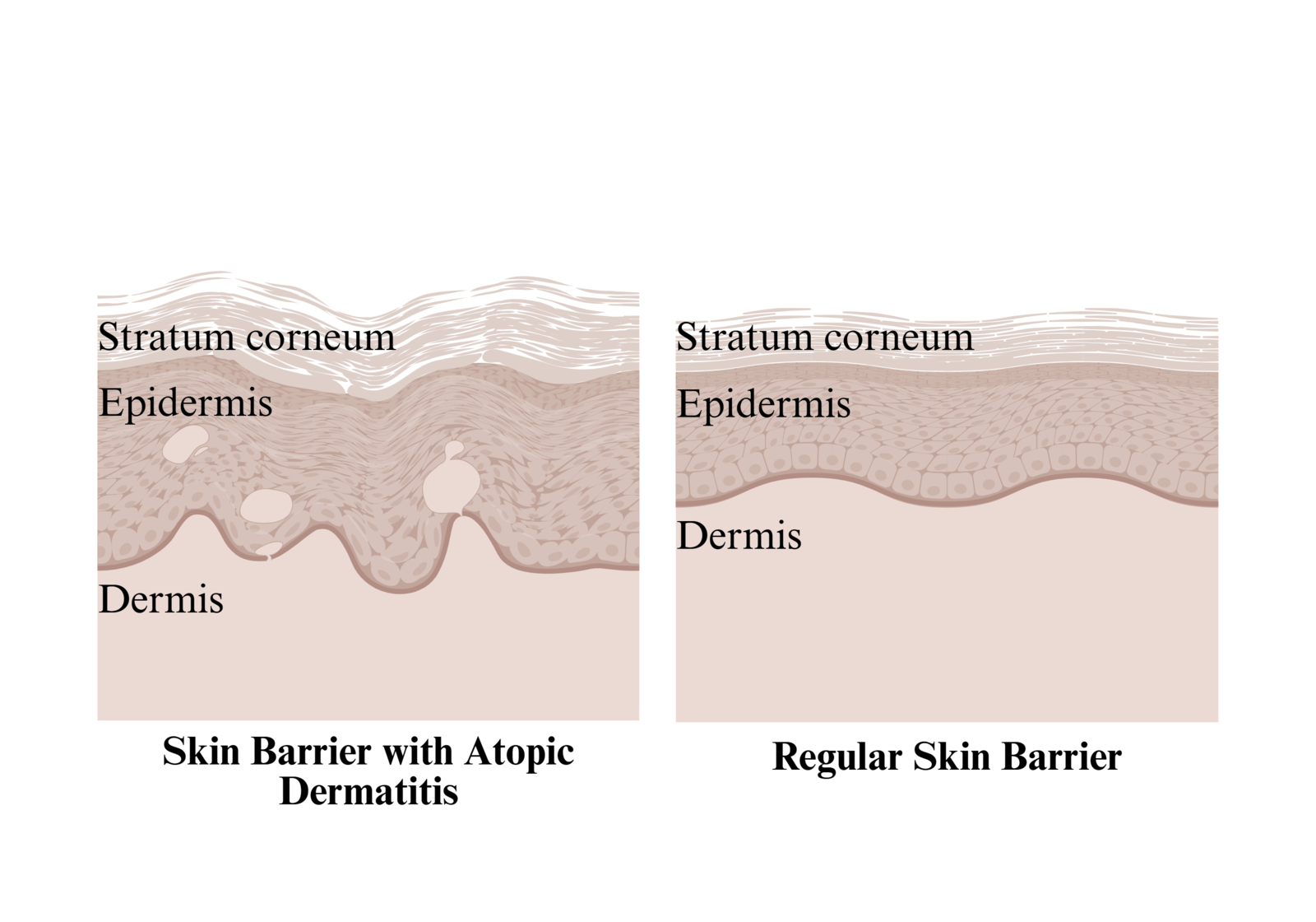 |
|---|
In particular, the colonization of S. aureus on eczema-affected skin is closely tied to flares and inflammation, as it disrupts the microbial balance by crowding out beneficial species, forming biofilms, and exacerbating disease activity (Ballard, 2022; Mayo Clinic, 2024). Globally, AD affects about 4.0% of children and 2.0% of adults, which is approximately 204 million people, and it is the leading cause of global burden from skin disease (Tian et al., 2023). Beyond physical symptoms, many patients experience sleep disturbance, stress, and a reduced quality of life (Cleveland Clinic, 2022). Although regular moisturization and medicated treatments can help manage itch and flares, AD has no cure; symptoms often return, and long-term management is usually necessary (Cleveland Clinic, 2022; Mayo Clinic, 2024).
Many of the cases of AD can be supported through treatment; however, more than 55% of adults with moderate to severe atopic dermatitis report inadequate disease control (Eczema Statistics, n.d.). Atopic dermatitis affects the quality of life of those who suffer from the condition. Patients with moderate to severe AD reveal the condition significantly limits their lifestyles and, in some extreme cases, leads to academic or career issues (Beckman et al., 2023).
This skin condition can lead to recurrent infections if left untreated. There is a strong genetic component to eczema, as people with a family history of eczema, allergies, and asthma are the more likely to develop the condition (Nemeth et al., 2024). In addition, it has been found that certain factors, such as age and environment, have heightened the chances of developing atopic dermatitis (Abraham & Roga, 2014).
Some approach treating eczema with antihistamines. Allergens stimulate the histamine-1 receptors located in your airways, blood vessels, stomach, and esophagus, which triggers histamine release, causing the symptoms of an allergic reaction. Antihistamines block the release of these histamines, alleviating the allergy symptoms they create (Antihistamine Drugs, 2023). Individuals with eczema condition experience itchiness, which prompts some to seek relief with an antihistamine. However, eczema is not histamine-driven, and therefore, antihistamines are not a practical treatment approach – though they may relieve inflammation caused by allergies that occur in conjunction with eczema (Gillette, 2023).
Corticosteroids are the most popular and effective treatment option for eczema today. This class of steroids is used to treat a variety of ailments, including eczema (Gallagher, 2023). The first in this class, hydrocortisone, was introduced in 1951 (Abraham & Roga, 2014). Its creation offered significant relief to eczema patients while also opening the door to further research on the condition, and their shortcomings prompted efforts to find non-steroidal treatments for eczema (Gallagher, 2023). Steroid creams and ointments remain the primary treatments for eczema today, but their flaws remain, underscoring the continued need for a non-steroid treatment (Cristol, 2024). The anti-inflammatory and anti-proliferative properties of such steroids allow them to treat various skin conditions successfully, but these very properties also lead to side effects (Abraham & Roga, 2014).
One side effect of topical steroids is skin thinning. Though this effect can typically be reversed after halting use, these treatments do not prevent eczema from being triggered; they merely remedy its effects. Therefore, many patients use these damaging creams and ointments for extended periods (Brannon, 2025). Figure 2 illustrates the progressive effects of topical corticosteroids on skin architecture across three sequential phases: pre-atrophy, atrophy, and tachyphylaxis (Abraham & Roga, 2014).
In Figure 2A (Pre-atrophy), the skin retains largely intact architecture, with only subtle early changes. The epidermis is mildly thinned compared to normal skin, reflecting steroid-induced inhibition of keratinocyte proliferation, while overall stratification remains preserved. The dermis appears structurally intact, with normal thickness and organization. Fibroblasts are present and exhibit typical spindle-shaped morphology, indicating preserved dermal cellular function. Collagen fibers (types I and III) are abundant and organized, maintaining extracellular matrix integrity. Hyaluronan is evenly distributed within the dermis, forming a continuous and organized matrix consistent with normal dermal hydration and elasticity (Abraham & Roga, 2014).
In Figure 2B (Atrophy), prolonged steroid exposure produces overt structural changes, particularly within the dermis. The epidermis shows further thinning relative to Panel A, consistent with sustained suppression of keratinocyte proliferation. The dermis is noticeably reduced in thickness, marking the onset of cutaneous atrophy. Collagen fibers are diminished in density and exhibit mild disorganization, reflecting inhibition of collagen I and III synthesis. Fibroblasts remain present but appear less active, with reduced prominence and diminished association with collagen fibers. Hyaluronan is still detectable within the dermal extracellular matrix but appears moderately reduced and less uniformly organized compared to Panel A, indicating early destabilization of dermal matrix composition (Abraham & Roga, 2014).
In Figure 2C (Tachyphylaxis), chronic corticosteroid exposure results in severe and global skin atrophy. The epidermis is notably thinned, with reduced cellular layering and minimal proliferative capacity. The dermis is profoundly attenuated, demonstrating extensive loss of structural integrity. Fibroblasts are sparse and hypoactive, appearing smaller and faint, consistent with functional suppression and steroid tachyphylaxis. Collagen fibers are scarce, fragmented, and disorganized, indicating advanced extracellular matrix degradation rather than simple inhibition of synthesis. Hyaluronan is markedly reduced and irregularly distributed, reflecting inhibition of hyaluronan synthase 3 (HAS3) and contributing to loss of dermal hydration and mechanical support. Together, these changes culminate in dermal atrophy and progressive degradation of skin tissue architecture (Abraham & Roga, 2014).
Prolonged use of corticosteroids can also impair the skin’s immune response to infections (Brannon, 2025). While these topical steroids’ mechanisms of action inhibit a broad range of immune responses, which supports their effect against inflammatory disorders such as atopic dermatitis, this also increases infection risk when use persists, as essential immune reactions are also inhibited (Chatham, 2025). As seen in Figure 3, steroids can also cause permanent stretch marks, or striae, on the skin (Brannon, 2025). This is due to cross-linking of immature collagen in the dermis, which results in irreversible intradermal tears (Abraham & Roga, 2014). Further risks with topical steroid use include steroid-induced telangiectasia (small, visibly dilated blood vessels), steroid acne, steroid rosacea, perioral dermatitis (red rash around the mouth), purpura (increased bruising), stellate pseudoscars (white, star shaped lesions), ulcerations, aggravation of cutaneous infections, delayed wound healing, contact sensitization to topical steroids, eyelid dermatitis, tachyphylaxis (decreasing response to the drug), trichostasis spinulosa, and post-peel erythema syndrome, which are also shown in Figure 3 (Abraham & Roga, 2014). All of these potential risks are intrinsic, as the traits that cause them are also the traits that aid eczema when using corticosteroids; therefore, a non-steroid remedy is necessary.
Our solution includes creating an eczema treatment embedded with S. epidermidis, a natural skin bacterium. S. epidermidis is one of the most abundant bacteria found on healthy human skin. It is not simply a passive inhabitant on skin, but rather an active contributor to maintaining balanced skin health, commonly known as skin homeostasis (Brown & Horswill, 2020). By helping sustain a healthy skin barrier, healing wounds, and supporting the immune system, S. epidermidis plays a key role in skin homeostasis. Additionally, it helps form a biofilm that prevents nutrient competition and antimicrobial substances by resisting pathogen colonization of the skin (Brown & Horswill, 2020).
Because S. epidermidis’s natural habitat is human skin, incorporating the bacteria into an eczema treatment allows the skin to naturally restore its barrier, rather than using harsh chemicals found in other therapies. This approach not only helps reduce eczema’s skin flare-ups and redness but also provides long-term support by increasing overall water retention and skin acidity (Brown & Horswill, 2023). Additionally, the bacteria's ability to produce antimicrobial peptides helps the skin suppress harmful bacteria and germs that often trigger flare-ups and secondary infections, such as S. aureus, thereby treating eczema at the source (Joshi et al., 2023).
In our design, increased icaADBC expression would increase PIA production, which would strengthen S. epidermidis biofilm formation and may help retain moisture at the skin surface, however, this increased biofilm formation would need to be carefully controlled for safety. Using the operon icaADBC, which is located in S. epidermidis, and is responsible for synthesizing polysaccharide intercellular adhesin (PIA). This process forms an essential element of a biofilm, which is a complex community of microorganisms in a matrix of polysaccharides, proteins, and nucleic acids, often helping fight pathogens and contributing to microbial health. However, biofilm colonizations create a shield in the body that can protect harmful bacteria, making them harder to eliminate (Brind’Amour, 2024). In this case, the biofilm is an important piece of our solution as it helps protect the components and bacteria we want to enhance in the skin barrier rather than unwanted bacteria (Brind’Amour, 2024). S. epidermidis helps retain moisture and create a healthy skin barrier through exopolysaccharide PIA, which contributes to the biofilm. Exopolysaccharides are sticky sugars that trap nutrients and water, helping keep the skin hydrated and balanced for longer (Fey & Olson, 2011).
By enhancing the natural components of S. epidermidis using an engineered plasmid carrying a second copy of the icaADBC operon, we can increase PIA production as the icaADBC operon encodes enzymes involved in PIA synthesis. This contributes to biofilm formation that may help retain moisture and support barrier function, however, increased biofilm formation also creates safety concerns that must be controlled. The modified gene would confer S. epidermidis with a stronger biofilm, strengthening the skin barrier and increasing water retention via PIA. Overall, the gene's increased expression of nutrients and lipids would be trapped by the increased expression of PIA and sticky sugars, ensuring that skin receives more nutrients and lipids and retains them for a longer amount of time. This engineered bacteria will be formulated into a cream that heals damaged skin directly and offer atopic dermatitis patients a natural solution.
Systems level
Our system engineers the bacterium, S. epidermidis to support the skin barrier in eczema-affected areas. S. epidermidis is naturally part of the skin microbiome and plays an important role in keeping the skin stable and protected (Severn & Horswill, 2022). Our design builds on this bacterium and relies on the role it already plays on healthy skin, helping maintain normal skin function (Landemaine et al., 2023). To improve the pre-existing benefits of S. epidermidis, our system is engineered to increase the production of polysaccharide intercellular adhesin (PIA), which plays a key role in biofilm formation by promoting intercellular adhesin between bacterial cells (Büttner et al., 2015). This results in stronger and more stable biofilms on the skin surface that are resistant to environmental stress. This biofilm acts as a protective layer on the skin, helping reduce moisture loss and blocking irritants, allergens, and harmful microbes that can cause eczema.
However, the role of S. epidermidis in eczema is complex, as different types can have either protective or potentially harmful effects on the skin, which makes it important to carefully control how this chassis is used in the engineered system (Landemaine et al., 2023). To deliver the engineered bacterium effectively, it will be applied through a topical cream or gel that serves to introduce S. epidermidis onto eczema-affected skin. This cream will maintain the bacteria’s viability before and during application. It will be used similarly to common skincare products, making it easy to incorporate into existing routines.
Once applied, the bacteria can remain on the skin surface and interact with its natural microbiome. As it produces increased levels of PIA, a stable biofilm forms, which reinforces the skin’s natural barrier, improves hydration, and reduces irritation over time. However, there are still important uncertainties, including how long the engineered bacteria can persist on eczema-affected skin, what level of PIA production is most effective without causing excessive biofilm buildup, and how different variants of S. epidermidis may affect the skin microbiome over time (Cau et al., 2021).
Device level
Key Functional Components:
The primary functional components of this system consist of the engineered chassis, genetic input, and biochemical output that together function to enhance the skin barrier and thereby heal the damage caused by eczema. Our chosen chassis, S. epidermidis, is Gram-positive and naturally inhabits human skin, playing a critical role in maintaining microbial homeostasis. It is typically considered a beneficial bacteria – despite the rare occurrence where it becomes an opportunistic pathogen – as it contributes to immune modulation and competitive exclusion of potentially dangerous pathogens. However, research is lacking surrounding its potential opportunistic pathogenicity in certain conditions, highlighting a necessary area of focus for future research surrounding this proposal (Brown & Horswill, 2020). While we selected this as our chassis for safety reasons, it also holds the icaADBC operon, which encodes for the synthesis of PIA, a major component of biofilms (Brind’Amour, 2024).
Input and Outputs:
The system’s inputs will consist of engineered plasmids containing an enhanced/overexpressed version of the icaADBC operon, introduced to upregulate PIA synthesis. This operon is tightly regulated by transcriptional repressors such as IcaR and TcaR, and its modulation allows for controlled increases in exopolysaccharide synthesis. The resulting output is elevated PIA production, which promotes formation of a structured biofilm matrix (Fey & Olson, 2011; Vuong et al., 2004). PIA is a key exopolysaccharide that facilitates intercellular adhesin and biofilm stability. These exopolysaccharides function as hydrophilic, adhesive polymers that retain water, lipids, and nutrients within the matrix, thereby enhancing skin hydration and barrier integrity (Fey & Olson, 2010; Flemming & Wingender, 2010). Consequently, increased PIA production is expected to improve retention of antimicrobial peptides, lipids, and other beneficial metabolites on the skin surface through enhanced biofilm production. This creates a positive feedback loop in which enhanced nutrient and lipid availability further supports microbial and skin health. However, a remaining research gap involves fully characterizing how increased biofilm formation impacts long-term skin homeostasis and whether unintended accumulation of materials could disrupt normal physiological processes.
Functional Pathways:
The functional pathway underlying PIA synthesis is governed by the icaADBC operon, which encodes enzymes responsible for the stepwise production and export of the polysaccharide matrix seen in Figure 4. The genes icaA, icaD, icaB, and icaC (seen in Figure 4) each play specific roles in PIA biosynthesis, while operon expression is modulated by regulatory proteins such as IcaR in response to environmental conditions. The engineered plasmid amplifies this naturally occurring pathway through increased operon expression, resulting in increased flux through the operon and elevated exopolysaccharide output (Flemming & Wingender, 2010).
| Figure 4: | 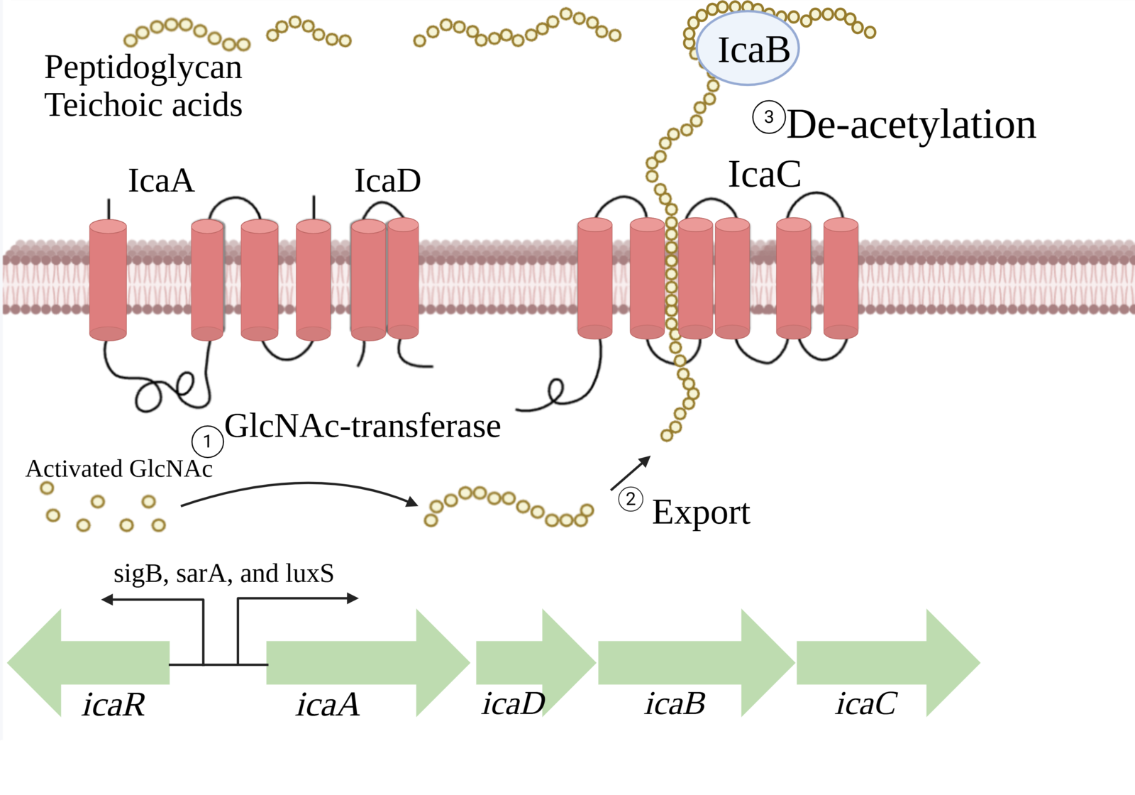 |
|---|
Biofilm
The product of this pathway, PIA, is a central structural component of biofilm – the complex, surface-associated microbial community embedded in a matrix of polysaccharides, proteins, and extracellular DNA that is shown in Figure 5 (Flemming & Wingender, 2010). Biofilms provide multiple advantages, including improved resistance to desiccation (drying), enhanced nutrient capture, and protection from environmental stressors and competing microbes (Brind’Amour, 2024; Costerton et al., 1999). Figure 5 displays the five crucial phases of biofilm formation, which involve the formation of a matrix. In the skin context, this matrix supports hydration by retaining water and lipids while stabilizing beneficial microbial populations. The increased lipid retention is particularly important, as sebum-derived lipids contribute to barrier integrity, antimicrobial defense, and prevention of transepidermal water loss.
| Figure 5: | 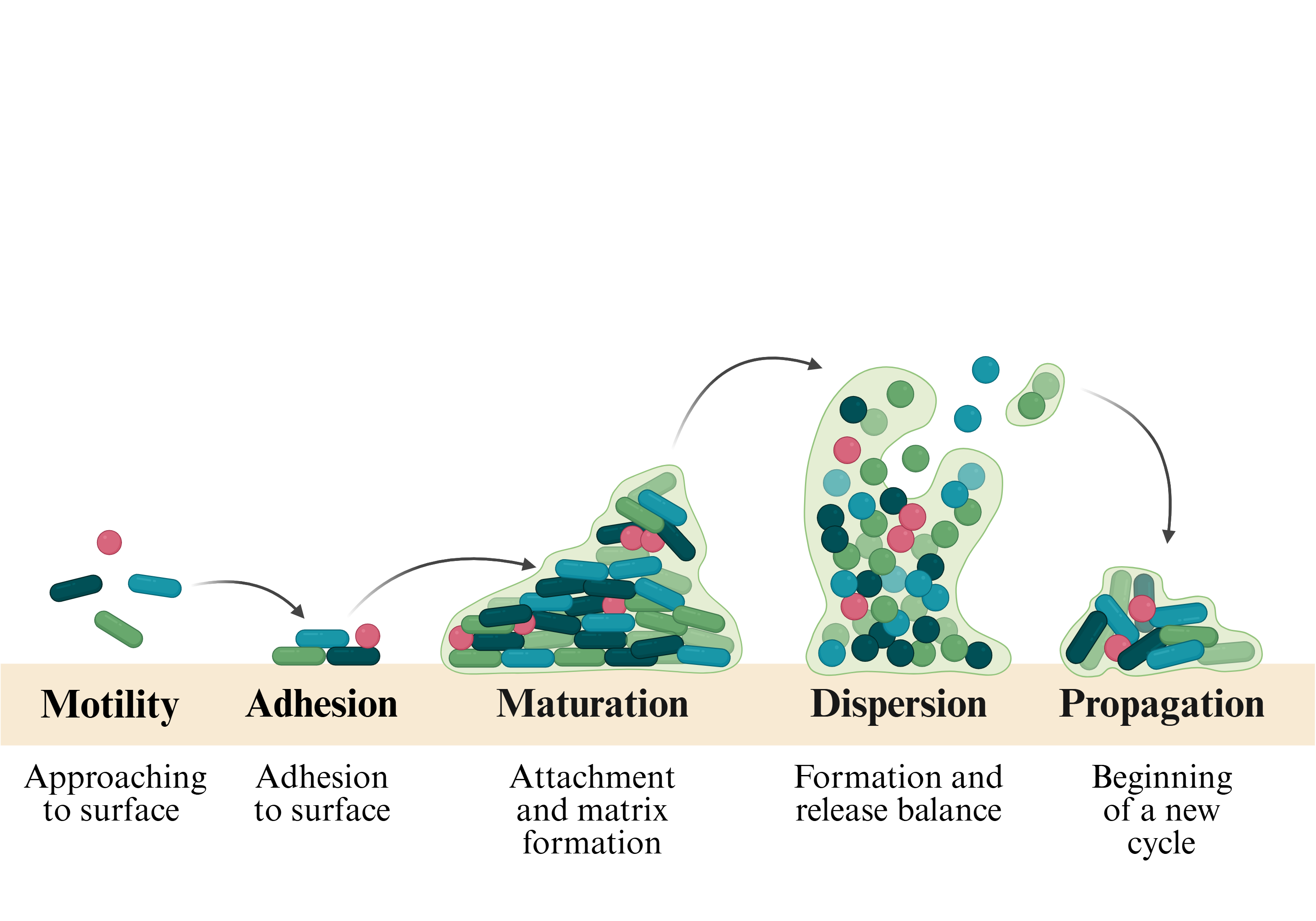 |
|---|
However, biofilms also present inherent risks, as their protective properties can shield pathogenic organisms and contribute to chronic infections (Costerton et al., 1999; Otto, 2009). This underscores a critical area which will require further research to ensure that enhanced biofilm formation can selectively benefit commensal organisms like S. epidermidis without facilitating colonization by harmful species. Addressing this will require precise regulatory control of operon expression, as well as potential integration of antimicrobial safeguards.
Parts level
Our project depends on the increased production of polysaccharide intercellular adhesin (PIA) and its contribution to the biofilm. An essential element of this is the matrix that is depicted in Figure 6. This dense extracellular matrix network of PIA embedded with staphylococcal cells acts as scaffolding and mediates the cell-to-cell adhesion that ultimately creates a biofilm that will provide short-term support and aid the skin barrier’s repair. This formation will protect from antimicrobial peptides (AMPs) and phagocytosis (the ingestion of bacteria), which is also demonstrated in Figure 6. The biofilm’s protection against AMPs and phagocytosis will allow the bacteria, S. epidermidis, to exist long-term within a host, leading to a biofilm/device-associated infection seen in Figure 6.
| Figure 6: PIA’s contribution to biofilm formation. | 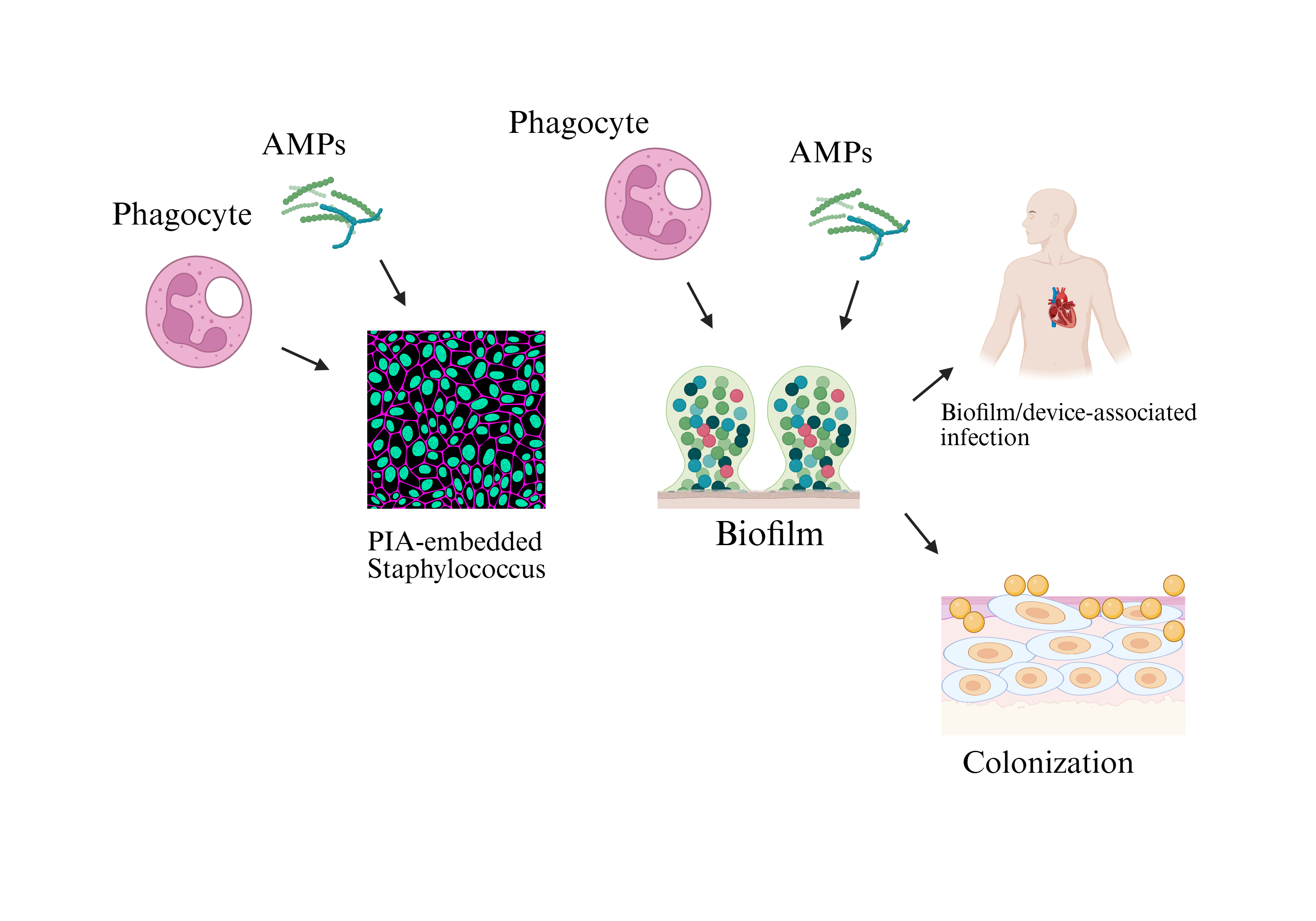 |
|---|
In Figure 2A (Pre-atrophy), the skin retains largely intact architecture, with only subtle early changes. The epidermis is mildly thinned compared to normal skin, reflecting steroid-induced inhibition of keratinocyte proliferation, while overall stratification remains preserved. The dermis appears structurally intact, with normal thickness and organization. Fibroblasts are present and exhibit typical spindle-shaped morphology, indicating preserved dermal cellular function. Collagen fibers (types I and III) are abundant and organized, maintaining extracellular matrix integrity. Hyaluronan is evenly distributed within the dermis, forming a continuous and organized matrix consistent with normal dermal hydration and elasticity (Abraham & Roga, 2014).
In Figure 2B (Atrophy), prolonged steroid exposure produces overt structural changes, particularly within the dermis. The epidermis shows further thinning relative to Panel A, consistent with sustained suppression of keratinocyte proliferation. The dermis is noticeably reduced in thickness, marking the onset of cutaneous atrophy. Collagen fibers are diminished in density and exhibit mild disorganization, reflecting inhibition of collagen I and III synthesis. Fibroblasts remain present but appear less active, with reduced prominence and diminished association with collagen fibers. Hyaluronan is still detectable within the dermal extracellular matrix but appears moderately reduced and less uniformly organized compared to Panel A, indicating early destabilization of dermal matrix composition (Abraham & Roga, 2014).
In Figure 2C (Tachyphylaxis), chronic corticosteroid exposure results in severe and global skin atrophy. The epidermis is notably thinned, with reduced cellular layering and minimal proliferative capacity. The dermis is profoundly attenuated, demonstrating extensive loss of structural integrity. Fibroblasts are sparse and hypoactive, appearing smaller and faint, consistent with functional suppression and steroid tachyphylaxis. Collagen fibers are scarce, fragmented, and disorganized, indicating advanced extracellular matrix degradation rather than simple inhibition of synthesis. Hyaluronan is markedly reduced and irregularly distributed, reflecting inhibition of hyaluronan synthase 3 (HAS3) and contributing to loss of dermal hydration and mechanical support. Together, these changes culminate in dermal atrophy and progressive degradation of skin tissue architecture (Abraham & Roga, 2014).
| Figure 2: A) Pre-atrophy Topical steroids initially suppress epidermal keratinocyte proliferation with minimal dermal structural change. B) Atrophy Continued steroid exposure reduces dermal collagen synthesis, resulting in skin thinning and early extracellular matrix disruption. C) Tachyphylaxis Chronic steroid use leads to fibroblast dysfunction, loss of hyaluronan, and severe dermal atrophy. | 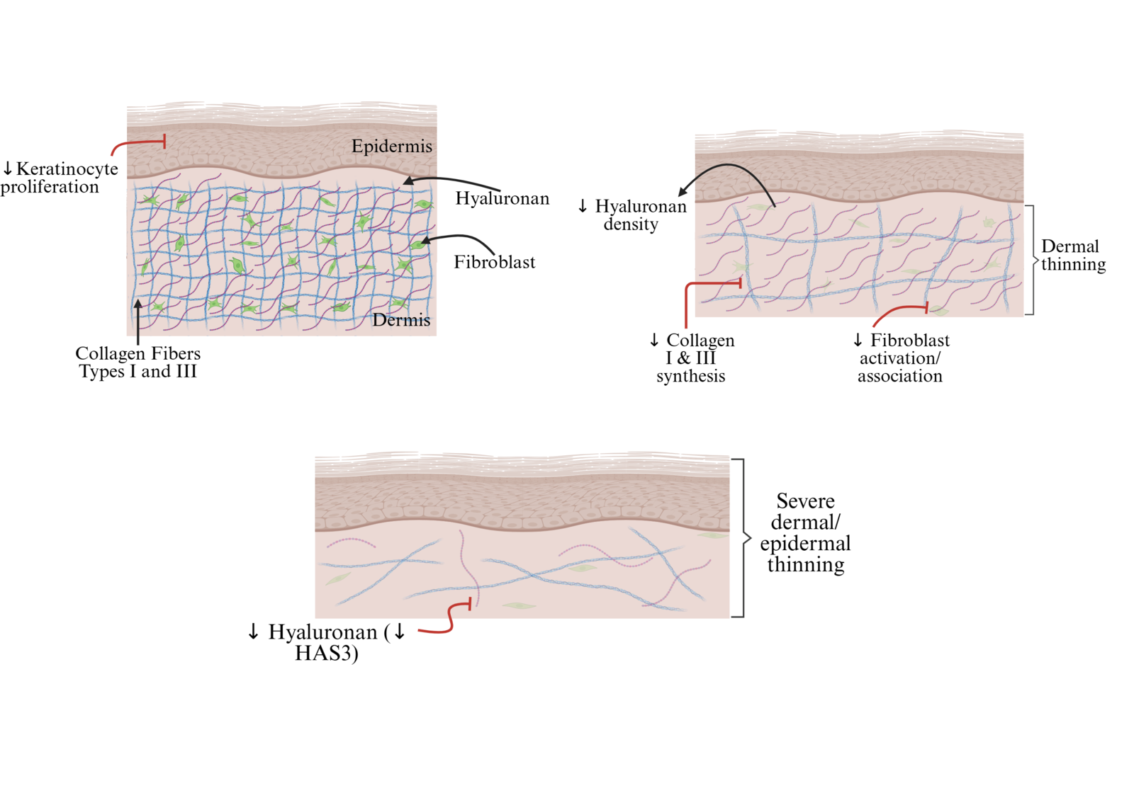 |
|---|
Prolonged use of corticosteroids can also impair the skin's immune response to infections (Brannon, 2025). While these topical steroids’ mechanisms of action inhibit a broad range of immune responses, which supports their effect against inflammatory disorders such as atopic dermatitis, this also increases infection risk when use persists, as essential immune reactions are also inhibited (Chatham, 2025). As seen in Figure 3, steroids can also cause permanent stretch marks, or striae, on the skin (Brannon, 2025). This is due to cross-linking of immature collagen in the dermis, which results in irreversible intradermal tears (Abraham & Roga, 2014). Further risks with topical steroid use include steroid-induced telangiectasia (small, visibly dilated blood vessels), steroid acne, steroid rosacea, perioral dermatitis (red rash around the mouth), purpura (increased bruising), stellate pseudoscars (white, star shaped lesions), ulcerations, aggravation of cutaneous infections, delayed wound healing, contact sensitization to topical steroids, eyelid dermatitis, tachyphylaxis (decreasing response to the drug), trichostasis spinulosa, and post-peel erythema syndrome, which are also shown in Figure 3 (Abraham & Roga, 2014). All of these potential risks are intrinsic, as the traits that cause them are also the traits that aid eczema when using corticosteroids; therefore, a non-steroid remedy is necessary.
| Figure 3: Schematic representation of genetic constructs for proposed sensor bacteria. | 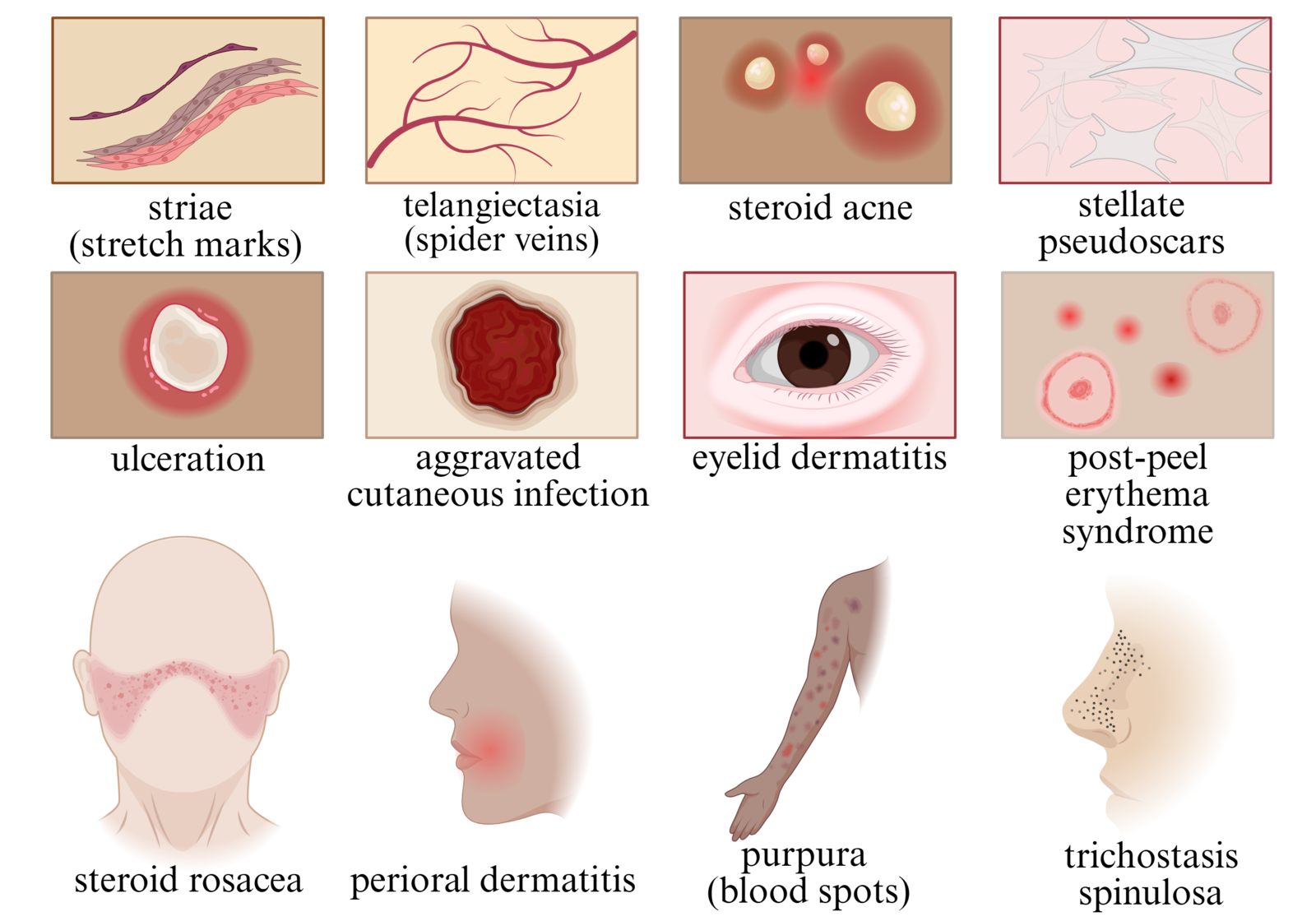 |
|---|
The biosynthetic machinery for PIA synthesis is encoded in the ica locus, which uses the icaA gene as a critical component of its composition. This makes it a critical target for aiding staphylococcus biofilms (Mack et al., 2000). However, the biosynthetic icaADBC operon mediates the production of PIA in the ica locus as it is contains regulatory genes, meaning expression is mediated (Nguyen et al., 2020). This operon consists of four different genes, the icaA, icaD, icaB, and icaC, which are arranged in the operon as represented in Figure 7. The icaADBC operon is controlled by the icaR gene, which is encoded and binds to two dimers in the icaADBC promoter regions, repressing transcription. To further break down the operon's characteristics, the icaA and icaD genes encode two membrane proteins that synthesize a growing poly-GlcNAc chain (a group of exopolysaccharides bonded together to support biofilm production) from activated precursor GlcNAc units (a donor sugar that is essential for modifying proteins) into oligomeric chains (Gerke et al., 1998; Mack et al., 1996; Ramos et al., 2019). More specifically, IcaA is an N-acetylglucosaminyltransferase, which means it is an enzyme that is good at branching complex glycans, which synthesizes PIA oligomers from UDP-N-acetylglucosamine (an essential nucleotide sugar) from activated precursor GlcNAc units (Lau et al., 2007; Paneque et al., 2023; Uridine Diphosphate, n.d.). IcaC facilitates elongation and translocation of the growing polysaccharide across the membrane, while IcaB modifies the polymer through partial deacetylation, increasing its positive charge and enhancing its adhesive properties (Vuong et al., 2004; Heilmann et al., 1996). These processes are vital for the surface attachment and functionality of PIA.
| Figure 7: The genetic encoding and biosynthesis of PIA. | 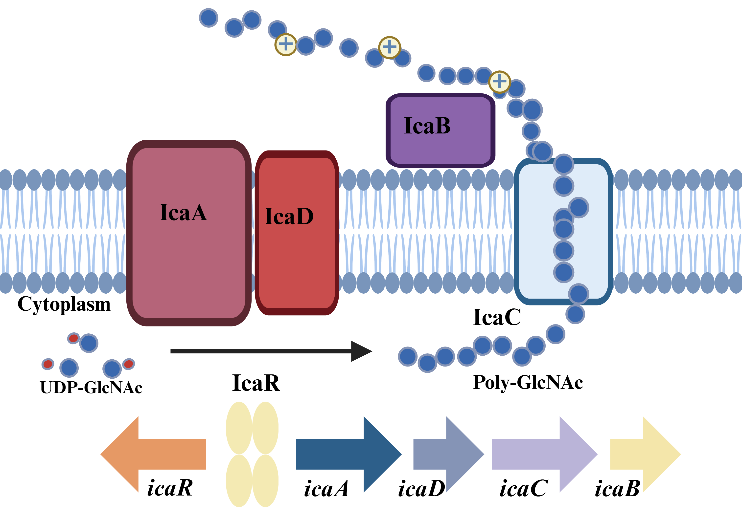 |
|---|
In order to increase the nutrients and lipid retention through the icaADBC operon, we need to incorporate an additional copy of this operon onto a plasmid. We plan to use the pTXicaADBC plasmid because of its compatibility with S. epidermidis and the existing icaADBC operon. This plasmid already contains the entire icaADBC operon, whereas other plasmids, such as pTXicaADB, pTXicaBC, and pTXicaADC, which we encountered in our research, only contain two or three out of the four genes needed to have the fully functional operon (Gerke et al., 1998). The pTXicaADBC plasmid is proven to work in S. epidermidis because the phenotype WW06 (a arlS gene deletion mutant of SE1457 [a strain of S. epidermidis]). Previous studies have demonstrated that the plasmid complements WW06 and research successfully continues after testing with the plasmid (Wu et al., 2012). The pTXicaADBC was cloned into pTX15 (a staphylococcal expression vector designed for high-level, xylose-inducible gene expression), which shows that the backbone of the pTXicaADBC is not only compatible but complementary to the Staphylococcus genus(Gerke et al., 1998). This confirms it will be able to enter, replicate, and persist in S. epidermidis. Naturally, the native plasmid is xylose inducible, meaning it only expresses icaADBC when xylose is present.
As seen in Figure 8, we developed a device to maximize the plasmid's expression of the operon that is inserted into the plasmid and will be initiated by a constitutive promoter. This way, PIA synthesis will be constantly overexpressed, rather than conditionally. However with this overexpression comes the concern of excessive biofilm formulation, which we would control by using a weaker constitutive promoter and adjusting the ribosome binding site’s strength. Skin with AD creates a suboptimal environment for S. epidermidis, so the constitutive promoter will guarantee PIA will make a significant impact when the bacteria is present. The ribosomal binding site (RBS) and coding sequence (CDS) seen in Figure 8 will ensure efficient and accurate mRNA translation that will allow for proper protein expression. This transcription will be ended by the double terminator displayed in Figure 8 to ensure it comes to a complete stop (Assembly: RBS-CDS, n.d.).
| Figure 8: | 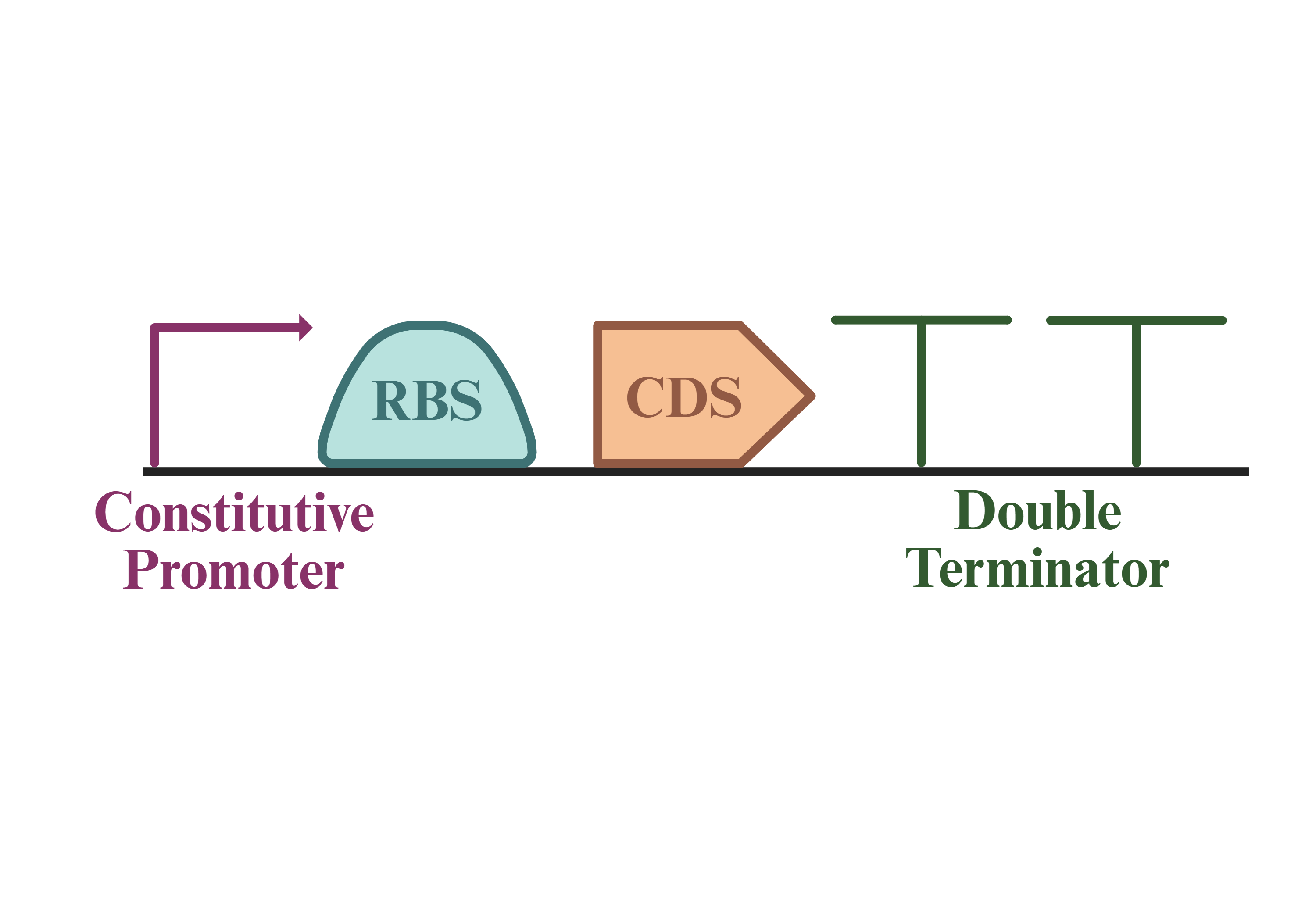 |
|---|
Safety
General Safety
S. epidermidis is generally considered a safe organism to engineer, however, general safety practices should always be followed when handling it in a lab. The bacteria is classified as Biosafety level 1 (BSL-1) as it is not associated with any disease in healthy adults (S. epidermidis, 2023). However, despite its low pathogenic risk, immunocompromised individuals are more susceptible to develop an opportunistic infection. These infections are often rooted in contamination of clinical samples or wrongful laboratory handling. To minimize this risk, we are prioritizing appropriate personal protective equipment (PPE) such as lab coats, goggles, gloves, and a clean workspace. On top of this, certain laboratory devices such as high energy-creating activities (centrifugation, sonication, high pressure systems, vortexing, tube cap popping) and splash/droplet creating-activities (shaking incubators, liquid culturing, mechanical pipetting) should be minimized or avoided as they create safety hazards and possible contamination of our bacteria (Staphylococcus Epidermidis, 2023). As a result, our procedure will avoid these activities, prioritize proper lab wear, and prevent contamination.
Government Restrictions
The production of any topical treatment faces problem areas that are addressed by the U.S. Food and Drug Administration (FDA) such as potency, active ingredient uniformity, physical characteristics, microbial purity and chemical purity. In addition to these drug pre-approval tests and requirements, our product, because it is a topical treatment, requires another set of drug pre-approval tests. In the transdermal drug pre-approval tests, uniformity and particle size are significant aspects of the product that require analysis. Viscosity, or the measure of a fluid’s resistance to flow, also needs to be tested, since it affects the absorption of the product. These parameters ensure appropriate investigational and developmental efforts of creating the topical cream product described in our project (Topical Drug, 2014). Additional consideration will play into the categorization of our product, and therefore the regulations that come with it, considering its use of a live, engineered bacteria.
Biofilm
While our project relies on PIA’s production of a biofilm, this comes with risks. During the adhesion phase of biofilm formation – which is depicted in Figure 5 – there is a risk of harmful bacteria joining the microcolony intended to only include the engineered S. epidermidis (What Is a Biofilm, 2025). In fact, the involvement of PIA in S. epidermidis biofilm formation both strengthens the matrix and creates an environment with varying oxygen levels and nutrient availability. This encourages a diverse array of bacteria to join the colony (S. epidermidis, 2025). Once one of these microorganisms joins this colony, it will continue to spread with it, and becomes highly resistant to antibiotic treatment given the strong three-dimensional matrix structure formed by EPS (What Is a Biofilm, 2025). However, S. epidermidis’ antibiotic resistance can function in favor of our project's safety. Given S. epidermidis’ slow division rate, it is particularly resistant to the antibiotic family known as beta-lactams which target fast dividing bacteria (Staphylococcus Epidermidis, 2025). We will incorporate a topical form of one of these antibiotics – specifically from the Monobactum family which mainly targets Gram-negative bacteria (which S. epidermidis is not) – into the cream (Memon, n.d.). This will not prevent the ability for S. epidermidis to form a biofilm, but we will inhibit potentially harmful bacteria from joining it. This of course comes with implications of its own, including the long term impacts of applying antibiotics to the skin.
Additionally, biofilms produced by S. epidermidis counteract some such harmful biofilms on their own. For example, they inhibit the formation of an S. aureus biofilm, which is one of the most commonly eczema associated pathogenic infections. On its own, S. epidermidis biofilms also support skin homeostasis and prevent further microcolony formation. Furthermore, the healthy barrier our engineered S. epidermidis treatment aims to restore on damaged AD skin also limits harmful biofilms for forming, as an intact skin barrier is less easily infiltrated by such pathogens. Regular skin care practices such as face washing and moisturizing help maintain barrier health beyond eczema repair, so doing so is crucial. Before applying the cream, individuals should ensure their face is clean (What Is a Biofilm, 2025).
Topical Application
Using S. epidermidis in a cream also comes with some risks. Although it normally helps maintain skin homeostasis and protect against pathogens. Because the cream directly delivers the bacteria onto the skin, it is important to ensure that the formulation controls how much bacteria and antibiotic is applied. In addition, the cream must be designed carefully to maintain the bacteria’s stability without promoting excessive growth after application. Repeated use of this treatment (especially considering its inclusion of an antibiotic) may also affect the balance of the natural skin microbiome, making it crucial to monitor long-term effects (Severn & Horswill, 2022).
Discussions
Possible Impacts
S. epidermidis is a common symbiont bacterium that has the ability to cause nosocomial infections once inside the human host. While this poses a harmful risk, it is a possible impact of our project. We recognize that the transmission risk could be serious to vulnerable patients however, by ensuring that proper safety measures are taken, this risk carries less consequences, making it more accessible to all patients. Because of this risk, we will ensure that aseptic technique is applied when dealing with the bacteria.
Marketing
In addition to making our product safe for use, we will face the challenge of gaining the public’s trust in our product. As the cream does contain live bacteria, something that may not necessarily be a positive aspect to the public’s perception, this will have to be addressed. We plan to combat this marketability issue by publishing clinical trial data and gaining the FDA approval and regulatory classification.
The first step of introducing a medical product to the public in the United States is to determine how the FDA has classified the device. The device can be categorized into three classes (I, II, III) based on the risk factor of the medical product. The higher the class, more risk exists and the requirement for regulatory controls increases (How to Study, 2026).
Because our product is not a life support device and rather a treatment to assist in naturally managing eczema symptoms, the topical cream our project aims to create would be classified as class I or class II. However, because a live engineered bacteria may not fit the same criteria as a simple device, we will re-evaluate and investigate after engineering. Products classified as class I or class II require a premarket notification of a 510k, where a sponsor will demonstrate the product is “substantially equivalent” to a predicate device in terms of intended use (How to Study, 2026).
Other Applications
The fundamental framework of our cream could be used to treat other conditions that impact the skin barrier, as its primary aim is revitalizing it. While slight alterations may be necessary to target traits specific to such conditions, the engineered system could be widely applied. Some conditions it might benefit include psoriasis, acne vulgaris, or rosacea.
Next steps
The next steps of our project will focus on turning our conceptual design into a safe, effective, and applicable product. A key priority is developing the actual topical cream, including identifying the most important components that allow it to be stable, easy to apply, and capable of delivering S. epidermidis effectively to the skin. This includes determining how to include an antibiotic component to prevent harmful biofilms while maintaining the beneficial PIA-producing system. This antibiotic inclusion is to be considered a safeguard against harmful biofilms, rather than an essential part of our project.
At the genetic level, we must confirm that our selected plasmid, pTXicaADBC, is the most optimal and compatible option, ensuring it functions efficiently with the existing icaADBC operon and meets all safety requirements for potential human use. This will involve testing plasmid compatibility, confirming that the icaADBC operon successfully increases PIA production, and identifying safety criteria for laboratory and future clinical settings.
We will also plan how we will bring our system into the lab. This includes designing the experiment, deciding on an appropriate number of trials, and making sure our results are consistent and reproducible. We also must calculate the optimal level of PIA production, since we know it supports biofilm formation but still need to determine how effective it is without causing unintended effects. Increasing moisture retention and reducing S. aureus growth all while not disrupting the normal skin microbiome balance is the criteria for recognizing the optimal level of PIA production. As mentioned in the system sections, we will also test to make sure that increased PIA synthesis truly strengthens the biofilm, as we hypothesized it would. Additional testing will help determine whether the bacteria can remain viable in different cream or gel formulations over time, because the success of the system depends on delivering live bacteria to the skin. In addition, comparing different strains of S. epidermidis will help us identify the safest and most effective option. Beyond the experimental side, we also need to consider how this would be manufactured in practice, making sure the cream can be produced safely, consistently, and in line with government regulations. This includes developing a base formula that meets those standards and ensuring it could realistically be approved for distribution. Overall, these steps help move our project from a strong concept toward something that could be safely and effectively used in the real world.
Author contributions
DP conceived the project concept and contributed to the Parts level. LL corresponded directly with our mentor, Ms. Jackie Thompson, and contributed to the Parts level. AK created and designed the majority of our figures and contributed to the Devices Level. AL contributed to the Systems level.
Acknowledgements
We would like to express our gratitude to Western Reserve Academy for providing us with the opportunity to study synthetic biology and move forward with our project design. We extend our appreciation to Ms. Vivien Marmerstein and Ms. Jackie Thompson (Takeda Pharmaceuticals) for their guidance throughout our research. Their mentorship has significantly contributed to the progress of our project and deepened our comprehension of this topic. Lastly, we would like to thank the BioTreks program for the opportunity to pursue our research. References
References
Abraham, A., & Roga, G. (2014). Topical steroid-damaged skin. National Library of Medicine. https://pmc.ncbi.nlm.nih.gov/articles/PMC4171912/
Antihistamine drugs. (2023, April 17). Drugs. Retrieved December 3, 2025, from https://www.drugs.com/drug-class/antihistamines.html
Assembly: RBS-CDS issues. (n.d.). Registry of Standard Biological Parts. Retrieved April 9, 2026, from https://parts.igem.org/Assembly:RBS-CDS_issues
Ballard, A. (2022, October 28). Staph & Eczema: A Skin-Duo You Need to Know About. National Eczema Association. Retrieved November 28, 2022, from https://nationaleczema.org/blog/staph-eczema-a-skin-duo-you-need-to-know-about/
Beckman, L., Hagquist, C., Svensson, Å., Langan, S. M., & Von Kobyletzki, L. (2023, June 5). Relationship between eczema and self-reported difficulties keeping up with school education: A cross-sectional study. National Library of Medicine. Retrieved October 29, 2025, from https://pmc.ncbi.nlm.nih.gov/articles/PMC10259463/
Bogaard, E. H. van D., Elias, P. M., & Goleva, E. (2024, August 26). Targeting skin barrier function in atopic dermatitis. National Library of Medicine. Retrieved December 4, 2025, from https://pmc.ncbi.nlm.nih.gov/articles/PMC11346348/
Brannon, H. L. (2025, October 10). Topical steroid side effects. Verywell Health. Retrieved October 24, 2025, from https://www.verywellhealth.com/steroids-topical-steroid-side-effects-1068831
Brind'Amour, K. (2024, September 23). Biofilms: The good, the bad & the groundbreaking. Pediatrics Nationwide. Retrieved January 13, 2026, from https://pediatricsnationwide.org/2024/09/23/biofilms-the-good-the-bad-the-groundbreaking/
Brown, M. M., & Horswill, A. R. (2020). Staphylococcus epidermidis—Skin friend or foe? PLOS Pathogens, 16(11), e1009026. https://doi.org/10.1371/journal.ppat.1009026
Büttner, H., Mack, D., & Rohde, H. (2015). Structural basis of staphylococcus epidermidis biofilm formation: Mechanisms and molecular interactions. Frontiers in Cellular and Infection Microbiology, 5. https://doi.org/10.3389/fcimb.2015.00014
Cau, L., Williams, M. R., Butcher, A. M., Nakatsuji, T., Kavanaugh, J. S., Chenge, J. Y., Shafiq, F., Higbee, K., Hata, T. R., Horswill, A. R., & Gallo, R. L. (2021, April 21). Staphylococcus epidermidis protease EcpA can be a deleterious component of the skin microbiome in atopic dermatitis. National Library of Medicine. Retrieved October 30, 2025, from https://pmc.ncbi.nlm.nih.gov/articles/PMC8058862/
Chatham, W. W. (2025). Glucocorticoid effects on the immune system. UpToDate. https://www.uptodate.com/contents/glucocorticoid-effects-on-the-immune-system
Cleveland Clinic. (2022). Eczema (atopic dermatitis): Symptoms, causes & treatment. In Cleveland Clinic. Cleveland Clinic. October25,2022 (Reprinted from Cleveland Clinic)
Costerton, J. W., Stewart, P. S., & Greenberg, E. P. (1999). Bacterial biofilms: A common cause of persistent infections. Science, 284(5418), 1318-1322. https://doi.org/10.1126/science.284.5418.1318
Cristol, H. (2024, March 12). Why eczema treatments fail. WebMD. Retrieved October 24, 2025, from https://www.webmd.com/skin-problems-and-treatments/eczema/eczema-treatments-fail
Dermis. (2022, February 10). Cleveland Clinic. https://my.clevelandclinic.org/health/body/22357-dermis
Eczema statistics. (n.d.). National Eczema Association. Retrieved October 28, 2025, from https://nationaleczema.org/eczema-facts/
Fey, P. D., & Olson, M. E. (2010). Current concepts in biofilm formation of Staphylococcus Epidermidis. Future Microbiology, 5(6), 917-933. https://doi.org/10.2217/fmb.10.56
Flemming, H.-C., & Wingender, J. (2010). The biofilm matrix. Nature Reviews Microbiology, 8(9), 623-633. https://doi.org/10.1038/nrmicro2415
Gallagher, M. (2023). The origin of eczema and the centuries-old history behind it. National Eczema Association. https://nationaleczema.org/blog/the-origin-of-eczema-and-the-centuries-old-history-behind-it/
Gerke, C., Kraft, A., Süßmuth, R., Schweitzer, O., & Götz, F. (1998). Characterization of theN-Acetylglucosaminyltransferase activity involved in the biosynthesis of the staphylococcus epidermidisPolysaccharide intercellular adhesin. Journal of Biological Chemistry, 273(29), 18586-18593. https://doi.org/10.1074/jbc.273.29.18586
Gillette, H. (2023, December 20). Can you take antihistamines for eczema? Healthline. Retrieved October 30, 2025, from https://www.healthline.com/health/eczema/antihistamines-for-eczema
Heilmann, C., Schweitzer, O., Gerke, C., Vanittanakom, N., Mack, D., & Götz, F. (1996). Molecular basis of intercellular adhesion in the biofilm-forming Staphylococcus epidermidis. Molecular Microbiology, 20(5), 1083-1091. https://doi.org/10.1111/j.1365-2958.1996.tb02548.x
How to study and market your device. (2026, January 30). U.S. Food and Drug Administration. Retrieved April 15, 2026, from https://www.fda.gov/medical-devices/device-advice-comprehensive-regulatory-assistance/how-study-and-market-your-device#:~:text=Medical%20devices%20marketed%20in%20the,assurance%20of%20safety%20and%20effectiveness.
Joshi, A. A., Vocanson, M., Nicolas, J.-F., Wolf, P., & Patra, V. (2023). Microbial derived antimicrobial peptides as potential therapeutics in atopic dermatitis. Frontiers in Immunology, 14. https://doi.org/10.3389/fimmu.2023.1125635
Landemaine, L., Da Costa, G., Fissier, E., Francis, C., Morand, S., Verbeke, J., Michel, M.-L., Briandet, R., Sokol, H., Gueniche, A., Bernard, D., Chatel, J.-M., Aguilar, L., Langella, P., Clavaud, C., & Richard, M. L. (2023). Staphylococcus epidermidis isolates from atopic or healthy skin have opposite effect on skin cells: Potential implication of the AHR pathway modulation. Frontiers in Immunology, 14. https://doi.org/10.3389/fimmu.2023.1098160
Lau, K. S., Partridge, E. A., Grigorian, A., Silvescu, C. I., Reinhold, V. N., Demetriou, M., & Dennis, J. W. (2007). Complex n-glycan number and degree of branching cooperate to regulate cell proliferation and differentiation. Cell, 129(1), 123-134. https://doi.org/10.1016/j.cell.2007.01.049
Mack, D., Fischer, W., Krokotsch, A., Leopold, K., Hartmann, R., Egge, H., & Laufs, R. (1996). The intercellular adhesin involved in biofilm accumulation of Staphylococcus epidermidis is a linear beta-1,6-linked glucosaminoglycan: Purification and structural analysis. Journal of Bacteriology, 178(1), 175-183. https://doi.org/10.1128/jb.178.1.175-183.1996
Mack, D., Rohde, H., Dobinsky, S., Riedewald, J., Nedelmann, M., Knobloch, J. K.-M., Elsner, H.-A., & Feucht, H. H. (2000). Identification of three essential regulatory gene loci governing expression of staphylococcus epidermidis polysaccharide intercellular adhesin and biofilm formation. Infection and Immunity, 68(7), 3799-3807. https://doi.org/10.1128/iai.68.7.3799-3807.2000
Memon, N. (n.d.). How do monobactams work? RxList. https://www.rxlist.com/how_do_monobactams_work/drug-class.htm
Nemeth, V., Syed, H. A., & Evans, J. (2024, March 1). Eczema. National Library of Medicine. Retrieved October 28, 2025, from https://www.ncbi.nlm.nih.gov/books/NBK538209/
Nguyen, H. T., Nguyen, T. H., & Otto, M. (2020). The staphylococcal exopolysaccharide PIA – biosynthesis and role in biofilm formation, colonization, and infection. Computational and Structural Biotechnology Journal, 18, 3324-3334. https://doi.org/10.1016/j.csbj.2020.10.027
Paneque, A., Fortus, H., Zheng, J., Werlen, G., & Jacinto, E. (2023). The hexosamine biosynthesis pathway: Regulation and function. Genes, 14(4), 933. https://doi.org/10.3390/genes14040933
Ramos, Y., Rocha, J., Hael, A. L., van Gestel, J., Vlamakis, H., Cywes-Bentley, C., Cubillos-Ruiz, J. R., Pier, G. B., Gilmore, M. S., Kolter, R., & Morales, D. K. (2019). PolyGlcNAc-containing exopolymers enable surface penetration by non-motile enterococcus faecalis. PLOS Pathogens, 15(2), e1007571. https://doi.org/10.1371/journal.ppat.1007571
Sawyer, T. (2023, June 27). What is the stratum corneum? Healthline. Retrieved December 4, 2025, from https://www.healthline.com/health/stratum-corneum
Severn, M. M., & Horswill, A. R. (2022, August 30). Staphylococcus epidermidis and its dual lifestyle in skin health and infection. Nature Reviews Microbiology. https://pmc.ncbi.nlm.nih.gov/articles/PMC9903335/
Staphylococcus epidermidis biofilm: Formation & significance. (2025, July 22). Biology Insights. https://biologyinsights.com/staphylococcus-epidermidis-biofilm-formation-significance/
Tian, J., Zhang, D., Yang, Y., Huang, Y., Wang, L., Yao, X., & Lu, Q. (2023, December 20). Global epidemiology of atopic dermatitis: a comprehensive systematic analysis and modelling study. National Library of Medicine. https://pubmed.ncbi.nlm.nih.gov/37705227/
Topical drug products (7/94). (2014, November 13). U.S. Food and Drug Administration. Retrieved April 15, 2026, from https://www.fda.gov/inspections-compliance-enforcement-and-criminal-investigations/inspection-guides/topical-drug-products-794
Uridine diphosphate N acetylglucosamine in subject area: Medicine and dentistry. (n.d.). Science Direct. Retrieved April 9, 2026, from https://www.sciencedirect.com/topics/medicine-and-dentistry/uridine-diphosphate-n-acetylglucosamine#:~:text=In%20subject%20area:%20Medicine%20and,How%20useful%20is%20this%20definition
Vuong, C., Voyich, J. M., Fischer, E. R., Braughton, K. R., Whitney, A. R., DeLeo, F. R., & Otto, M. (2004). Polysaccharide intercellular adhesin (PIA) protects staphylococcus epidermidis against major components of the human innate immune system. Cellular Microbiology, 6(3), 269-275. https://doi.org/10.1046/j.1462-5822.2004.00367.x
What is a biofilm on skin and how do you manage it? (2025, July 27). Biology Insights. https://biologyinsights.com/what-is-a-biofilm-on-skin-and-how-do-you-manage-it/
Wu, Y., Wang, J., Xu, T., Liu, J., Yu, W., Lou, Q., Zhu, T., He, N., Ben, H., Hu, J., Götz, F., & Qu, D. (2012). The two-component signal transduction system arlrs regulates staphylococcus epidermidis biofilm formation in an ica-Dependent manner. PLoS ONE, 7(7), e40041. https://doi.org/10.1371/journal.pone.0040041