
Anna Barger, Madison Glass, Collin Si, and Lauren Smeltzer, Western Reserve Academy, Hudson, Ohio, United States of America
Reviewed on 9 May 2026; Accepted on 22 June 2026; Published on 26 October 2026
With help from the 2026 BioTreks Production Team.
A primary risk associated with surgery comes not from the operation itself, but the risk of infection afterward. The sutures left in place give pathogens easy access to the wound, and they often form hard-to-remove shells around themselves known as biofilms. To combat the risk of infection, most sutures are coated in anti-microbial agents such as Triclosan. While these agents may reduce pathogenic colonization for a short time, they struggle to act against the biofilms often formed by the bacteria. Beyond the financial burden placed on healthcare systems, surgical site infections (SSIs) negatively impact patients' quality of life: extending hospital stays, delaying recovery time, and increasing morbidity rates. Biofilms are not entirely impervious to attack. Bacteriophages (phage) use an enzyme known as depolymerase to kill bacteria by breaking down the biofilms and the capsules that protect them. Our project proposes transforming the genetic code for a phage-derived depolymerase, DpoMK34, into Escherichia coli for protein production. The protein would then be purified using histidine tagging before being used to coat the sutures, thereby preventing biofilm formation. By targeting the mechanisms underlying biofilm formation, rather than relying on traditional antibiotics, enzyme-based antimicrobial sutures can reduce SSIs while minimizing cost and health risks.
Keywords: Biofilm, surgical site infection, depolymerase, Escherichia coli
Authors are listed in alphabetical order. Cisil Köksaldi mentored the group. Please direct all correspondence to mailto:marmersteinv@wra.net.





Background
Postoperative infections are estimated to affect 500,000 annually within the United States alone (Perencevich et al., 2003). Although prevalence has slightly decreased over time, it is not statistically significant (see Figure 1), and surgical site infections (SSIs) remain far too common (Seidelman et al., 2022). These infections are often severe and may even necessitate wound debridement (the removal of dead or damaged tissue), which significantly slows the healing process (Steed, 2004). It is imperative to find a safer alternative to prevent and eliminate SSIs.
| Figure 1: SSI rates have plateaued in the last several years. The Bar chart shows the percentage of surgeries that resulted in an infection from 2013 to 2018. While surgical site infections in the United States are relatively rare, it is important to note that these fractions of a percent represent thousands of infections. Though there have been recent decreases in their prevalence, those decreases are not statistically significant and represent a plateau in ability to combat these infections (Seidelman et al., 2022). | 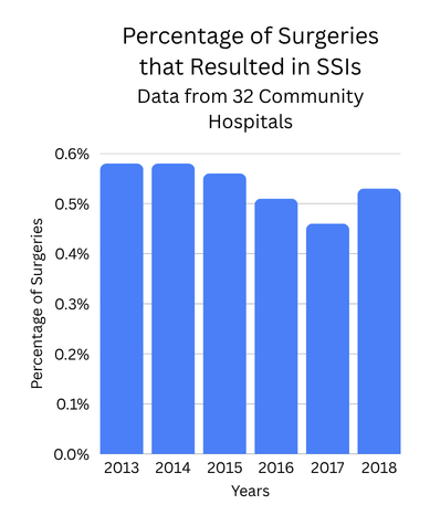 |
|---|
SSIs are defined by the Centers for Disease Control and Prevention as wound infections occurring within 30 days of an operation, or up to one year if an implant is left in place (National Healthcare Safety Network Center for Disease Control, 2026). When treating SSIs, healthcare professionals may struggle because the infection is commonly characterized by biofilm formation (Hrynyshyn et al., 2022). SSIs represent a significant challenge in healthcare, accounting for up to 15% of all hospital-acquired infections (Reichman & Greenberg, 2009). The consequences of an SSI are severe, including prolonged hospitalization and drastically escalated expenses. On average, SSIs extend hospital stay by over 9 days and increase cost by about $20,842 per admission (Reichman & Greenberg, 2009). Importantly, an infection occurring within the first 30 days post-surgery can be associated with significant long-term harm, correlating with over 3-fold higher risk of subsequent infection and nearly a 2-fold increased risk of mortality within the following year (O’Brien et al., 2020).
A major contributing factor to the persistence of SSIs is biofilm formation. Biofilms are large groups of microorganisms that attach to surfaces, for example, the suture in case of a surgery, and are involved in 80% of SSIs (Hrynyshyn et al., 2022). Biofilms form in several steps (see Figure 2). First step is the surface colonization, during which microorganisms attach to the surface (in the case of SSIs this is commonly the suture and wound). Attachment is reversible, so that if conditions become adverse, the bacteria can detach. Next, the microorganisms change their orientation and begin to lie flat. For example, with a vortex structure, bacteria move in concentric circles in order to create a flow of nutrients towards the inside and to expel waste towards the outside (Yazdi & Ardekani, 2012). After this, multiplication and aggregation occur, forming microcolonies. Maturation follows, during which the biofilm forms a structure similar to a mushroom or tower. Finally, dispersion occurs, and cells are released from the mature biofilm, which primes another cycle of biofilm formation (Abebe, 2020).
| Figure 2: The five steps of biofilm formation. Beginning with surface colonization, continuing with the change of orientation, then multiplication and aggregation occur, followed by maturation, and finally dispersion. Created in BioRender. (2026) https://BioRender.com/lhvz6oy | 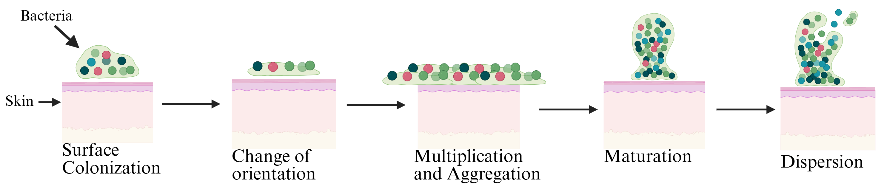 |
|---|
Biofilms are encased in extracellular polymeric substances (EPS) which include polysaccharides, proteins, lipids, nucleic acids, extracellular enzymes, and metal ions. All of these components help the biofilm obtain nutrients, provide structural support, and protect it from harsh environments (Hrynyshyn et al.). Within the biofilm, bacterial cell growth is gradual, but the cells are persistent. Biofilms also have multiple ways to resist antibiotics, including physical, physiological, and gene-related factors, further complicating infection treatment (Abebe, 2020). Biofilms can potentially lead to chronic infections as well, due to their ability to survive different conditions and adapt (Høiby et al., 2010).
In order to minimize the risk of SSI formation, the current generation of antimicrobial sutures mainly relies on antibiotic coatings. Currently, the most widespread solution involves coating sutures with triclosan, a broad-spectrum antiseptic introduced in 2002. Studies comparing these treated sutures to standard materials demonstrate effectiveness in reducing microbial colonization. For instance, a randomized clinical study involving lower third molar removal found that the Monocryl® Plus suture showed a statistically significant lower presence of microorganisms compared to silk sutures, achieving mean bacterial reductions of 83% at 72 h and 65% at 7 days post-surgery. The most pronounced antibacterial effect in this setting occurred during the first three days (Sala-Perez et al., 2016). Furthermore, a large second-order meta-analysis encompassing data from numerous studies reported a statistically significant reduction in SSI rates attributed to antimicrobial-coated sutures, presenting an aggregated relative risk of 0.68 (Suleiman et al., 2024). While coating sutures in triclosan has proven to be a fairly effective way to prevent bacterial colonization after surgery, it remains a controversial topic assailed by conflicting research claims about how effective or safe it truly is, as discussed below (Leaper et al., 2011). One of the main arguments against the drug is that if a biofilm forms around a suture, triclosan is likely to fail (Abebe, 2020).
Despite the evidence supporting initial bacterial reduction with the use of triclosan, several significant drawbacks necessitate the development of alternative solutions. A major concern includes the potential for triclosan’s widespread use to contribute to increased antibiotic resistance in microbial populations as they adapt to its near universal presence (Suleiman et al., 2024). Triclosan operates as a general antimicrobial agent, prompting the suggestion that a more targeted approach focusing on specific problematic bacteria (such as Streptomyces, Pseudomonas aeruginosa, or Enterobacteriaceae) might yield improved results. The efficacy of existing solutions is often short-term and generally shows only a limited capacity to resolve or treat established infections at the surgical site. Furthermore, some meta-analyses focusing specifically on abdominal closures found no conclusive evidence to support the routine use of antibiotic-impregnated sutures (Elsolh et al., 2017). Critically, implanted sutures serve as foreign bodies easily colonized by pathogenic bacteria to form biofilms, which are highly resistant to conventional antibiotics and represent a proven cause of persistent chronic SSIs (Kathju et al., 2014). The presence of antimicrobial agents on the suture material can also be cytotoxic to local cells, negatively affecting the natural wound healing process.
Researchers are actively investigating advanced alternatives to overcome these limitations. Nanotechnology represents one promising avenue; for instance, nanostructured silver coatings have been successfully deposited onto polyglactin 910 braided sutures using a photochemical process. Silver is valued for its broad-spectrum antimicrobial activity against Gram-positive and Gram-negative bacteria, including antibiotic-resistant strains. This silver-treated material demonstrated strong antibacterial efficiency (ranging from 83% to 94% reduction in bacterial viability) and successfully inhibited biofilm formation by S. aureus and antibiotic-resistant P. aeruginosa in laboratory testing, while proving non-hemolytic, or not damaging red blood cells, in vitro. (Paladini et al., 2025). Furthermore, enzyme-based therapeutics are being researched for combating established biofilms, with studies showing that combining a bacteriophage endolysin (LysK) and a polysaccharide depolymerase (DA7) demonstrates a synergistic ability to remove static and dynamic S. aureus biofilms in experimental models (Olsen et al., 2018). Beyond infection control, next-generation sutures may also be designed to incorporate bioactive molecules intended to regulate local cellular behavior, such as promoting cell proliferation and migration, crucial for optimal wound healing.
With the goal of trying to prevent biofilm formation, we propose coating the sutures with an enzyme called depolymerase. Depolymerases are enzymes produced by bacteriophages in order to degrade bacterial surface proteins so they can enter the cell; they are characterized by their high fidelity to targets (i.e. a depolymerase used by a phage that hunts E. coli will only function against E. coli) (Guo et al., 2023). There are numerous varieties, each specialized for the bacteriophage that uses it. This fidelity would allow for a targeted treatment of the specific bacteria that commonly attempt to colonize surgical sites without worry about affecting other non-harmful bacteria. We propose transforming a depolymerase gene into the microbe E. coli DH5α because it is a model organism with a fully sequenced genome. E. coli has also been shown to be an excellent chassis for synthetic biology projects due to its ability to amplify plasmid DNA (Chen et al., 2018). In addition to this, obtaining the bacteria would be relatively simple, as it is rated Biosafety Level 1 (Safety Data, 2023).
We would transform the genes for a depolymerase into E. coli, our selected chassis, with the aim of developing a depolymerase that will be most effective against bacteria that commonly colonize wound sites. In studies researching the effects of depolymerases on harmful skin bacteria, research was done on a common bacterium known as A. baumannii (Chen et al., 2022). This bacterium is an opportunistic pathogen that commonly affects patients in hospitals and could reasonably be the first target of the project (Howard et al., 2012). Throughout the study, the depolymerase DpoMK34 saw a significant decrease in A. baumannii populations in only the first few hours after exposure (Guo et al., 2023). The depolymerase produced by the phage vB_AbaP_PMK34, and the genes for it can be isolated and inserted into a plasmid for E. coli. Using this transformation, we would essentially turn the chassis (E. coli) into a factory. In order to purify proteins produced by the bacteria and separate them from the rest of the bacteria, a histidine tag would be used. As seen in Figure 3, histidine tagging involves coding the vector plasmid in order to attach a histidine tag to the tail end of the target protein (in this case, the depolymerase). After producing the protein, the bacteria are lysed, and the proteins inside are placed in a tube containing immobilized metal ions, such as nickel, cobalt, and copper. The histidine (specifically its imidazole ring) will be attracted to and will bind to the metal beads, while any other proteins will pass through during the wash. Then the tagged proteins are eluted (extracted using a solvent) and purified depolymerase is produced (His-tagged Proteins).
| Figure 3: Purification of depolymerase using histidine tagging. In order to successfully purify the depolymerase proteins, a code for a histidine tag is added to the plasmid. When the bacteria are lysed, the depolymerase with a histidine tag will bind to metal ions in the tube while non-tagged proteins are washed out, purifying the depolymerase. Created by BioRender. (2026) https://BioRender.com/jrxoj4a | 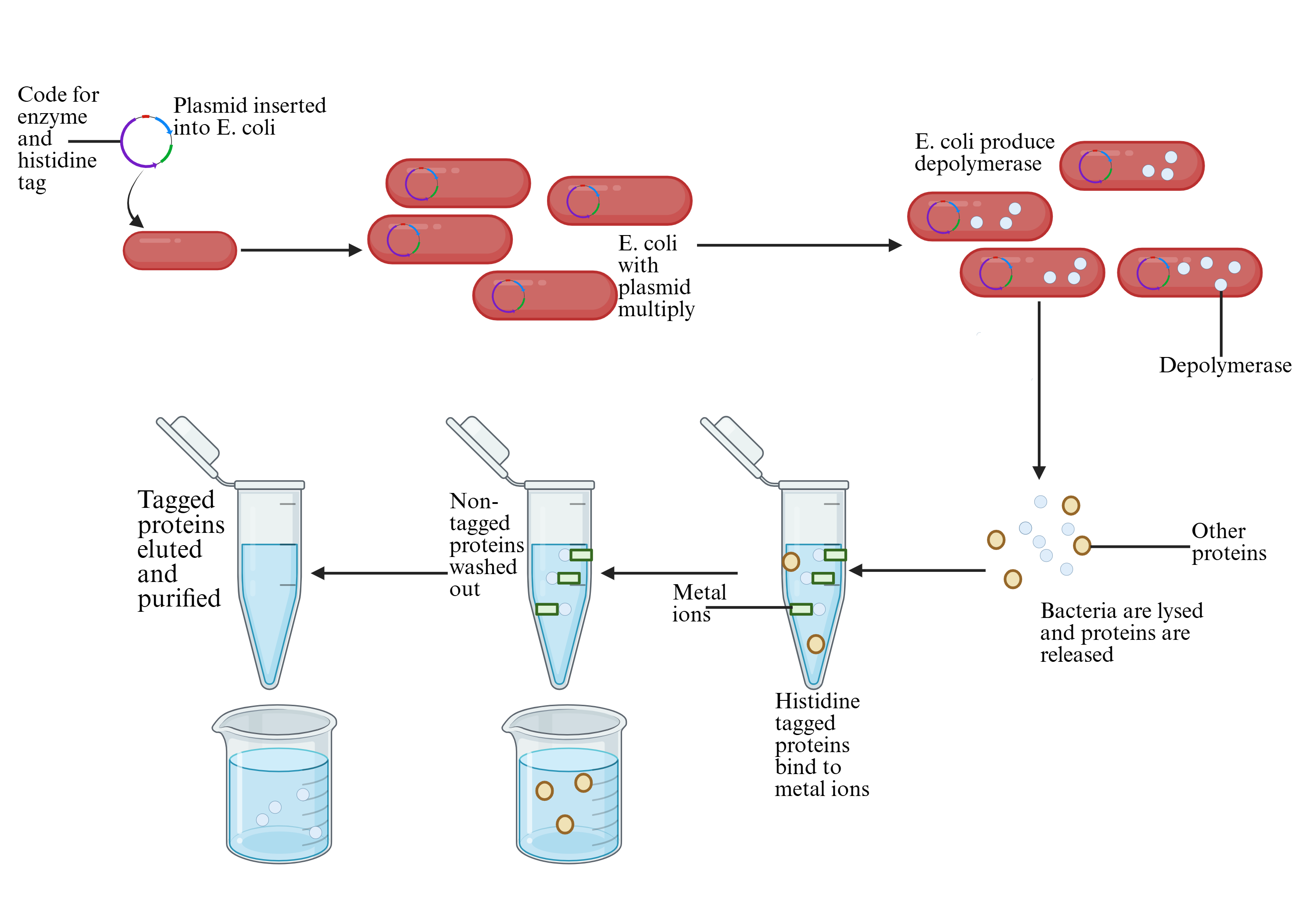 |
|---|
SSIs massively impact the healthcare industry each year, and they will continue to do so for the foreseeable future—limiting the number of SSIs reduces the factors patients need to manage after their surgery when they re-enter the world. Around 47% to 84% of SSIs occur after discharge outside of the hospital setting, demonstrating the vulnerability of patients once they leave the care of a provider (Perencevich et al., 2003). The use of alternative biofilm—preventing sutures would reduce the risks of SSIs and all the complications that come with them.
Systems level
The chassis will be modified using an inserted plasmid to express a phage-derived depolymerase. This depolymerase will then be used to coat sutures with the aim of preventing the buildup of biofilms created by pathogens taking advantage of suture presence. Because of the depolymerase’s close contact with skin, it will need to have little effect on the skin biome. The protein will also need to be easily produced by the chassis, as well as easily purifiable using histidine tagging. Most importantly, the depolymerase will need to successfully breakdown biofilms fast enough to allow antibiotics already in place on the suture to act. E. coli has been shown to be highly efficient and controlled especially in protein expression efficiency (Jiang et al., 2021). However, it is imperative that it be easily maintained and is able to reproduce at a fast rate. Using these constraints, our team determined a phage-derived depolymerase to be the ideal protein, and E. coli to be a suitable chassis.
Device level
E. coli as a chassis will give access to a straightforward approach with a fully sequenced genome and the ability to amplify plasmid DNA (Chen et al., 2018). Using the plasmids, we are then able to produce large amounts of protein compared to other bacteria. Because E. coli is non-pathogenic and has a biosafety level 1 rating, its use would be safe in lab and industrial production (Safety Data, 2023). In addition, our depolymerase will play a key role in the prevention of biofilm to break down the extracellular polymeric substance. Its effects will significantly reduce the population of skin-dwelling pathogens that take advantage of sutures. Depolymerase also directly dismantles biofilm by fully penetrating it and making the bacteria more vulnerable to treatment compared to other methods (Guo et al., 2023). When no biofilm remains on the sutures there will be nothing for the depolymerase to target due to its high specificity.
Parts level
We have designed a standard plasmid, including a promoter and an open reading frame (ORF) that work together to encourage rapid protein production. BBa_K206000, an inducible promoter, will allow the bacteria to continuously produce the depolymerase in the presence of arabinose (Schleif, 2000), as shown in Figure 4. The use of an inducible promoter with a common trigger will allow the team to control how much depolymerase is produced and when. The promoter is also native to E. coli, allowing for easy insertion. The ORF, ORF45, encodes a depolymerase known as DpoMK34 that has been isolated from phage vB_AbaP_PMK34 and can break down A. baumannii biofilms (Abdelkader et al., 2022; Guo et al., 2023). Because the device will be functioning independently of any other biological systems, a standard terminator will be used, specifically BBa_K2549053, a terminator native to E. coli to ease plasmid insertion. After treatment, the sutures coated in depolymerase are removed, and any small amounts of depolymerase remaining will not be harmful to humans (Chen et al., 2022).
| Figure 4: A diagram of the plasmid plan. The plasmid will contain parts mostly derived from E. coli to ease transition into the bacteria, with promoter, ribosome binding site, and terminator optimized to maximize protein production. The ORF will code for DpoMK34 and the specific ORF used will be ORF45 as described by Abdelkader et al. Note that the diagram does not include the code for histidine as further experimentation is needed to determine its precise location. Created in BioRender. (2026) https://BioRender.com/ji5kwsf | 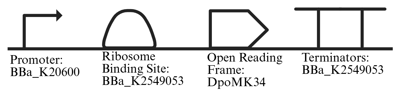 |
|---|
Safety
Our chassis (E. coli) is a biosafety level one organism, meaning the pathogen being used is not typically known to cause disease. Safety will be ensured by following standard microbial practices, and no special equipment will be required (Biosafety Level, n.d.). The E. coli will also be kept inside a controlled environment for the entire time it is producing the target protein before the bacteria are lysed and the protein isolated. A kill switch will not be used as the bacteria are kept in the lab, and the modified gene will be controlled by an inducible promoter that limits protein production to only environments where arabinose is present (Schleif, 2000), a protein that the team will deploy or remove as needed to manage protein production.
Depolymerase also poses very little risk to the environment and to patients. This safety comes from the high specificity to target bacterial capsules (Islam et al., 2024), giving confidence that they will have little to no effect on human skin. This high specificity not only limits the negative effects on the human skin cells, but also the effects on the beneficial bacteria of the skin microbiome. Each depolymerase targets specific formations of capsules or EPS, leaving non-harmful bacteria that we aren’t targeting unaffected. This specificity cannot be claimed by traditional antibiotics, which often kill all bacteria in their path regardless of impact, making a depolymerase based solution even safer than an antibiotic one (Oliveira et al., 2019). Along with its low impact on other cells, depolymerase has been shown to have very low impact on the environment and variants have even been used in bioremediation (Jiao et al., 2026; Mishra et al., 2022).
The depolymerase will coat surgical sutures, a commonly used and accepted medical device throughout the world. When testing the depolymerase’s effectiveness in preventing biofilm, it will be important to follow how other products that purport to remove biofilms are tested and regulated. The EPA (Environmental Protection Agency) requires antimicrobial products that claim to control biofilms to undergo standardized efficacy testing and submit data demonstrating effectiveness before approval. These regulations ensure that any biofilm-targeting solution is both effective and safe for public health use (United States Environmental Protection Agency, 2025). Overall, the use of depolymerase to prevent surgical site infections presents a complex issue when it comes to ensuring patient and environmental safety; however, by following standard practices and using caution in developing the product, our team believes that the benefits will greatly outweigh those minimal risks presented by this project.
Discussions
Although the project has the potential to positively impact patients in post-operative recovery, some challenges may arise. Examples of possible challenges include the following: issues with integrating histidine tagging on complex proteins, lack of research regarding depolymerase and its effects on skin, struggles with preserving depolymerase within a solution, E. coli being made to accept an ORF that comes from a phage, and the specificity of depolymerase.
Histidine tagging could be a challenge within the project as we do not yet understand how the protein will fold and how the attachment of the histidine tag will affect the folding of the protein. The histidine tag must remain on the outside of the protein, but on a nonessential exterior part in order to keep it functioning. An additional area of consideration is the lack of research regarding depolymerase and its effects on skin can lead to fear of possible harm, even though it is highly unlikely. Its interactions with other solutions on sutures, such as antibiotics, are currently unknown, requiring further testing. The exact concentration needed to best preserve and store the depolymerase is also unknown, but common storage solutions include phosphate-buffered saline, Tris-HCl, and storage buffers with 10% glycerol (Gispert & Elani, 2025). These solutions are used to stabilize the molecules (Tchiakam et al., 2025). Regardless of the solution’s makeup it will likely need to be stored at cold temperatures, with some sources noting temperatures from 4°C to −20°C and −80°C (Gispert & Elani, 2025).
Another issue arises when working with E. coli and DNA from a phage: the difficulty of E. coli accepting the ORF. The E. coli chromosome carries numerous −35 and −10 sequences with weak promoter activity due to other features used to enhance promoter activity and the absence of transcript stability (Kawano et al., 2005). E. coli has also evolved antiphage systems that stop phage replication inside the bacteria. This system has the potential to damage the host cell and destroy the phage (Nagy et al., 2026). While this project would not be using a full phage genome, it is possible that the chassis could recognize the plasmid ORF as extraneous and attack it, necessitating further experimentation with disabling the E. coli’s protective systems.
A key limitation of the DpoMK34 depolymerase is its strong specificity, which restricts its effectiveness to only a small subset of bacterial strains (Guo et al., 2023). Similar to other phage-derived depolymerases, DpoMK34 targets the polysaccharide capsule that surrounds certain bacteria, but it can only recognize and degrade one specific capsule type (Oliveira et al., 2019). This means that even closely related strains of the same species may remain unaffected if their capsule structure differs (Lu et al., 2025). As a result, this narrow host range presents a significant challenge for therapeutic use, since effective treatment would require precise identification of the infecting strain prior to application (Guo et al., 2023). In the context of this project, the specificity of DpoMK34 limits its ability to function as a broad-spectrum biofilm treatment (Oliveira et al., 2019). To overcome this, our future approaches may involve combining multiple depolymerases together to expand the coverage and improve the efficacy against diverse bacterial populations (Guo et al., 2023; Lu et al., 2025). Over 60% of SSIs are caused by only 6 types of organisms, indicating that by targeting only a few varieties of pathogens a great decrease in prevalence could be achieved (Seidelman et al., 2022). However, by targeting these specific varieties openings could be made for less common pathogens to attack wounds.
Despite potential struggles, depolymerase has been used before to successfully destroy biofilms. Chen et al. attempted to express Dpo71 in E. coli and found it to be successful in treating A. baumannii that was susceptible to the depolymerase, but it was largely ineffective against non-susceptible strains, even of the same species (2022), reflecting our apprehensions around the depolymerase’s specificity. It has also been found that for greater effectiveness, a multi-phage cocktail could be used to counter specificity issues among the phages (Guo et al., 2023). Finally, Islam et al. in an experiment specifically targeting A. baumannii using a variety of phage-derived depolymerases found them to be largely effective against the bacteria, but also discovered that phage resistance could develop with high usage (2024). Overall testing of depolymerase on common pathogenic biofilms has been limited and only done in a lab environment, but it provides an encouraging basis for the further development of depolymerase techniques like those in this project.
Next steps
Additional experimentation needs to be conducted regarding the effects of the depolymerase and our ability to manufacture and purify it. As such, in the continuation of the project, much experimentation will be needed on the purification strategy, the depolymerase’s effects on biofilms, the interactions with skin cells and antibiotics, and the storage of the solution.
First will be the insertion of the recombinant plasmid into the chassis. In order to do this, we plan to follow standard protocols for the creation of a plasmid and its insertion into the plasmid. For the creation of the plasmid, the base plasmid we plan to use will be cut open using restriction enzymes in order to create sticky ends that will allow us to easily insert the gene of interest (Creating the pARA-R, 2024).
After the plasmid is created, an important step of our project includes determining how to ensure the histidine tag is on the outside of the protein (Spriestersbach et al., 2015). In order to do so, we would need to identify how the protein folds so the histidine tag can be inserted in the correct place. . A model may be utilized in this next step to visualize the protein folding, allowing us to more precisely determine where to insert the histidine tag. The biofilm needs to avoid binding to the histidine, but still on the surface for our accessibility. Along with this, the histidine will need to be on the same plasmid, and attached to the tail of the depolymerase. Because of these requirements, it remains unconfirmed whether the ends will be exposed on the surface, or if they will ball up, pointing to another consideration we need to make when attempting to place the histidine tag on the surface.
Obviously the most important function of the system will be the depolymerase’s ability to break down biofilms and bacterial capsules. In order to determine its effectiveness, we would first need to successfully determine the composition of the A. baumannii biofilms and reconstruct them in the lab as the bacteria is biosafety level 2. Afterwards, the depolymerase could be tested on different thicknesses of the biofilm and variations in composition to determine how its specificity might affect its ability to destroy the natural variations that occur in biofilm.
A next step in our project is to test how the depolymerase enzyme affects human skin cells, so that further evaluation can be done to see how safe it is for real-world medical use. Since our application does not involve a living organism directly, the focus shifts to how the purified protein interacts with human tissue. Once the depolymerase is purified, it can be applied to cultured human skin cells to observe any harmful effects, such as cell damage, or interference with normal body processes like blood clotting. In addition, we would test the enzyme under conditions similar to the human body, including body temperature, different pH levels, and the presence of bodily fluids (depending on the area of the wound) to see if it remains stable and effective. This step also connects to how the depolymerase might work alongside antibiotics or coatings on sutures, since interactions between these components could impact treatment success. Overall, these experiments would help determine whether the depolymerase is not only effective against biofilms, but also its safety and reliability in realistic medical conditions.
As stated in our discussion section, it is unclear the best temperature and pH needed to store the depolymerase. However, some sources suggest that specific types of depolymerase do best in a pH of 6, and at 25° Celsius, but can remain functioning in a pH of 3-9 and a temperature of 25-70° Celsius. (Wu et al., 2019.). Another source suggests the stability of a protein with the use of a pH within 5-12, and temperatures of 25 to 55° Celsius, but claims a pH of 7-12 is the most efficient (Wang et al., 2019). Within both of these sources, similarities between the different pH levels and temperatures can be noted, especially regarding the functioning between a pH of 5-12, creating a commonality. This allows us to have a baseline temperature to experiment with since research has been done successfully within the temperature and pH ranges discussed. A next step would be to replicate this environment and see how our depolymerase reacts, providing data on whether or not the temperature and pH are sufficient for our depolymerase to thrive.
Author contributions
Initial concept researched and proposed by MG Biofilm formation figure created by MG, and edited by AB Biofilm statistics, protein purification, and plasmid diagram made by AB Writing of Abstract, Background, Systems, Device, Parts, Safety, Discussion, and Next steps shared equally between LS, CS, AB, and MG Editing done by LS, CS, AB, and MG Figures 2, 3, and 4 created in Biorender.
Acknowledgements
Our teacher, Ms. Marmerstein, acted as both a mentor and a part of our team. She provided us with valuable feedback and helped us to find sources that were very beneficial to our paper. She also helped us to understand the scientific terms embedded in our project. She taught us about ORF, plasmid maps and insertions, genes of interest, etc. By doing this, we were able to create more accurate figures, such as our ORF figure. Along with this, Ms. Marmerstein provided feedback on our wording and grammar to help with being concise and clear.
Our mentor, Cisil Köksaldi, helped us immensely. She allowed us to discuss our ideas with her, and provided us with her experience-based insight. She also helped us troubleshoot and come up with new, innovative thoughts. She was especially helpful with figuring out where to place our histidine tag. Another issue she helped us work through includes recreating biofilm in a BSL 1 lab, as most biofilm only contains bacteria that can be worked with in a BSL 2 lab.
We want to also give thanks to our school Western Reserve Academy for the opportunity to pursue this project through the CL Synthetic Biology 1 class. Thank you to BioTreks for making our project’s publication possible.
References
Abdelkader, K., Gutiérrez, D., Latka, A., Boeckaerts, D., Drulis-Kawa, Z., Criel, B., Gerstmans, H., Safaan, A., Khairalla, A. S., Gaber, Y., Dishisha, T., & Briers, Y. (2022). The specific capsule depolymerase of phage pmk34 sensitizes acinetobacter baumannii to serum killing. Antibiotics, 11(5), 677. https://doi.org/10.3390/antibiotics11050677
Abebe, G. M. (2020). The role of bacterial biofilm in antibiotic resistance and food contamination. International Journal of Microbiology, 2020, 1-10. https://doi.org/10.1155/2020/1705814
Biosafety level requirements. (n.d.). Administration for Strategic Preparedness and Response. Retrieved April 13, 2026, from https://aspr.hhs.gov/S3/Pages/Biosafety-Level-Requirements.aspx
Chen, J., Li, Y., Zhang, K., & Wang, H. (2018). Whole-Genome sequence of phage-resistant strain Escherichia coli DH5α. Genome Announcements, 6(10). https://doi.org/10.1128/genomea.00097-18
Chen, X., Liu, M., Zhang, P., Xu, M., Yuan, W., Bian, L., Liu, Y., Xia, J., & Leung, S. (2022). Phage-Derived depolymerase as an antibiotic adjuvant against multidrug-resistant Acinetobacter baumannii. Frontiers in Microbiology, 13. https://doi.org/10.3389/fmicb.2022.845500
Creating the pARA-R recombinant plasmid. (2024, February 9). LabXchange. Retrieved April 23, 2026, from https://www.labxchange.org/library/items/lb:LabXchange:7c2b6682:html:1
Elsolh, B., Zhang, L., & Patel, S. V. (2017). The Effect of Antibiotic-Coated Sutures on the Incidence of Surgical Site Infections in Abdominal Closures: a Meta-Analysis. J Gastrointest Surg., 5. https://doi.org/10.1007/s11605-017-3357-6
Gispert, I., & Elani, Y. (2025). Synthetic cell preservation strategies enable their storage and activation at the point of use. Chemical Communications, 61(46), 8359-8362. https://doi.org/10.1039/d5cc00826c
Guo, Z., Liu M, M., & Zhang, D. (2023). Potential of phage depolymerase for the treatment of bacterial biofilms. Virulence, 14(1). https://doi.org/10.1080/21505594.2023.2273567
His-tagged proteins – production and purification. (n.d.). ThermoFisher Scientific. Retrieved December 5, 2025, from https://www.thermofisher.com/us/en/home/life-science/protein-biology/protein-biology-learning-center/protein-biology-resource-library/pierce-protein-methods/his-tagged-proteins-production-purification.html
Howard, A., O'Donoghue, M., Feeney, A., & Sleater, R. (2012). Acinetobacter baumannii. Virulence, 3(3). https://doi.org/10.4161/viru.19700
Høiby, N., Bjarnsholt, T., Givskov, M., Molin, S., & Ciofu, O. (2010). Antibiotic resistance of bacterial biofilms. International Journal of Antimicrobial Agents, 35(4), 322-332. https://doi.org/10.1016/j.ijantimicag.2009.12.011
Hrynyshyn, A., Simões, M., & Borges, A. (2022). Biofilms in surgical site infections: Recent advances and novel prevention and eradication strategies. Antibiotics, 11(1), 69. https://doi.org/10.3390/antibiotics11010069
Islam, M. M., Mahbub, N. U., Shin, W. S., & Oh, M. H. (2024). Phage-encoded depolymerases as a strategy for combating multidrug-resistant acinetobacter baumannii. Frontiers in Cellular and Infection Microbiology, 14. https://doi.org/10.3389/fcimb.2024.1462620
Jiang, N., Ding, X., & Lu, Y. (2021). Development of a robust escherichia coli-based cell-free protein synthesis application platform. Biochemical Engineering Journal, 165, 107830. https://doi.org/10.1016/j.bej.2020.107830
Jiao, H., Al-Tohamy, R., Xiong, M., Schagerl, M., Reinthaler, T., Al-Zahrani, M., Sun, J., & Ali, S. S. (2026). Microplastic biodegradation and environmental safety: From microbial mechanisms to engineered systems and circular bio-based implementation. Ecotoxicology and Environmental Safety, 313, 120016. https://doi.org/10.1016/j.ecoenv.2026.120016
Kathju, S., Nistico, L., Tower, I., Lasko, L.-A., & Stoodley, P. (2014). Bacterial biofilms on implanted suture material are a cause of surgical site infection. Surgical Infections, 15(5), 592-600. https://doi.org/10.1089/sur.2013.016
Kawano, M., Storz, G., Rao, B. S., Rosner, J. L., & Martin, R. G. (2005). Detection of low-level promoter activity within open reading frame sequences of escherichia coli. Nucleic Acids Research, 33(19), 6268-6276. https://doi.org/10.1093/nar/gki928
Leaper, D., Assadian, O., Hubner, N.-O., McBain, A., Barbolt, T., Rothenburger, S., & Wilson, P. (2011). Antimicrobial sutures and prevention of surgical site infection: Assessment of the safety of the antiseptic triclosan. International Wound Journal, 8(6), 556-566. https://doi.org/10.1111/j.1742-481x.2011.00841.x
Lu, Y., Fang, C., Xiang, L., Yin, M., Qian, L., Yan, Y., Zhang, L., & Li, Y. (2025). Characterization and therapeutic potential of three depolymerases against k54 capsular-type klebsiella pneumoniae. Microorganisms, 13(7), 1544. https://doi.org/10.3390/microorganisms13071544
Mishra, S., Huang, Y., Li, J., Wu, X., Zhou, Z., Lei, Q., Bhatt, P., & Chen, S. (2022). Biofilm-mediated bioremediation is a powerful tool for the removal of environmental pollutants. Chemosphere, 294, 133609. https://doi.org/10.1016/j.chemosphere.2022.133609
Nagy, T. A., Gersabeck, G. W., Conte, A. N., & Whiteley, A. T. (2026). A phage protein screen identifies triggers of the bacterial innate immune system. Nature Microbiology, 11(2), 597-609. https://doi.org/10.1038/s41564-025-02239-6
National Healthcare Safety Network Center for Disease Control. (2026, January). Surgical site infection event (SSI). https://www.cdc.gov/nhsn/pdfs/pscmanual/9pscssicurrent.pdf#:~:text=The%20CDC%20healthcare%2Dassociated%20infection%20(HAI)%20prevalence%20survey,categories%20combined%20compared%20to%20the%20previous%20year2.
O'Brien, W. J., Gupta, K., & Itani, K. M. F. (2020). Association of postoperative infection with risk of long-term infection and mortality. JAMA Surgery, 155(1), 61. https://doi.org/10.1001/jamasurg.2019.4539
Oliveira, H., Mendes, A., Fraga, A. G., Ferreira, A., Pimenta, A. I., Mil-Homens, D., Fialho, A. M., Pedrosa, J., & Azeredo, J. (2019). K2 capsule depolymerase is highly stable, is refractory to resistance, and protects larvae and mice from acinetobacter baumannii sepsis. Applied and Environmental Microbiology, 85(17). https://doi.org/10.1128/aem.00934-19
Olsen, N. M. C., Thiran, E., Hasler, T., Vanzieleghem, T., Belibasakis, G. N., Mahillon, J., Loessner, M. J., & Schmelcher, M. (2018). Synergistic removal of static and dynamic staphylococcus aureus biofilms by combined treatment with a bacteriophage endolysin and a polysaccharide depolymerase. Viruses, 10(8), 438. https://doi.org/10.3390/v10080438
Paladini, F., Panico, A., Masi, A., Russo, F., Sannino, A., & Pollini, M. (2025). Silver-Treated sutures for the prevention of biofilm-associated surgical site infections. Antibiotics, 14(1), 49. https://doi.org/10.3390/antibiotics14010049
Perencevich, E. N., Sands, K. E., Cosgrove, S. E., Guadagnoli, E., Meara, E., & Platt, R. (2003). Health and economic impact of surgical site infections diagnosed after hospital discharge. Emerging Infectious Diseases, 9(2), 196-203. https://doi.org/10.3201/eid0902.020232
Reichman, D. E., & Greenberg, J. A. (2009). Reducing surgical site infections: A review. Reviews in Obstetrics and Gynecology, 212-221. https://pmc.ncbi.nlm.nih.gov/articles/PMC2812878/
Safety data sheet [Fact sheet]. (2023, December). New England Biolabs. Retrieved January 13, 2026, from https://www.neb.com/-/media/4992168dd50343dba078555fdd1520e2.pdf?srsltid=AfmBOopskWOD-aHoAT0AIzIVYVJXEzetGZKmiXiTh48-uVZU0rdUfKI_
Sala-Perez, S., Lopez-Ramirez, M., Quinteros-Borgarello, M., Valmaseda-Castellon, E., & Gay-Escoda, C. (2016). Antibacterial suture vs silk for the surgical removal of impacted lower third molars. A randomized clinical study. Medicina Oral Patología Oral Y Cirugia Bucal, 21(1), e95-e102. https://doi.org/10.4317/medoral.20721
Schleif, R. (2000). Regulation of the l-arabinose operon of escherichia coli. Trends in Genetics, 16(12), 559-565. https://doi.org/10.1016/s0168-9525(00)02153-3
Seidelman, J. L., Baker, A. W., Lewis, S. S., Advani, S. D., Smith, B., & Anderson, D. (2022). Surgical site infection trends in community hospitals from 2013 to 2018. Infection Control & Hospital Epidemiology, 44(4), 610-615. https://pmc.ncbi.nlm.nih.gov/articles/PMC10194399/
Spriestersbach, A., Kubicek, J., Schäfer, F., Block, H., & Maertens, B. (2015). Purification of his-tagged proteins. Methods in Enzymology, 1-15. https://doi.org/10.1016/bs.mie.2014.11.003
Steed, D. L. (2004). Debridement. The American Journal of Surgery, 187(5), S71-S74. https://doi.org/10.1016/s0002-9610(03)00307-6
Suleiman, A. S., Abbass, M., Hossain, M., Choudhary, P., Bhattacharya, P., & Islam, A. (2024). Impact of antibiotic-coated sutures on surgical site infections: A second-order meta-analysis. International Journal of Surgery, 1. https://doi.org/10.1097/JS9.0000000000000822
Tchiakam, S. B., Berger, S. L., Southall, J., Walden, H., & Gabrielsen, M. (2025). Stabilising proteins in solution using affordable and readily available small molecules. Biophysical Reviews, 17(4), 1093-1104. https://doi.org/10.1007/s12551-025-01341-7
United States Environmental Protection Agency. (2025, July 21). Efficacy Test Methods, Test Criteria, and Labeling Guidance for Antimicrobial Products with Claims Against Biofilm on Hard, Non-Porous Surfaces. https://www.epa.gov/pesticide-analytical-methods/efficacy-test-methods-test-criteria-and-labeling-guidance
Wang, C., Li, P., Niu, W., Yuan, X., Liu, H., Huang, Y., An, X., Fan, H., Zhangxiang, L., Mi, L., Zheng, J., Liu, Y., Tong, Y., Mi, Z., & Bai, C. (2019). Protective and therapeutic application of the depolymerase derived from a novel kn1 genotype of klebsiella pneumoniae bacteriophage in mice. Research in Microbiology, 170(3), 156-164. https://doi.org/10.1016/j.resmic.2019.01.003
Wu, Y., Wang, R., Xu, M., Liu, Y., Zhu, X., Qiu, J., Liu, Q., He, P., & Li, Q. (2019). A novel polysaccharide depolymerase encoded by the phage sh-kp152226 confers specific activity against multidrug-resistant klebsiella pneumoniae via biofilm degradation. Frontiers in Microbiology, 10. https://doi.org/10.3389/fmicb.2019.02768
Yazdi, S., & Ardekani, A. M. (2012). Bacterial aggregation and biofilm formation in a vortical flow. Biomicrofluidics, 6(4). https://doi.org/10.1063/1.4771407