
Evan Campbell, William (Liam) Curry, Jonathan Fanikos, Alexander Hall, Anthony Issa, Maddox Lund, Panagiotis (Peter) Matheos, Shubh Mehrotra, Nolan Miley, Jack O'Neil, Zachary Parlon, Gabriel Perez, Max Richards, and Zachery Skutt, Xaverian Brothers High School, Westwood, Massachusetts, United States of America
Reviewed on 9 May 2026; Accepted on 22 June 2026; Published on 26 October 2026
With help from the 2026 BioTreks Production Team.
The increasing concentration of carbon dioxide (CO2) in the atmosphere is a major contributor to climate change. We hypothesize that the production of flv3, a flavodiiron protein that dissipates excess electrons during photosynthesis, in Spirulina will accelerate photosynthesis and reduce atmospheric CO2 levels. As the expression of the introduced flv3 gene would result in the synthesis of flavodiiron proteins to balance electron flow in photosystem I, this would ultimately provide extra ATP and NADPH electron carriers necessary to drive the Calvin Cycle faster, thereby increasing photosynthetic rate. We have designed a plasmid based on the pDV044 backbone to contain the flv3 gene from Synechocystis nigrescens, a model photosynthetic cyanobacterium. We identified the gene using NCBI BLAST (National Center for Biotechnology Information, 2018), then used the Benchling program to incorporate it into our plasmid and design primers for gene amplification. To measure the rate of photosynthesis, our procedure uses changes in water pH to detect the efficiency of the light reactions during photosynthesis. This method ensures the system remains open, with gasses readily available to spirulina without risking damage or death. It uses the principle that removing CO2 from water raises the pH by reducing the formation of carbonic acid, making the water more basic and because the light reactions occur more efficiently. The hope is that, as the transformed and flv3-containing spirulina removes CO2 faster than spirulina without the plasmid, the pH will rise faster. We selected spirulina for our study because of its global abundance and its potential for use in bioengineering efforts to reduce atmospheric CO2. Future work will focus on optimizing flv3 expression and testing additional environmental conditions to directly assess whether this engineered spirulina is viable for improved photosynthetic efficiency and CO2 reduction.
Keywords: Bacterial transformation, CO2 removal, gene cloning, spirulina.
Authors are listed in alphabetical order. Jason Boock mentored the group. Please direct all correspondence to mailto:hosborne@xbhs.com.



Background
Since the Industrial Revolution, CO2 levels have climbed from roughly 280 ppm to over 420 ppm, a 50% spike that is directly driving climate change, ocean acidification, and increasingly devastating weather events worldwide (Lindsey, 2025). Most proposed solutions to mitigate high CO2 levels are expensive, energy-intensive, and difficult to scale.Cyanobacteria have been fixing carbon through photosynthesis for billions of years. Spirulina (Arthrospira platensis) is an attractive l organism to work with due to its fast growth, genetic accessibility, and well-documented safety (Figure 1). However, under high light conditions, spirulina's photosynthetic efficiency is low. This is due to the build up of NADPH that accumulates after photosystem I (Figure 2). Organisms such as S. nigrescens have developed pathways that can recycle NADPH to NADP+ to prevent its build up. This work seeks to determine if the protein, flv3, that permits NADPH to NADP+ recycling from Synechocystis will improve photosynthetic efficiency in Spirulina.
| Figure 1. Photo of spirulina culture that we grew under a microscope. | 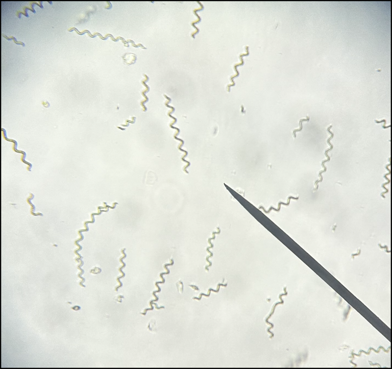 |
|---|
| Figure 2. Diagram of the light reactions with flv3 protein present. | 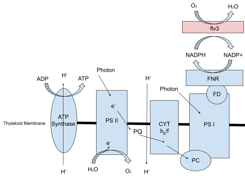 |
|---|
Our research pointed us toward Flavodiiron proteins, specifically flv3, as a potential solution. flv3 acts as a drain for electrons, reducing the harmful buildup of ROS (Reactive Oxygen Species). flv3 does this by forming water from oxygen, hydrogen, and electrons from NADPH buildup, protecting PSI (Photosystem I), and keeping photosynthesis running efficiently (Allahverdiyeva et al., 2013; Hasunuma et al., 2014). We hypothesize that boosting flv3 expression could meaningfully reduce photorespiration, thereby increasing CO2 fixation. To test this, we identified the flv3 gene from Synechocystis sp. PCC 6803 with the intent to introduce it into spirulina using the pDV044 plasmid backbone with the ultimate hope of engineering a fast-growing, low-cost biological carbon sink capable of contributing to real climate solutions.
Materials and methods
Plasmid design
We used a program called Benchling to design a unique plasmid that includes the pDV044 backbone with our gene of interest, flv3. We found the sequence of our plasmid backbone from Addgene and the sequence of flv3 from NCBI. We designed our own primers to insert the gene of interest into the backbone and put them together in the Benchling program.
| Figure 3. Map of the design plasmid with the gene of interest. | 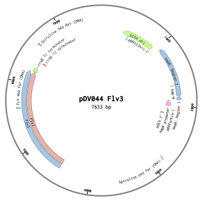 |
|---|
The gene of interest, flv3, was obtained from S. nigrescens. The flv3 gene was amplified using polymerase chain reaction (PCR) with New England Biolabs (NEB) PCR Master Mix, Ipswich, Massachusetts, and specific primers. We attempted to isolate the plasmid backbone, pDV044, from Escherichia coli using the Monarch Spin Plasmid Miniprep kit (NEB, Ipswich, Massachusetts). We then ran a plasmid digest using the restriction enzyme BsaI.
Photosynthetic rate analysis
Photosynthetic rate was measured by monitoring changes of pH, using Vernier pH probes (Vernier Science Education, Beaverton, Oregon), of the culture medium as an indirect indicator of carbon fixation (Measuring the Rate of Photosynthesis, 2024). Multiple cultures of wild-type A. platensis samples were exposed to identical environmental conditions with the exception of light presence. As a negative control, a sample was enclosed in a beaker covered with aluminum foil and kept away from light, so that the spirulina would not have access to light. The other sample was exposed to high intensity light. pH was recorded over time to measure the change due to photosynthesis.
Safety
All procedures were conducted in accordance with standard microbiological safety practices (Vanderbilt University Medical Center, n.d.). These include the use of protective equipment such as gloves and goggles, sterile tools and techniques to prevent contamination, and proper waste disposal. Both organisms used in experimentation, spirulina and synechocystis, are Bio-Safety Level 1 (BSL-1) organisms, widely recognized as safe (BacDive, n.d).
Results
After running a PCR to isolate and amplify the flv3 gene from S. nigrescens, the gel electrophoresis showed extremely faint bands far past the ladder and no other bands. Expected results were one band at 1752 bp, which would have been the gene and attached primers. The faint bands past the ladder are the primers that had no gene to attach themselves to. No other DNA was present, meaning the PCR to amplify the gene was unsuccessful.
| Figure 4. Gel electrophoresis of the attempted PCR of flv3. | 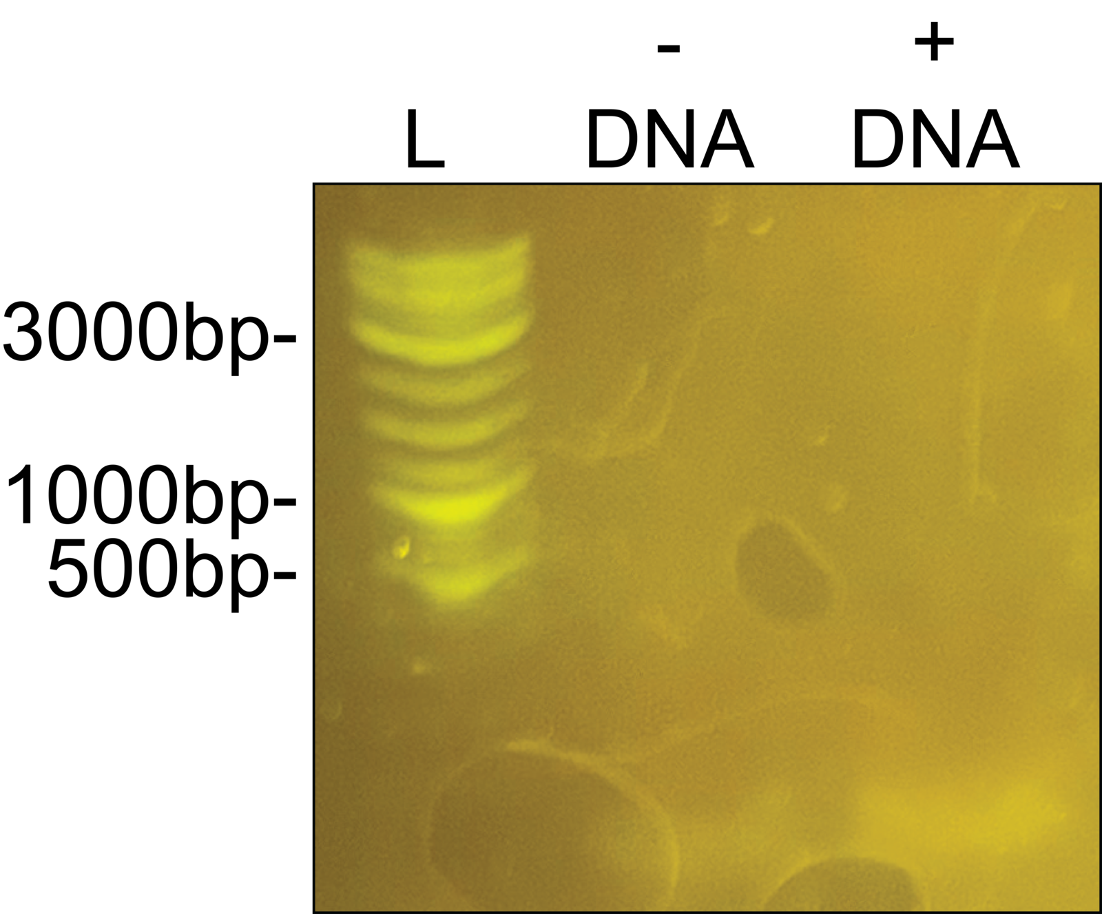 |
|---|
After attempting to isolate the pDV044 plasmid backbone from E. coli and running the digest, the gel electrophoresis showed no bands at all. The expected results were a single band at about 5910 bp, which would have been the plasmid backbone. Because no DNA was present, the plasmid isolation was unsuccessful.
| Figure 5. Gel electrophoresis of the attempted pDV044 isolation. | 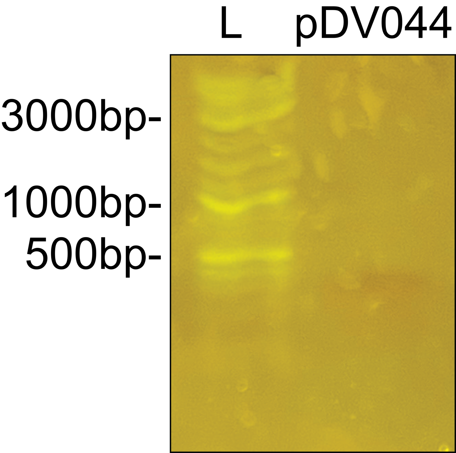 |
|---|
A pH test on a culture of wild-type A. plantensis (spirulina), was run. When photosynthesis occurs, it is expected for the pH to increase because CO2 levels decrease, and therefore carbonic acid, (CO2 dissolved in water) decreases. This causes a decrease in acidity, and an increase in pH levels.Results showed a successful proof of concept. pH in the culture of spirulina exposed to light had an increase in pH over time and the culture of spirulina in darkness had a decrease in pH over time.
| Figure 6. Graph of pH in the light and dark trials over time. | 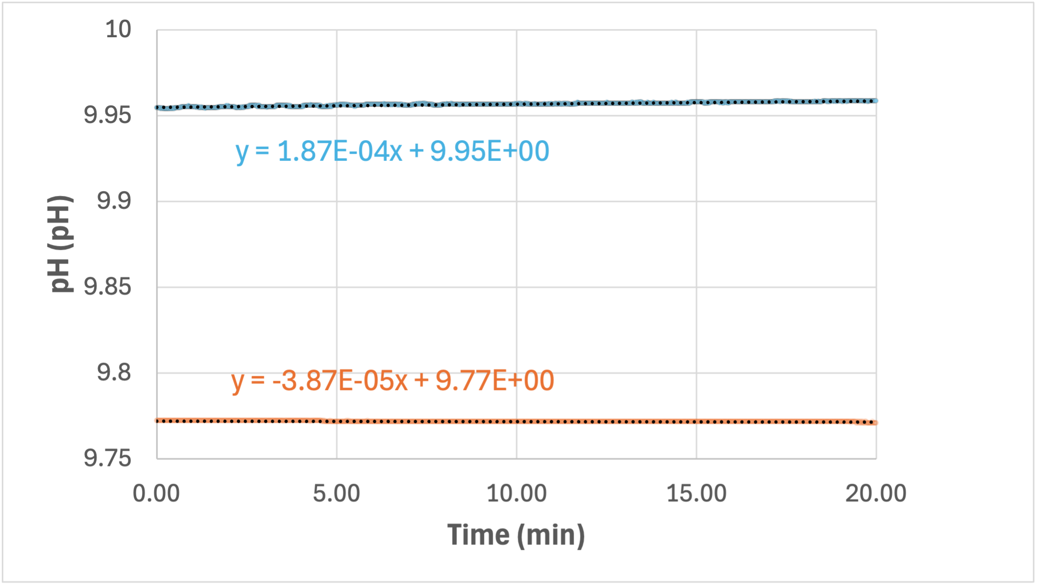 |
|---|
Discussions
The absence of any full bands on the gel electrophoresis (figure 4) illustrates a failure of our PCR. The faint bands past the ladder represented the primers, as they failed to attach to absent DNA. Because our PCR was unsuccessful, we believe the flv3 gene sequence that we found online was not the same as the one in the bacteria we used for the PCR. We found the gene sequence for Synechocystis sp. PCC 6803, a slightly different version of synechocystis than the one we could afford. We designed our primers for the exact version we found online. If the experimental gene we obtained and PCR-amplified had a different gene sequence, the PCR would have been unsuccessful. This leaves us with two options: either sequence the genome of S. nigrescens or purchase one of the three (very expensive) sequenced bacterial strains with flv3 in order to successfully isolate the flv3 gene.
Analysis of the plasmid isolation results suggests that a mistake was made (figure 5). This error was most likely due to a procedural error during the miniprep protocol rather than flaws in the experiment design. This mistake varied the DNA recovery and led to the absence of detectable plasmid DNA. This outcome highlights the complexity of plasmid isolation protocols, and the importance of avoiding even small deviations in technique. In future experiments, more careful attention to protocol steps and improved handling during the miniprep procedure will be implemented to enhance the likelihood of a successful isolation.
The pH test results (figure 6) show that our concept is functional and ready for future testing. The initial pH testing shows that pH can be used to indicate changes in photosynthetic rates. This means that this test could be used to determine increases in photosynthetic rate of transformed bacteria in future trials.
Next steps
Our team aims to obtain a strain of synechocystis with a sequenced genome including flv3. . Once , We will isolate and amplify the flv3 gene through PCR. The amplified gene will then be inserted into a plasmid backbone using Golden Gate cloning with the BsaI restriction enzyme and ligase (NEB, Ipswich, Massachusetts). The recombinant plasmid will be transformed into competent E. coli, and successful insertion will be verified through restriction digest analysis. Verified plasmids containing the gene of interest will then be introduced into A. platensis cells, which will be cultured under controlled conditions to allow the stable expression of our gene (Tabakh et al., 2023). If transformation is successful, pH testing will be used to determine success of photosynthetic improvements.
Author contributions
ZP led the pH data collection, monitoring and analysis. EC led everything regarding the isolation of the plasmid and the cloning of the gene of interest. EC, MR, and ZP were responsible for drafting and finalizing the abstract. All authors collaborated on the research regarding the Spirulina cultivation together. The entire team contributed to the completion of our labs, which include PCR and transformation, as well as the design and assembly of a final presentation poster and this paper
Acknowledgements
We would like to express our overwhelming gratitude to our moderator, Ms. Heather Osborne, for her leadership, mentorship, and support throughout this project. Her feedback and guidance throughout this process have been invaluable. We would also like to thank our mentors, Dr. Jason Boock and Dr. Hiroko Kaczmarek, for their continued support and expertise throughout this project.
Thank you to Xaverian Brothers High School for providing funding and a lab space for our project.
Thank you to New England Bio Labs for providing free materials in support of our project.
Thank you to all of the Xaverian Brothers High School students who attended our club and have contributed to this project in any form.
Finally, we would like to thank and acknowledge the whole BioBuilder organization for supporting us and creating an environment for learning and growth in the field of biochemistry.
References
A guide on how to grow Synechocystis. (n.d.). Retrieved February 13, 2026, from https://static.igem.org/mediawiki/2016/1/11/T–Chalmers_Gothenburg–Protocols_Synechocystis_growth_guide.pdf
AlFadhly, N. K. Z., Alhelfi, N., Altemimi, A. B., Verma, D. K., & Cacciola, F. (2022). Tendencies Affecting the Growth and Cultivation of Genus Spirulina: An Investigative Review on Current Trends. Plants, 11(22), 3063. https://doi.org/10.3390/plants11223063
Algae Research Supply: Algae Culture, Arthrospira platensis (Spirulina). (2017). Algae Research Supply. https://algaeresearchsupply.com/products/spirulina-culture-arthrospira-platensis
Allahverdiyeva, Y., Mustila, H., Ermakova, M., Bersanini, L., Richaud, P., Ajlani, G., Cournac, L., & Aro, E.-M. (2013). Flavodiiron proteins Flv1 and Flv3 enable cyanobacterial growth and photosynthesis under fluctuating light. 110(10), 4111–4116. https://doi.org/10.1073/pnas.1221194110
Arthrospira platensis NIES-39 DNA, complete genome – Nucleotide – NCBI. (2026). Nih.gov. https://www.ncbi.nlm.nih.gov/nuccore/AP026945
BacDive. (n.d.). Spirulina sp. (DSM 101359, CCY 1205, RCC 4300). https://bacdive.dsmz.de/strain/140177#section_1
Basics of Biosafety Level 1 | Vanderbilt Environmental Health and Safety. (n.d.). Www.vumc.org. https://www.vumc.org/safety/bio/biosafety-basics-level-1
BG-11 Algae Culture Medium Concentrate- Great for Feed or Science Projects! (2024). Wild Chemicals. https://wildchemicals.com/products/bg-11-algae-culture-medium-concentrate-great-for-feed-or-science-projects
DDBJ-Search. (2026). Nig.ac.jp. https://ddbj.nig.ac.jp/resource/sra-submission/DRR411675
Hasunuma, T., Matsuda, M., Senga, Y., Aikawa, S., Toyoshima, M., Shimakawa, G., Miyake, C., & Kondo, A. (2014). Overexpression of flv3 improves photosynthesis in the cyanobacterium Synechocystis sp. PCC6803 by the enhancement of alternative electron flow. Biotechnology for Biofuels, 7(1). https://doi.org/10.1186/s13068-014-0183-x
Iijima, H., Nakaya, Y., Kuwahara, A., Hirai, M. Y., & Osanai, T. (2015). Seawater cultivation of freshwater cyanobacterium Synechocystis sp. PCC 6803 drastically alters amino acid composition and glycogen metabolism. Frontiers in Microbiology, 6. https://doi.org/10.3389/fmicb.2015.00326
KEGG GENOME: Limnospira platensis. (2026). Genome.jp. https://www.genome.jp/kegg-bin/show_organism?org=arp
KEGG ORTHOLOGY: K21860. (2026). Genome.jp.
https://www.genome.jp/entry/K21860
KEGG ORTHOLOGY: K21861. (2026). Genome.jp.
https://www.genome.jp/entry/K21861
Lindsey, R. (2025, May 21). Climate change: Atmospheric carbon dioxide. NOAA Climate.gov. https://www.climate.gov/news-features/understanding-climate/climate-change-atmospheric-carbon-dioxide
Measuring the rate of photosynthesis. (2024). Science & Plants for Schools. https://www.saps.org.uk/teaching-resources/resources/157/measuring-the-rate-of-photosynthesis/
National Center for Biotechnology Information. (2018). Protein BLAST: search protein databases using a protein query. Nih.gov. https://blast.ncbi.nlm.nih.gov/Blast.cgi?PROGRAM=blastp&PAGE_TYPE=BlastSearch&LINK_LOC=blasthome
Poljsak, B. (2011). Strategies for Reducing or Preventing the Generation of Oxidative Stress. Oxidative Medicine and Cellular Longevity, 2011. https://doi.org/10.1155/2011/194586
Shiraishi, H., & Nishida, H. (2023). Complete Genome Sequence of the Edible Filamentous Cyanobacterium Arthrospira platensis NIES-39, Based on Long-Read Sequencing. Microbiology Resource Announcements, 12(1). https://doi.org/10.1128/mra.01139-22
“Synechocystis nigrescens, Living, Tube.” (2019). Carolina Biological Supply. https://www.carolina.com/cyanobacteria/synechocystis-nigrescens-tube-culture-living/151910.pr
Tabakh, H., Jester, B. W., Zhao, H., Kuestner, R., Khuong, N., Shanitta, C., Takeuchi, R., & Roberts, J. (2023). Protocol for the transformation and engineering of edible algae Arthrospira platensis to generate heterologous protein-expressing strains. STAR Protocols, 4(1), 102087. https://doi.org/10.1016/j.xpro.2023.102087