
Julian Guevara Castellanos, Thanat Jarukulvanich, Tanya Jay, Tasheen Kashem, Lakshya Kesanakurti, Yeseo Kim, Nathaniel Lawrence, Aarav Phadnis, Charvi Talupuru, and Annabel Volpe, Lexington High School, Lexington, Massachusetts, United States of America
Reviewed on 9 May 2026; Accepted on 22 June 2026; Published on 26 October 2026
With help from the 2026 BioTreks Production Team.
Whilst advancements in burn wound care have improved healing outcomes, a major unmet challenge in burn treatment is preventing burn conversion—the process by which superficial burns deepen within the first few hours of injury due to factors such as inflammation, oxidative stress, and infection. Our solution is to bioengineer a biodegradable hydrogel bandage that releases burn-stabilizing agents to prevent burn conversion during the progression period. By using a hydrogel scaffold design, we aim to regulate the delivery of chitosan, a burn-stabilizing agent and inflammation suppressor,directly to the burn site. Our project will integrate a base hydrogel with an alginate-chitosan matrix that will serve as a structural and microbial barrier, retaining moisture while enabling oxygen diffusion. For tissue preservation, curcumin-loaded nanoparticles will be incorporated into biodegradable microspheres that are embedded throughout the hydrogel, providing controlled, sustained release of stabilizing agents. To enable high-yield and consistent curcumin production, E. coli will be genetically engineered via an artificial curcumin synthesis pathway derived from microbial and plant-derived enzymes: 4-courmarate-CoA ligase (4CL), diketide-CoA synthase (DCS), and curcumin synthase (CURS). The synthesis pathway will be employed to generate curcumin, which will then be extracted and incorporated into microspheres. This delivery system supports antioxidant activity, reduces inflammation, and helps preserve microvascular integrity within the zone of stasis. To maximise the beneficial properties of the hydrogel complex, we aim to replace the patch every 24-48 hours, providing a stable burn intervention solution that works alongside standard treatment and potentially reduces the need for surgical intervention in certain instances for deep partial-thickness burns.
Keywords: Second-degree burn, hydrogel patch, curcumin, microsphere.
Authors are listed in alphabetical order. Reginald Hobbs. Lexington High School mentored the group. Please direct all correspondence to mailto:rhobbs@lexingtonma.org.




Background
First and second-degree burns represent a significant challenge due to their complexity and susceptibility to complications. These burns often heal slowly, are prone to infection and pain, and can result in scarring. These wounds occur when thermal energy destroys skin cells and denatures proteins, with first-degree burns affecting only the epidermis and second-degree burns extending into the dermis layer. Common causes of first and second-degree burns include sunburn, scalds from hot liquids or steam, brief contact with hot surfaces, flame exposure, and minor chemical or radiation injuries (Mayo Clinic, 2022). In children, scald injuries are especially prevalent, while adults often experience burns through workplace or household accidents. Globally, burns account for approximately 180,000 deaths per year and hundreds of thousands of non-fatal injuries, making them a leading cause of disability (World Health Organization, 2023). In the United States alone, estimates suggest hundreds of thousands of burn injuries require medical attention annually (World Health Organization, 2023). First-degree burns typically heal within 3–10 days with minimal risk of scarring, while second-degree burns vary significantly, with superficial forms healing in 1–3 weeks and deeper partial-thickness burns taking up to 3–8 weeks and often resulting in scarring or requiring surgical intervention (Warby & Maani, 2023). While current treatment options, such as traditional gauze dressings and topical antimicrobials like silver sulfadiazine, are widely used and generally effective at preventing infection, they are largely passive in function. They do not actively promote tissue regeneration, modulate the wound microenvironment, or provide controlled delivery of wound-healing factors. Additionally, these treatments can introduce drawbacks such as delayed healing, pain during application or removal, skin irritation, and, in some cases, contribute to antibiotic resistance. Silver sulfadiazine, while effective at reducing microbial growth, has been shown to slow epithelial regeneration and may cause adverse side effects such as skin discoloration or allergic reactions (Silver Sulfadiazine: MedlinePlus Drug Information, n.d.). Traditional gauze dressings can adhere to wounds, causing trauma and pain during removal, and often fail to maintain the moist environment necessary for optimal healing (Progress in Wound Management, 2026).
A critical limitation of existing therapies is their inability to prevent burn wound conversion, a phenomenon in which initially superficial burns worsen over time, progressing into deep partial-thickness or full-thickness injuries. This process is driven by a series of events, including progressive ischemia, inflammation, reactive oxygen species, and cell death in the surrounding region of tissue surrounding the most severely damaged central zone, known as the zone of stasis (see Figure 1). Unlike the central zone, which is irreversibly damaged, the zone of stasis is initially viable but highly vulnerable. Without immediate and effective intervention, this region could undergo irreversible damage and potentially necrosis within 48-72 hours post-injury (Salibian et al., 2016). Thus, a treatment to salvage this region has the potential to limit burn depth, reduce the need for surgical interventions such as grafting, and improve recovery time and outcomes.
| Figure 1: Schematic of burn wound pathophysiology (). | 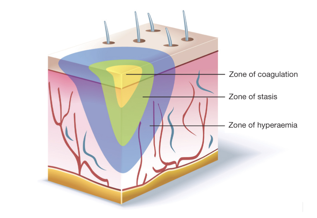 |
|---|
However, current treatments do not effectively target the underlying mechanisms of burn conversion, such as oxidative stress and inflammatory signaling, nor do they provide sustained, localized delivery of therapeutic agents. This gap in treatment is particularly critical because early intervention can significantly reduce burn severity, prevent deeper tissue damage, lower infection risk, minimize scarring, and accelerate healing (Ji et al., 2024) . Without such intervention, partial-thickness burns may progress into more severe injuries that require invasive procedures and prolonged recovery.
To address these limitations, our team proposes the development of a bioengineered hydrogel bandage designed to release burn-stabilizing agents that specifically target the mechanisms underlying burn conversion after injury. Hydrogels have emerged as promising platforms due to their high water content, biocompatibility, and ability to mimic the extracellular matrix (Cook et al., 2022). These materials can be engineered to deliver therapeutic compounds, including antioxidants, anti-inflammatory agents, and growth factors, in a controlled and gradual manner directly to the wound site (Pintilei et al., 2025). By reducing oxidative stress, minimizing inflammation, and promoting cell survival within the zone of stasis, this system aims to preserve viable tissue and prevent burn progression. If successful, our project could speed up burn healing, reduce pain and infection, and minimize scarring, improving recovery for patients. By directly delivering growth factors to the wound, the hydrogel bandage could contribute to more localized care for first and second-degree burns.
Systems level
To address the issue of second-degree burn conversion, our team has designed a biological system that operates within an alginate-based hydrogel scaffold. Our goal is to simultaneously protect the wound site from infection and oxidative damage, reduce inflammation, create a tissue-regeneration-favoring environment, and directly promote wound healing through direct delivery of wound healing factors. Our main wound-stabilizing agent is curcumin, a natural compound derived from turmeric whose strong anti-inflammatory and antioxidant properties make it ideal for burn wound healing applications (Phan et al., 2001; Fu et al., 2024; Salibian et al., 2016 ). Curcumin is especially relevant during burn wound conversion, which is driven by ischemia, inflammation, and oxidative stress in the zone of stasis. Interventions during this period target tissue salvage by reducing inflammatory signaling and oxidative damage before the injury becomes irreversible (Salibian et al., 2016; Palackic et al., 2022).
Our system integrates biomaterial and genetic engineering by producing curcumin through bioengineered microbial biosynthesis, harvesting the curcumin from the engineered bacteria, and incorporating it into a hydrogel scaffold for controlled delivery. We will introduce the key curcumin-biosynthesis genes 4-coumarate-CoA ligase (At4CL), diketide-CoA synthase (DCS), and curcumin synthase (CURS) into an engineered E. coli plasmid with the goal of synthesizing curcumin experimentally. Following the extraction and harvesting steps of the Azevedo et al. (2025) study, we will then incorporate the harvested curcumin into the microspheres in the hydrogel patch network, extending their approach beyond biosynthesis to include a direct delivery platform for the prevention of burn conversion. Encapsulation in microspheres is critical due to the hydrophobic properties of curcumin, which will be prevented from breaking down upon contact with the moist hydrogel environment by the microspheres. The microspheres, embedded with curcumin, will be incorporated in the hydrogel matrix prior to crosslinking, also assisting in controlled and gradual delivery to the wound site (Lei et al., 2022). Crosslinking is the hydrogel formation technique in which polymer chains are linked by calcium ions to form a stable three-dimensional gel network. Calcium ions act as bridges between alginate chains, connecting them into a three-dimensional porous network that forms the final gel patch.
| Figure 2: Schematic of hydrogel patch construction, microsphere incorporation, and ionic crosslinking. Curcumin-loaded microspheres are first homogeneously dispersed within an alginate polymer solution, forming a preliminary composite matrix in which therapeutic agents are physically embedded. Following incorporation, chitosan is introduced to enhance biological functionality through its intrinsic antimicrobial and anti-inflammatory properties. Ionic cross-linking is subsequently incorporated through the addition of calcium ions, forming a three-dimensional hydrogel network. Embedded microspheres are distributed throughout this network, allowing for controlled diffusion of curcumin over time. | 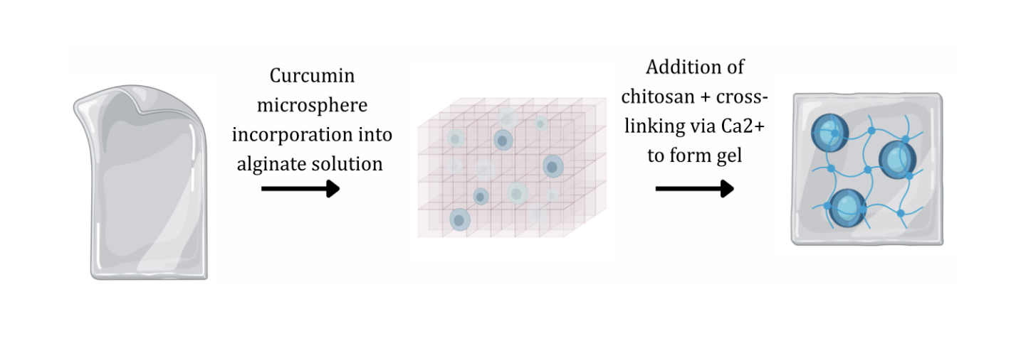 |
|---|
The hydrogel matrix itself acts as a structural scaffold and platform for targeted and controlled drug release. Its porous structure mimics the extracellular matrix, creating an environment favoring cell-regeneration. The alginate base is reinforced with the addition of chitosan, a natural antimicrobial and anti-inflammatory agent derived from chitin, which actively promotes wound healing by enhancing cell migration and collagen deposition (Eski et al., 2012). The finished, crosslinked matrix is designed to be placed directly over the burn site in the first 24-48 hours after injury, because this is when burn wound conversion risk and wound fluid changes are the greatest, and when stabilizations are most likely to influence the progression of the wound (Salibian et al., 2016; Cook et al., 2022). Dressing change timing depends on exudate and dressing stability rather than a fixed interval across all patients and dressings, since conversion from superficial to deeper injury may not be immediately apparent (Cook et al., 2022). Delayed application is expected to reduce the effect because progressive microvascular failure and inflammatory injury become less reversible over time, increasing the likelihood that tissue loss has already occurred by the time treatment begins (Salibian et al., 2016). This bandage allows for moisture retention, and wound-stabilizing factor diffusion into the site during this critical time, reducing oxidative stress and inflammation to help prevent burn conversion and promote more effective healing.
As a whole, the system operates by integrating curcumin, chitosan, and alginate into a composite hydrogel matrix to stabilize the zone of stasis, reduce inflammation and infection risk, and ultimately prevent burn conversion. This results in faster healing and better outcomes because it preserves microvascular integrity, limits oxidative damage, and creates optimal conditions for tissue regeneration.
Device level
The hydrogel scaffold consists of an alginate and chitosan matrix formed by calcium-mediated ionic crosslinking. Ionic crosslinking creates junctions between polymer chains and sets a network in water-based conditions (Goh et al., 2024). Crosslinking density and polymer ratio control how a hydrogel behaves when it is wet on the wound. Higher crosslinking density creates more junction points and a tighter mesh. This improves cohesion and wet-state integrity, but it also lowers permeability and slows diffusion because the water channels are smaller. On the other hand, lower crosslinking density creates a looser mesh, which increases swelling and permeability, but it can reduce mechanical persistence under fluid exposure and movement (Goh et al., 2024). Polymer ratio also changes how strongly the network holds together in water, affecting electrostatic association and therefore cohesion and particle trapping, which influences whether embedded microspheres remain fixed in place or shift and detach during wear (Straccia et al., 2015).
The pore structure of the hydrogel controls transport within the scaffold. The cone shaped pores with a gradient channel diameter maximize oxygen diffusion and vascularization, which is key for healing second degree burns (Ahn et al., 2010). To optimize functionality, pore sizes will vary across the scaffold, mimicking the layers of the skin. Smaller pore sizes replicate the epidermal layer, promoting cell attachment. Moderate pore sizes emulate the dermal layer, allowing for proper cell migration and nutrient transportation. Large pore sizes promote vascularization, supporting specialized structures such as blood vessels (Fariza Mukasheva et al., 2024). This gradient can be produced through layer-by-layer casting and verified by sectioning the scaffold and measuring pore sizes across layers (Ahn et al., 2010). This gradient would also be tested experimentally before being implemented into the hydrogel.
used in the formulation of hydrophobic curcumin. Because curcumin is hydrophobic, nanoparticle formulation improves its dispersion in a hydrogel and reduces clumping or rapid precipitation that can create uneven local release. These particles set the physical form that determines how curcumin interacts with the hydrated network. They increase the effective surface area in contact with wound fluid, which supports more uniform distribution through the scaffold and more reproducible release behavior. Nanoparticles can be incorporated directly into the hydrogel for faster availability, or they can be loaded into microspheres for slower, sustained delivery. In the combined approach, microspheres control residence time and reduce early burst release at the wound interface, while nanoparticles improve compatibility with the hydrated environment and can protect curcumin from early degradation (Goh et al., 2024). To quickly summarize the parts of the system: curcumin is formulated into nanoparticles, nanoparticles are loaded into microspheres, and microspheres are embedded in the hydrogel matrix for sustained release.
| Figure 3: Schematic of microparticles (microspheres) dispersed in a hydrogel matrix for controlled delivery (Carrêlo et al., 2021). | 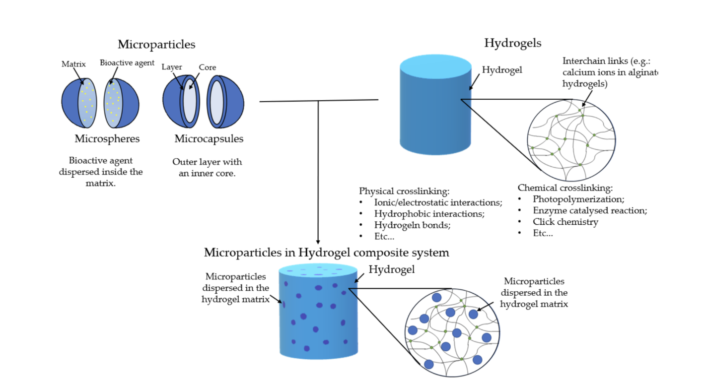 |
|---|
Another key component of the hydrogel is microspheres, which are useful for delivering systems for curcumin to the wound. Microspheres act as a local reservoir inside the hydrogel and reduce early loss of payload into wound fluid. This adds a second barrier to transport because the payload exits the microsphere matrix before it crosses the hydrogel network. Microsphere properties help build the stable release system. Particle diameter changes diffusion distance, and polymer composition changes permeability and degradation rate. Diffusion drives early release from the microsphere surface and near-surface regions, while polymer breakdown increases internal porosity and supports later release by opening additional transport pathways. A wound delivery study that incorporated protein-loaded microspheres into a hydrogel dressing reported improved wound healing outcomes, which supports the feasibility of combining a hydrogel contact layer with microspheres for sustained local exposure (Lei et al., 2022). Microspheres also support spatial stability. A cohesive hydrogel network limits particle movement, which keeps the local concentration field more consistent over the wound region.
The exterior surface of the hydrogel is designed as a barrier layer because burn wounds face high contamination risk and to ensure that the dressing remains hydrated. A surface layer with higher density can reduce bacterial entry and wash-off while the interior region remains more permeable for diffusion from the microspheres. A chitosan-coated alginate surface increases antibacterial performance and alters transport across the dressing boundary (Straccia et al., 2015). This boundary layer can reduce particle loss at the surface and limit rapid exchange with external fluid, which improves consistency of local delivery during wear. Importantly, this barrier layer is not designed to be fully occlusive; rather, it is a selectively permeable membrane that allows for sufficient oxygen diffusion across the site, given that oxygen transport is critical for cellular metabolism, angiogenesis, and tissue regeneration. A semi-permeable design is facilitated through the incorporation of a hydrated polymer network that facilitates oxygen permeability even as bacteria passage is restricted (Narayanan & Bhaskar, 2025).
Parts level
Curcuminoids, such as curcumin, are naturally synthesized in Curcuma longa rhizomes via the polyketide pathway. In microbial systems, an artificial synthesis pathway for curcumin can be created using microbial and plant-derived enzymes (Katsuyama et al., 2008).
The study by Azevedo et al. (2025) evaluates the ability of E. coli to produce curcumin through the expression of a biosynthetic pathway involving three genes: 4-coumarate-CoA ligase (At4CL), diketide-CoA synthase (DCS), and curcumin synthase (CURS). To assess the yield of curcumin created through the aforementioned biosynthetic pathway, researchers tested varying experimental conditions. Firstly, varying temperature conditions (26 and 30°C) were assessed. Secondly, differences between planktonic and biofilm cell growth conditions were examined. The results of this study demonstrated that the biofilms produced up to 10-fold higher curcumin levels compared to the planktonic counterparts. However, no significant differences were observed in curcumin yield between differing temperatures (Azevedo et al., 2025).
Our proposal aims to integrate the biosynthetic curcumin production approach based on the Azevedo et al. (2025) study, with our hydrogel bandage. Curcumin will be synthesized using a 3-gene biosynthetic pathway expressed in E. coli, comprising of At4CL from Arabidopsis thaliana, DCS, and CURS from Curcuma longa (Azevedo et al., 2025; Katsuyama et al., 2008).
Plasmid design
To successfully express curcumin in the biosynthetic pathway of E. coli, we will design a plasmid in accordance with the Azevedo et al. (2025) study (see Figure 4). This plasmid design incorporates: a lac promoter (lacI); a lactose operator; a transcriptional promoter from the T7 phage (T7 promoter); a T7 transcriptional terminator (T7 terminator); a kanamycin resistance gene (KanR); and the At4CL, DCS, and CURS genes designed for the curcumin biosynthetic pathway (Azevedo et al., 2025).
| Figure 4: At4CL_DCS_CURS plasmid designed in the Azevedo et al. study and assembled by NZYTech (Lisboa, Portugal) for experimental testing (Azevedo et al., 2025). | 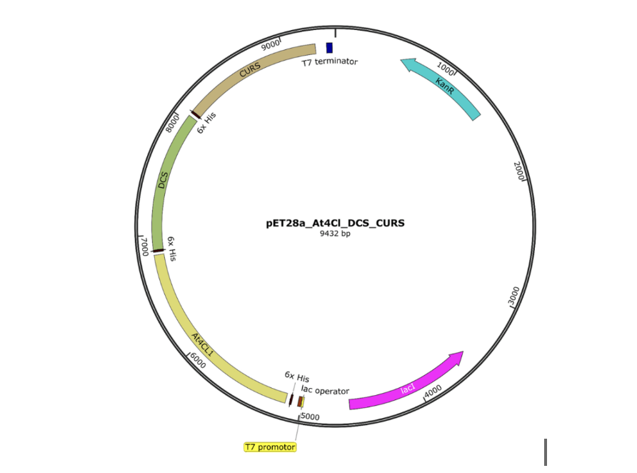 |
|---|
The 4CL enzyme converts the precursor ferulic acid into feruloyl-CoA (Azevedo et al., 2025). Ferulic acid is a phenolic compound that has been shown to be effective for curcumin production in E. coli, and was subsequently selected as the optimal precursor compound for this proposal (Rainha et al., 2022). Next, Feruloyl-CoA is converted to feruloyl-diketide-CoA by DCS. Finally, the CURS enzyme condenses the diketide, producing the final curcumin molecule (see Figure 5). This expression system is well suited for high-yield curcumin production because the strong T7 promoter enables robust transcription of the biosynthetic genes, enabling high and consistent biosynthetic yields (Azevedo et al., 2025; Katsuyama et al., 2008).
| Figure 5: Curcumin biosynthetic pathway (Azevedo et al., 2025). | 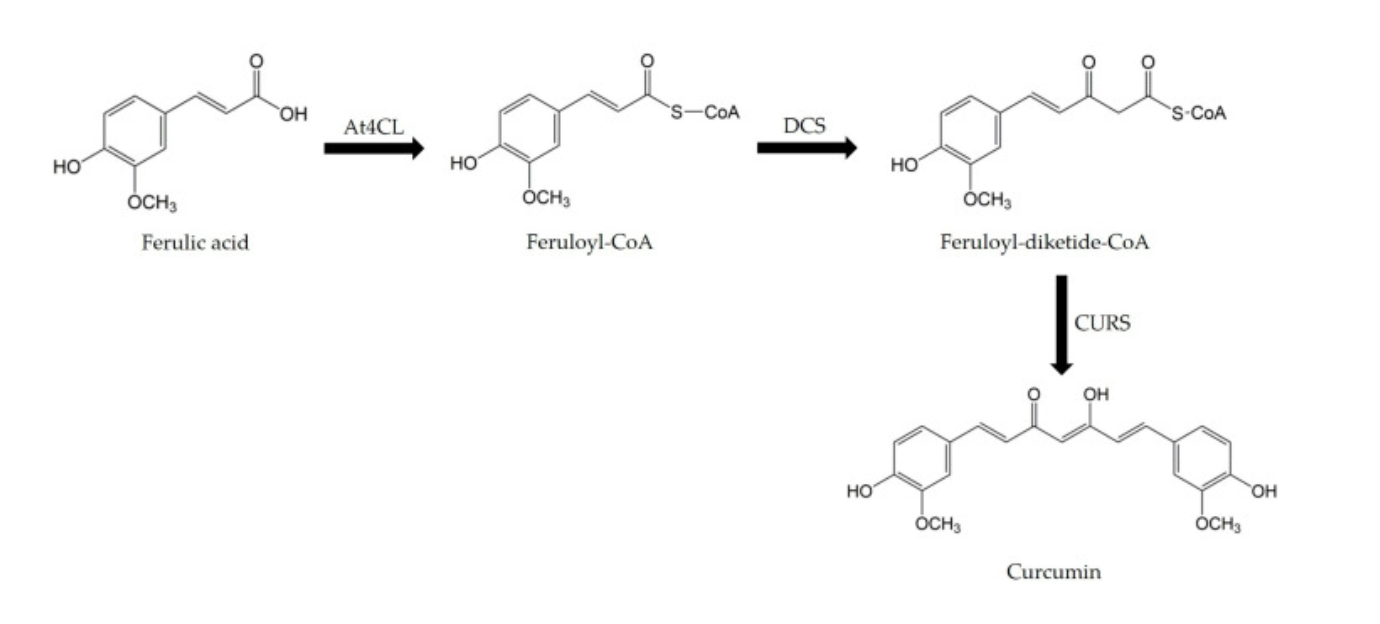 |
|---|
Bacterial growth conditions
In order to produce curcumin, Azevedo et al. (2025) performed a heat-shock transformation on the E. coli strain JM109(DE3) (Promega, Madison, WI, USA). To attain similar transformation results, our proposal aims to execute heat shock on the E. coli strain with the At4Cl_DCS_CURS plasmid (Azevedo et al., 2025). Transformation of a plasmid onto E. coli consists of a short incubation on ice, after which the chemically competent bacteria mixture and plasmid are placed at 42°C for 45 s and then returned to the ice bath. After applying the heat shock, SOC media is added to the mixture, and the transformed cells are transferred to a 37°C incubator for 30 min with agitation (Froger & Hall, 2007).
To achieve optimal curcumin yield, this proposal will mimic the biofilm formation methodology from Azevedo et al. (2025), which established that biofilm conditions were most effective in ensuring high curcumin production.
Curcumin extraction
At high concentrations, curcumin could settle in the supernatant following transformation and be located within the E. coli cells. Consequently, it is optimal for the broth to be mixed with equal volume of ethyl acetate. Subsequently, the mixture will be vortexed and centrifuged (2 min, 15,000 x g). This process will be repeated until the pellet becomes colorless. To concentrate the extracts, the solvent will be completely evaporated in a fume hood and dried, leaving behind a curcumin residue with minimal residual ethyl acetate that will be scraped and extracted using a spatula. In order to examine the concentration and purity of the produced curcumin, the sample will be analysed by ultra-high-performance liquid chromatography (Rodrigues et al., 2020), detecting any bacterial contaminants. Additionally, as E. coli is a Gram-negative bacterium, the presence of endotoxin-like substances will be assessed using a Limulus amebocyte lysate (LAL) test to ensure safety for wound application (Bolden et al., 2016). Finally, the extracted curcumin will be converted to a powder residue via rotary evaporation followed by lyophilization (freeze-drying) to remove residual solvent and the remaining moisture, for integration into the microspheres (Rodrigues et al., 2020). Contamination will be prevented by conducting all post-extraction steps under aseptic conditions, including the use of sterile equipment and storage in airtight containers (Froger & Hall, 2007).
Safety
While this study focuses on developing a growth factor-releasing hydrogel bandage for second-degree burn wound treatment, safety is an important consideration throughout the design, production, and usage phases. Safety concerns cover many aspects, including biological handling of engineered bacteria, chemical and material composition, risks to the patients, and the disposal of the genetically modified organisms and bandages.
The production of this bandage relies entirely on bacteria to create curcumin. To minimize risks, the hydrogel should use safe, already researched, and well-characterized lab strains of bacteria. These strains will lack the virulence factors found in E. coli and are incapable of colonizing the human gut if accidentally ingested. During all stages of bacterial growth and growth factor extraction, the organisms must strictly be contained within a certified laboratory environment. Per Biosafety Level 1 and 2 (BSL-1/BSL-2) protocols, this includes the use of closed bioreactors, laminar flow hoods, autoclaving all biological waste, and prohibition of live culture near assembly areas (CDC & NIH, 2020). Outside exposure risks include cross-contamination of the bacteria or exposure of the patient to disease or other harmful agents. Even more critically, the bandage must not contain any live bacteria of any kind. Burn wounds are very open injuries, often with compromised skin layers, which makes them very susceptible to infection. Even lab strains pose a risk of serious infection, inflammation, or other side effects. Additionally, live bacteria can overall produce proteins and molecules that can worsen the wound. Only hydrogels with purified and non-bacterial growth factors will be produced, thus eliminating the danger of a potential infection. All bacteria should be fully inactivated before the hydrogel product is assembled, which is essential for patient safety.
All materials that contact the wound or surround the skin should be biocompatible and not provoke toxic, cytotoxic, or unhealthy immune responses. The hydrogel bandage components, including alginate and chitosan, are natural polymers with pre-existing safety profiles that have already been cleared by the Food and Drug Administration (FDA) for wound care products. Curcumin-filled hydrogel bandages made from these natural biomaterials demonstrate good biocompatibility as well, and have sustained release suitable for wound healing (Sideek et al. 2023). Curcumin is included in this design as an antimicrobial and anti-inflammatory agent, and even though it is beneficial at low doses, it can be cytotoxic at high concentrations. Curcumin-loaded hydrogel delivery systems provide sustained, localized release that will speed up wound healing (Sideek et al. 2023). The hydrogel release rate will be carefully calibrated to keep curcumin at a non-cytotoxic range throughout the entire wear period. As the hydrogel degrades, the particles it breaks down must also be non-toxic and metabolically processable by the body. Alginate, chitosan, cellulose, and collagen all break down into natural things that may be metabolized by the body like amino acids and guluronic acids. However, given that the burn wound will have increased permeability, it is important that all agents used in fabrication do not have any wound-relevant concerns.
All engineered bacteria used in the production of the growth factors must be entirely inactivated before disposal. Recommended inactivation methods could include treatment with a 10% sodium hypochlorite, or autoclaving, with either being disposed of as biohazardous material in compliance with institutional biosafety protocols (CDC & NIH, 2020). Improper disposal could have adverse effects on the natural environments where genetically modified organisms are unpredictable and potentially harmful. As another extra safeguard against environmental release during production, the bacteria should have a kill switch, specifically an autotrophic dependency on a synthetic substance like an amino acid that is not found in nature. Should any bacteria escape the laboratory environment still viable, they would rapidly die in the absence of this substance, which would prevent colonization of outside environments and eliminate the risk of horizontal gene transfer.
Certain hydrogel materials might also carry allergy risks that must be disclosed or tested for prior to usage. Chitosan is a natural fiber derived typically from the shells of crustaceans, so people with shellfish and crustacean allergies might not be able to use the hydrogel. Similarly, animal derived collagen may cause reactions in patients with allergies. An allergic reaction could significantly worsen outcomes for the patient and delay healing. Thus, clear labelling of all components and pre-application protocols is essential. The bandage must also be manufactured under sterile conditions and sealed in single-use packaging.
The hydrogel bandage, being a device-like wound dressing matrix with genetically modified biological growth factors, would likely be classified as a combination product by the FDA, which states that wound dressings that contain antimicrobial or active agents are subject to 510(k) clearance.
Discussions
First and second-degree burns are painful, slow to heal, and can lead to scarring and infections. Current treatments for first-degree burns are limited but include cold water, pain medicine/medication, and petroleum jelly. These solutions do not actively promote healing, and most have side effects. NSAIDs such as ibuprofen and Naproxen, which are used for milder burns over the long term, can cause stomach discomfort, kidney issues, and reduced platelet function. Furthermore, the Topical Agent Silver Sulfadiazine, which is used to prevent infection of burns, can cause skin discoloration, temporary burning/itching, and slow healing (Maciel et al., 2019). Additional problems arise with non-stick dressings, which are uncomfortable and leave patients at risk of infection, while advanced dressings are expensive and inaccessible. Finally, current treatments do not sufficiently target the biological response mechanisms driving burn conversion and focus more largely on directly supporting burn healing. This lack of treatments that actively prevent burn progression is what we aim to alleviate. Our solution is a bioengineered hydrogel bandage designed to stabilize the burn site, prevent burn conversion, and optimize the healing environment. Unlike conventional single-purpose treatments, this integrated system is multifunctional, combining multiple therapeutic effects into one platform. It promotes active healing rather than passive protection and provides sustained therapeutic benefits, outperforming single-application solutions currently available on the market. Burn conversion happens due to inflammation, oxidative stress, and microvascular damage (Palackic et al., 2022). Our system mechanistically prevents burn conversion by using a sustained curcumin release strategy which reduces inflammation and oxidative stress. The moist environment created by the hydrogel system preserves tissue viability while its porous structure facilitates tissue oxygenation. Unlike conventional treatments, this system actively targets the biological processes responsible for burn progression rather than only addressing surface-level symptoms.
A key strength of this system is its multifunctional design. In particular, the hydrogel system is beneficial because it maintains a moist environment, which is ideal for burn healing (Choi et al., 2024). A moist environment accelerates tissue repair, reduces pain, and minimizes scarring compared to dry, scabbed environments (Goh et al., 2024). By keeping the wound hydrated, cells can migrate faster across the wound bed while promoting the growth of new blood vessels and collagen synthesis, unlike in a dry wound, where cells must travel deep under the scab to find moisture to move, counterproductively slowing healing (Cook et al., 2022). Moisture also helps the body’s own enzymes dissolve autolytic debridement, resulting in faster cleaning of the wound. Studies have found that moisture can lead to increased angiogenesis, which brings necessary nutrients and oxygen to the area. Finally, it helps maintain a lower and slightly acidic pH, which protects against bacteria and helps wounds heal more efficiently. The hydrogel system also mimics the extracellular matrix that is used to support cell proliferation and tissue regeneration. The alginate-chitosan matrix is used to set up a moist environment. Chitosan is compatible with alginate systems because it is positively charged and forms polyelectrolyte complexes with alginate, which can be blended and layered into hydrogels. Alginate is derived from seaweed and forms hydrogels in the presence of divalent ions such as calcium via cross-linking (Solomiia Kapatsila et al., 2025); This component provides structure and moisture retention. Chitosan works well with alginate hydrogel patches for various reasons. Chitosan contains strong antimicrobial properties against Gram+ bacteria (Staphylococcus, Streptococcus), Gram- bacteria (E. coli, Pseudomonas), and Fungi (Candida) (Barkat et al., 2025). Furthermore, it promotes cell migration and collagen deposition, and is also non-cytotoxic. The combined system of the alginate-chitosan complex is advantageous because of its increased mechanical strength, sustained antimicrobial activity, and enhanced wound healing. The structural design includes a cone-shaped pore gradient. The small pores mimic the epidermis, the medium pores mimic the dermis, and the large pores help support complex structures (Fariza Mukasheva et al., 2024). The pores maximize cell proliferation, tissue organization, and cell viability (Ahn et al., 2010). The drug delivery system contains microspheres loaded with curcumin which is antioxidative, and helps preserve microvascular integrity (Singer et al., 2011). We chose curcumin because it is naturally abundant, cost-effective, and has a scalable production potential. The microspheres are more effective than single-dose delivery because of their sustained release system, which keeps therapeutic agents at the wound site. Therefore, this function maintains consistent therapeutic levels and prevents underdosing or toxic spikes. However, it is important to note that release variability depends on polymer composition as well as environmental conditions. The functional outcome altogether supports moisture retention, oxygen diffusion, infection control, and reduced inflammation.
If this hydrogel system works as intended, our design could have a substantial clinical impact on burn care. Instead of just treating symptoms, it aims to slow and even stop burn progression, which is a large issue in determining how severe a burn becomes (Żwierełło et al., 2023). By keeping the wound moist, restricting pathogen growth, and delivering anti-inflammatory compounds over time, our solution could improve healing and lower the risk of infection (Żwierełło et al., 2023). This might also reduce the need for more serious treatments, such as surgical procedures. While these ideas still need to be tested, they are based on current knowledge and experience with burns and wound healing. In the long term, this kind of approach could help move wound care closer to more advanced, regenerative treatments instead of basic protection.
Next steps
Our proposed hydrogel system is based on empirical findings from previous scientific papers and experimental research conducted by professional laboratories. Individual components of said system were inspired by distinct studies and subsequently integrated into a theoretical framework. In the eventual case that this project advances beyond its conceptual stage towards prototype development, numerous elements would require iterative testing.
Developing and refining an effective hydrogel bandage for second-degree burn care begins with evaluating the pros and cons of various fabrication strategies. Fibrous hydrogels, which can mimic the extracellular matrix (ECM), are created via electrospinning, a process unique to lab settings, as explained in the Li et al. (2022) study. Electrospinning is executed in specialized machines that utilise electric force to overcome the surface tension of liquid droplets supplied from solutions with dissolved bioactive agents and polymers. The suspended droplets are positioned on a traditional needle-based system, where they then form a Taylor cone with a jet at the apex. The jet stretches into nanofibers that accumulate into a fibrous mat, which must undergo cross-linking to become a fully-functional hydrogel. Electrospun nanofiber dressings have a multitude of advantages, including fibers fashioned with a larger surface area, which allows for more adhesion and growth sites for cells (S. Zhang et al., 2024). Electrospun hydrogels are also intended for drug delivery, which explains their tendency to have high bioactivity loading, which leads to faster healing (S. Zhang et al., 2024). Lastly, the customizable structure of said hydrogels makes them ideal for the incorporation of microspheres and their ability to control the rate and time of drug release aligns with our idea of periodically releasing curcumin (S. Zhang et al., 2024). However, electrospinning faces several limitations that make large-scale production difficult, as traditional needle-based systems produce only small amounts of material, and multi-nozzle setups can reduce fiber quality (Li et al., 2022). The process also struggles with achieving uniform fiber diameter and alignment alongside defects like beading, which contribute to a final product with less physical integrity. Additionally, electrospinning is constrained by the need for specific, soluble materials and often relies on toxic or expensive solvents, which are highly sensitive to environmental conditions such as temperature and humidity (Li et al., 2022). The moist environment our theoretical hydrogel plans to employ would conflict with said solvents, resulting in a non-functional hydrogel. Operational challenges further complicate electrospinning, and the resulting fibers are bound to have technical faults (Li et al., 2022).
As an alternative, sprayable hydrogels, particularly those which are based on alginate- a seaweed-derived polymer, offer a far more accessible and efficient alternative to electrospinning (Rungrod et al., 2025). This is due to the fact that they can be mass-produced, rapidly applied to irregular wound surfaces, and instantly form a conforming, moist, three-dimensional polymer network that supports healing (Choi et al. 2024). Sprayable hydrogels are liquid or powder formulas with dissolved bioactive ingredients, polymers, and growth factors that instantly form a gel upon contact with tissue. Advanced sprayable systems are more resistant to changes in humidity and temperature and can include additives such as microspheres, although the microspheres must be tailored to certain characteristics, as sprayable methods have certain complications that do not apply to electrospinning (W. Zhang et al. 2024). When the infused spray meets calcium ions in the blood or a secondary sprayed solution, it initiates cross-linking and creates a 3D polymeric network (Rungrod et al., 2025). As detailed in the Cao et al. (2020) study, the G-units present in the polysaccharide alginate interact with calcium ions or other divalent cations to form a stable high-affinity cross-linking structure often referred to as the “egg-box” (Cao et al., 2020). This form of in situ cross-linking has been successfully demonstrated by research conducted by Korea Advanced Institute of Science and Technology (KAIST), a lab that developed a spray-on AGCL powder capable of stopping severe and intense bleeding within seconds (Son et al., 2025). The AGCL powder was also shown to promote angiogenesis, collagen deposition, and re-epithelialization in skin wound models (Son et al., 2025). Although these systems require optimization to address drawbacks such as lower mechanical integrity than electrospun hydrogels and a lack of inherent antimicrobial properties, such sprayable hydrogels are streamlined for rapid, low-cost, and sustainable mass production.
General future work should begin with the experimentation of distinct methods of cross-linking and their inherent properties to assess the structural integrity of the produced hydrogel in differing environmental conditions. Once it is ensured that the alginate-chitosan matrix can maintain its 3D structure through harsh circumstances, it would be appropriate to calibrate curcumin to stay at a non-cytotoxic range to guarantee the safety of future users. To follow, measuring the release rate of curcumin as detailed in Sayyar et al. (2023) and matching it to the time frame of tissue repair to confirm that the doses are delivered consistently and at non-toxic concentrations. Practical experimentation would occur through the use of in vitro release assays and cytotoxicity tests with skin cells to minimize hazards. Additionally, performing in vitro antimicrobial assays is vital to make sure the produced hydrogel prevents bacterial colonization, which could further harm the burn wound. Finally, once experimental validation has proved that the hydrogel prototype is harmless and viable in the real world, conducting investigations to maximize the capabilities of the hydrogel would be ideal. Adjusting cross-linking densities and polymer ratios within the alginate-chitosan gelatin matrices allows customizations to permeability, which contributes to the behavior of a hydrogel when moist, and optimizes structural design and healing efficiency (Goh et al., 2024). Other focus areas include the layout of differing pore structures and their gradient channel diameters throughout the hydrogel, in order to more accurately mimic the extracellular matrix to promote tissue regeneration (Mukasheva et al., 2024). Lastly, experimentation concerning the characteristics of microspheres pertaining to size and structure could also affect curcumin release kinetics, which could prove to be useful. Once the hydrogel meets all performance criteria and is fully optimized, we will proceed to conduct ex vivo simulated burn models to observe the functionality and efficiency of the hydrogel patch and fine-tune its design. Additionally, incorporating feedback from medical professionals and patients will be essential to ensure that our design is practical, comfortable, and feasible for real-world applications.
Author contributions
All authors made contributions to the conceptualization, design, and execution of this project, led by TaJ and AV. Background by TaJ, TeJ, and LK; Systems by TaJ; Device by YK, JGC; Parts by AV; Discussions by CT, NT; Next steps by TK; Safety by AP; Figures by AV; Editing by TaJ, AV, TK.
Acknowledgements
We would like to thank AoPS Lexington for sponsoring our participation in the BioBuilder Club program and making this project possible for our team. We are especially grateful to Ms. Susana Donkor for her valuable guidance in developing our project idea and helping us design our experiment. We also deeply appreciate Mr. Hobbs for his continuous support throughout the process, assisting us in our project, keeping us on track, and teaching us the foundational concepts in biology and scientific communication we needed to succeed. We would also like to thank the BioBuilder Foundation for their support and assistance in navigating the design process and for connecting us with our mentor.
References
Ahn, G., Park, J. H., Kang, T., Lee, J. W., Kang, H.-W., & Cho, D.-W. (2010). Effect of pore architecture on oxygen diffusion in 3D scaffolds for tissue engineering. Journal of Biomechanical Engineering, 132(10), 104506. https://doi.org/10.1115/1.4002429
Amlika Rungrod, Arthit Makarasen, Suwicha Patnin, Supanna Techasakul, & Runglawan Somsunan. (2025). Design and Development of a Sprayable Hydrogel Based on Thermo/pH Dual-Responsive Polymer Incorporating Azadirachta indica (Neem) Extract for Wound Dressing Applications. Polymers, 17(15), 2157–2157. https://doi.org/10.3390/polym17152157
Azevedo, A., Teixeira-Santos, R., Gomes, L. C., Duarte, S. O. D., Monteiro, G. A., & Mergulhão, F. J. (2025). Engineering Escherichia coli Biofilms for Curcumin Production. Molecules, 30(9), 2031. https://doi.org/10.3390/molecules30092031
Barkat, H., Abuzar, Md., Asiri, Y. I., Hassan, Mohd. Z., Alanezi, A. A., Hadi, H., & Barkat, Md. A. (2025). Chitosan-based nanotherapeutics in burn wound management: Clinical insights and healing potential. International Journal of Biological Macromolecules, 328, 147639. https://doi.org/10.1016/j.ijbiomac.2025.147639
Bolden, Jay S., et al. “Endotoxin Recovery Using Limulus Amebocyte Lysate (LAL) Assay.” Biologicals, vol. 44, no. 5, Sept. 2016, pp. 434–440, https://doi.org/10.1016/j.biologicals.2016.04.009. Accessed 4 May 2021.
Cao, L., Lu, W., Mata, A., Nishinari, K., & Fang, Y. (2020). Egg-box model-based gelation of alginate and pectin: A review. Carbohydrate Polymers, 242, 116389. https://doi.org/10.1016/j.carbpol.2020.116389
CDC. (2024, May 17). Biosafety in Microbiological and Biomedical Laboratories (BMBL) 6th Edition. CDC Laboratories. https://www.cdc.gov/labs/bmbl/index.html
Choi, J. Y., Joo, Y.-J., Kang, R. J., Jeon, H. K., & Hong, G. S. (2024). Effect of Spray-Type Alginate Hydrogel Dressing on Burn Wounds. Gels, 10(2), 152. https://doi.org/10.3390/gels10020152
Cook, K. A., Martinez-Lozano, E., Sheridan, R., Rodriguez, E. K., Nazarian, A., & Grinstaff, M. W. (2022). Hydrogels for the management of second-degree burns: currently available options and future promise. Burns & Trauma, 10. https://doi.org/10.1093/burnst/tkac047
Eski, M., Ozer, F., Firat, C., Alhan, D., Arslan, N., Senturk, T., & Işik, S. (2012). Cerium nitrate treatment prevents progressive tissue necrosis in the zone of stasis following burn. Burns, 38(2), 283–289. https://doi.org/10.1016/j.burns.2011.08.008
Fariza Mukasheva, Adilova, L., Aibek Dyussenbinov, Bota Yernaimanova, Madi Abilev, & Akilbekova, D. (2024). Optimizing scaffold pore size for tissue engineering: insights across various tissue types. Frontiers in Bioengineering and Biotechnology, 12. https://doi.org/10.3389/fbioe.2024.1444986
Fu, Z., Zou, J., Zhong, J., Zhan, J., Zhang, L., Xie, X., Zhang, L., Li, W., & He, R. (2024). Curcumin-Loaded Nanocomposite Hydrogel Dressings for Promoting Infected Wound Healing and Tissue Regeneration. International Journal of Nanomedicine, Volume 19, 10479–10496. https://doi.org/10.2147/ijn.s479330
Froger, A., & Hall, J. E. (2007). Transformation of Plasmid DNA into E. coli Using the Heat Shock Method. Journal of Visualized Experiments : JoVE, 6(6), 253. https://doi.org/10.3791/253
Goh, M., Du, M., Peng, W. R., Saw, P. E., & Chen, Z. (2024). Advancing burn wound treatment: exploring hydrogel as a transdermal drug delivery system. Drug Delivery, 31(1), 2300945. https://doi.org/10.1080/10717544.2023.2300945
Henrique Carrêlo, Paula, João Paulo Borges, & Maria Teresa Cidade. (2021). Injectable Composite Systems Based on Microparticles in Hydrogels for Bioactive Cargo Controlled Delivery. Gels, 7(3), 147–147. https://doi.org/10.3390/gels7030147
Ji, S., Xiao, S., & Xia, Z. (2024). Consensus on the treatment of second-degree burn wounds (2024 edition). Burns & Trauma, 12. https://doi.org/10.1093/burnst/tkad061
Katsuyama, Y., Matsuzawa, M., Funa, N., & Horinouchi, S. (2008). Production of curcuminoids by Escherichia coli carrying an artificial biosynthesis pathway. Microbiology (Reading, England), 154(Pt 9), 2620–2628. https://doi.org/10.1099/mic.0.2008/018721-0
Lei, L., Wang, X., Zhu, Y., Su, W., Lv, Q., & Li, D. (2022). Antimicrobial hydrogel microspheres for protein capture and wound healing. Materials & Design, 215, 110478. https://doi.org/10.1016/j.matdes.2022.110478
Li, T., Sun, M., & Wu, S. (2022). State-of-the-Art Review of Electrospun Gelatin-Based Nanofiber Dressings for Wound Healing Applications. Nanomaterials, 12(5), 784. https://doi.org/10.3390/nano12050784
Maciel, A. B. da S., Ortiz, J. F., Siqueira, B. S., & Zanette, G. F. (2019). Tissue healing efficacy in burn patients treated with 1% silver sulfadiazine versus other treatments: a systematic review and meta-analysis of randomized controlled trials. Anais Brasileiros de Dermatologia, 94(2), 204–210. https://doi.org/10.1590/abd1806-4841.20198321
Mayo Clinic. (2022, August 13). Burns – Symptoms and causes. Mayo Clinic. https://www.mayoclinic.org/diseases-conditions/burns/symptoms-causes/syc-20370539
Narayanan, K. B., & Bhaskar, R. (2025). Innovations in Designing Hydrogels for Advanced Wound Dressing Applications: An Editorial Review. Gels (Basel, Switzerland), 11(5), 332. https://doi.org/10.3390/gels11050332
Palackic, A., Jay, J. W., Duggan, R. P., Branski, L. K., Wolf, S. E., Ansari, N., & El Ayadi, A. (2022). Therapeutic Strategies to Reduce Burn Wound Conversion. Medicina, 58(7), 922. https://doi.org/10.3390/medicina58070922
Phan, T.-T., See, P., Lee, S.-T., & Chan, S.-Y. (2001). Protective Effects of Curcumin against Oxidative Damage on Skin Cells In Vitro: Its Implication for Wound Healing. The Journal of Trauma: Injury, Infection, and Critical Care, 51(5), 927–931. https://doi.org/10.1097/00005373-200111000-00017
Pintilei, P. S., Binaymotlagh, R., Chronopoulou, L., & Palocci, C. (2025). The Role of Natural Hydrogels in Enhancing Wound Healing: From Biomaterials to Bioactive Therapies. Pharmaceutics, 17(10), 1243. https://doi.org/10.3390/pharmaceutics17101243
Progress in Wound Management – Innovative Methods and Technologies for Recovery. (2026). Nyrequirements.com. https://nyrequirements.com/blog/progress-in-wound-management-innovative-methods-and-technologies-for-recovery
Rainha, J., Rodrigues, J. L., Faria, C., & Rodrigues, L. R. (2021). Curcumin biosynthesis from ferulic acid by engineered Saccharomyces cerevisiae. Biotechnology Journal, 17(3), 2100400. https://doi.org/10.1002/biot.202100400
Rodrigues, J. L., Gomes, D., & Rodrigues, L. R. (2020). A Combinatorial Approach to Optimize the Production of Curcuminoids From Tyrosine in Escherichia coli. Frontiers in bioengineering and biotechnology, 8, 59. https://doi.org/10.3389/fbioe.2020.00059
Salibian, A. A., Rosario, A. T. D., Severo, L. D. A. M., Nguyen, L., Banyard, D. A., Toranto, J. D., Evans, G. R. D., & Widgerow, A. D. (2016). Current concepts on burn wound conversion—A review of recent advances in understanding the secondary progressions of burns. Burns, 42(5), 1025–1035. https://doi.org/10.1016/j.burns.2015.11.007
Sayyar, Z., Mahdavinia, G. R., & Khataee, A. (2023). Dual-drug (Curcumin/Ciprofloxacin) loading and release from chitosan-based hydrogels embedded with magnetic Montmorillonite/Hyaluronic acid for enhancing wound healing. Journal of biological engineering, 17(1), 66. https://doi.org/10.1186/s13036-023-00385-1
Sideek, S. A., El-Nassan, H. B., Fares, A., Aliaa Nabil ElMeshad, & Elkasabgy, N. A. (2022). Different Curcumin-Loaded Delivery Systems for Wound Healing Applications: A Comprehensive Review. Pharmaceutics, 15(1), 38–38. https://doi.org/10.3390/pharmaceutics15010038
Silver Sulfadiazine: MedlinePlus Drug Information. (n.d.). Medlineplus.gov. https://medlineplus.gov/druginfo/meds/a682598.html
Singer, A. J., Taira, B. R., Lin, F., Lim, T., Anderson, R., McClain, S. A., & Clark, R. A. F. (2011). Curcumin Reduces Injury Progression in a Rat Comb Burn Model. Journal of Burn Care & Research, 32(1), 135–142. https://doi.org/10.1097/bcr.0b013e318203337b
Solomiia Kapatsila, Taras, R., Varchuk, D., Nataliia Nosova, Serhii Varvarenko, & Volodymyr Samaryk. (2025). Influence of Calcium Crosslinker Form on Alginate Hydrogel Properties. Gels, 11(11), 885–885. https://doi.org/10.3390/gels11110885
Son, Y., Pak, K., Lee, T., Prayogo, M. C., Choi, J., Kang, S., Kang, M., Oh, B., Yu Sun, S., Kim, S., Im, S. G., Jon, S., & Park, S. (2025). An Ionic Gelation Powder for Ultrafast Hemostasis and Accelerated Wound Healing. Advanced Functional Materials. https://doi.org/10.1002/adfm.202523910
Straccia, M. C., D’Ayala, G. G., Romano, I., Oliva, A., & Laurienzo, P. (2015). Alginate Hydrogels Coated with Chitosan for Wound Dressing. Marine Drugs, 13(5), 2890–2908. https://doi.org/10.3390/md13052890
Warby, R., & Maani, C. V. (2023). Burns Classification. Nih.gov; StatPearls Publishing. https://www.ncbi.nlm.nih.gov/books/NBKNBK539773/
World Health Organization. (2023, October 13). Burns. WHO; World Health Organization: WHO. https://www.who.int/news-room/fact-sheets/detail/burns
Zhang, S., Yang, W., Gong, W., Lu, Y., Yu, D.-G., & Liu, P. (2024). Recent progress of electrospun nanofibers as burning dressings. RSC Advances, 14(20), 14374–14391. https://doi.org/10.1039/d4ra01514b
Zhang, W., Jing, H., Niu, Q., Wu, Z., Sun, Y., Duan, Y., & Wang, X. (2024). Sprayable, thermosensitive hydrogels for promoting wound healing based on hollow, porous and pH-sensitive ZnO microspheres. Journal of Materials Chemistry B, 12(31), 7519–7531. https://doi.org/10.1039/d4tb00961d
Żwierełło, W., Piorun, K., Skórka-Majewicz, M., Maruszewska, A., Antoniewski, J., & Gutowska, I. (2023). Burns: classification, pathophysiology, and treatment: a review. International Journal of Molecular Sciences, 24(4), 3749. https://doi.org/10.3390/ijms24043749