
Anoushka Dechatterjee, Tanya Jay, Tasheen Kashem, Audrey Lu, Xiying Lu, Patrick Noonan, Aarav Phadnis, Erin Sim, Charvi Talupuru, Miyara Tokushige, Annabel Volpe, Jessie Xie, and Annie Zhao, Lexington High School, Lexington, Massachusetts, United States
Reviewed on 9 May 2026; Accepted on 22 June 2026; Published on 26 October 2026
With help from the 2026 BioTreks Production Team.
Anthropogenic greenhouse gas emissions are increasing atmospheric carbon dioxide concentrations, which in turn accelerate ocean acidification through the formation of carbonic acid in seawater. This process lowers seawater pH and reduces carbonate ion availability, which impairs calcification and places growing stress on coral ecosystems. Our proposal aims to bioengineer calcium carbonate-promoting bacteria using strains naturally found living symbiotically in coral tissues, to mitigate the effects of ocean acidification on coral reefs. By increasing calcium carbonate availability, our bioengineered bacterial system will create a localized microenvironment that stabilizes pH and chemical conditions immediately surrounding the fragile coral tissue. The engineered P. rubra will have several components, all working together to ultimately alleviate the impact of ocean acidification on corals. The bacteria will use the CadC/pCadBA system to detect and increase the surrounding pH. They will also secrete enzymes Carbonic anhydrase and urease, encoded by the taCA and ureA/B/C gene, into the coral mucus layer to create a buffer zone against the acidic ocean environment. Carbonic anhydrase converts CO2 into bicarbonate which can precipitate as calcium carbonate in the presence of carbon ions, resulting in the stabilization of pH and calcification. Urease catalyzes the production of ammonia which can neutralize the proton byproducts, further reducing acidity. To prevent the spread of engineered bacteria beyond the coral ecosystem, a regulatory kill switch will be implemented via the mazF and mazE genes.
Keywords: Coral bleaching, ocean acidification, Pseudoalteromonas rubra, calcium carbonate
Authors are listed in alphabetical order. Reginald Hobbs mentored the group. Please direct all correspondence to mailto:rhobbs@lexingtonma.org.



Background
Ocean acidification is a major and urgent ecological problem in which excess atmospheric carbon dioxide, driven by human-induced carbon dioxide emissions, dissolves in seawater and reacts to form carbonic acid (H₂CO₃), which then dissociates into hydrogen ions (H⁺) and bicarbonate ions, increasing ocean acidity (NOAA, 2025). Ocean acidification poses a significant threat to corals because the released hydrogen ions not only lower pH, but also bind with carbonate ions (CO₃²⁻), reducing their availability. These carbonate ions are essential for corals to produce calcium carbonate (CaCO3), a primary component of coral skeletons. As a result, decreased carbonate availability due to ocean acidification leads to slower coral growth and more brittle skeletal structures (Doney et al., 2009). Acting as a crucial habitat, the weakening of corals poses a threat to the complex microbiome inhabiting its tissues and skeleton. Moreover, corals have a mucus layer that consists primarily of water and mucin glycoproteins, which have high carbohydrate content, and is influenced by environmental conditions, such as fluctuating temperatures and nutrient availability (Phartade Vilas Bhagwat et al., 2024). The mucus layer functions as protection in response to exposure of unfavorable environmental conditions, such as ultraviolet (UV) radiation and desiccation, and will prevent the dispersal of microbes to mucin glycans, which controls the surrounding microbial community to prevent infection of the coral (Wright et al., 2019).
To address this crisis, the National Oceanic and Atmospheric Administration (NOAA) devised ways of intervention, one of which is Physiological Interventions, where scientists attempt to manipulate the internal mechanism of the coral or its symbiotic organisms in favor of the coral’s survival in ways such as providing probiotics or nutrients to boost the coral's natural immune system (Vardi et al., 2020). Our solution presents a novel addition to the Physiological Intervention with the goal of creating a biological buffer system that stabilizes the chemical conditions, namely pH, in a zone within the coral mucus layer that directly surrounds fragile coral tissue.
Our design will employ a marine bacterium, Pseudoalteromonas rubra (P. rubra), that lives symbiotically within the mucus membrane of coral (Fiegel, 2025). Evidence suggests P. rubra plays a significant role in defending coral from Gram-positive pathogens (Shnit-Orland et al., 2012). Genetically engineering a bacterium with symbiotic properties may increase the likelihood of more seamless integration within the coral microbiome. To reduce the adverse effects of acidification on coral, we aim to design a genetically engineered P. rubra that consists of a sensor that detects environmental stress and a response system that produces molecules that promote calcium carbonate formation. We will achieve this by designing an engineered E. coli plasmid that will later be transformed into the P. rubra chassis.
Systems level
To address the global challenge of ocean acidification, this proposal aims to design a synthetic biological system that functions within the coral microenvironment. Instead of attempting to alter ocean chemistry on a global level, this system engineers a bacteria to create a localized buffering zone within the coral mucus layer. By stabilizing chemical conditions directly at the corals’ surface, this approach aims to preserve calcium carbonate formation and maintain reef integrity under acidic conditions. Though this proposal focuses on combatting ocean acidification as a whole, specifically, it targets extreme conditions in which ocean water would become mildly acidic and drop to a pH below 6.0. These situations can occur in many ways, however our proposal targets two in particular, which include severe coastal eutrophication, and the release of high concentrations of carbon dioxide and sulfur into the ocean from shallow marine volcanoes (Sydney I. Louden et al., 2023). The system is composed of two modules: a sensing module and a response module. The sensing module detects environmental signals associated with ocean acidification, primarily increased hydrogen ion concentration and decreased pH. In response to these signals, the response module is activated to counteract acidification, causing pH buffering and calcium carbonate formation. When ocean acidification occurs, increased atmospheric CO2 dissolves into seawater, leading to the formation of carbonic acid and a subsequent decrease in pH (NOAA, 2025). This environmental shift is detected by the engineered bacteria through a pH-sensitive promoter system pCadBA, which is activated by CadC, a transcriptional activator bound to the inner membrane with a disulfide bond. For example, when exposed to mildly acidic conditions (approximately pH 4.8-6.0) these disulfide bonds are unable to form which, coupled with the encounter with lysine from lysine permease, fully activates CadC. Then CadC binds to Cad1 and Cad2 in the CadBA promoter resulting in the transcription of the CadBA gene, after going through translation, the CadBA gene secretes proteins CadA (lysine decarboxylase) and CadB (lysine/cadaverine antiporter) which converts the abundant extracellular lysine to cadaverine, raising and regulating pH (Haneburger et al., 2011). Upon activation, the bacteria also begin producing key enzymes, including carbonic anhydrase and urease. Carbonic anhydrase catalyzes the conversion of CO2 into bicarbonate, increasing the availability of the precursors necessary for calcium carbonate formation. Simultaneously, urease activity generates ammonia, which helps raise the local pH. These enzymes are directed to the extracellular environment via secretion signals, allowing them to directly modify the chemical conditions surrounding the coral.
The specific selection of Carbonic anhydrase is supported by two factors. For one, CA is found in plants, animals, and bacteria catalyzing the interconversion of CO2 and bicarbonate at a rate about 107 times more rapidly than the uncatalyzed rate which makes it function effectively on the vast expansion of the coral reef (Wang et al., 2017). Another factor is that CA is already being used by various marine species such as the Pacific Oyster Crassostrea gigas to respond to ocean acidification and participate in acid-base regulation, showing that CA can still effectively function in the salinity and temperature of sea water where our chassis, P. rubra is going to be released. The specific selection of a certain form of Urease is supported by the fact that Urease can function and remain highly stable within environments with high salinity. On the contrary, Urease is often used in biochemical labs at high concentration (6-8 M) of salt to break hydrogen bonds and denature proteins (Wang et al., 2017). Additionally, Urease also remains stable within a wide range of pH (typically pH 4.0 to 8.0)(Liu et al., 2020). This shows that Urease will serve as an effective component of our buffer as it can effectively function in sea water.
As a result, the local environment experiences increased bicarbonate concentration and elevated pH, promoting the precipitation of CaCO3 and restoring conditions required for coral skeletal growth. The coral mucus layer serves as a critical region between the coral tissue and the surrounding seawater, making it an ideal location for this engineered system. This layer is naturally rich in nutrients and supports diverse microbial communities, providing a stable microenvironment for the engineered bacteria to colonize and function (Phartade Vilas Bhagwat et al., 2024). This localized specificity improves efficiency compared to large-scale environmental interventions and minimizes unintended consequences to the broader marine ecosystem. In addition to buffering pH, the selected chassis organism, P. rubra, has been shown to support coral health by promoting larval settlement and providing protection against pathogens (Shnit-Orland et al., 2012). By enhancing the natural microbial community rather than replacing it, this system integrates into existing biological processes, reinforcing coral resilience under environmental stress. To ensure environmental safety, the system incorporates multiple layers of biocontainment that restrict bacterial survival to the coral mucus environment. The bacteria are engineered with a knockout of the glmS gene which produces N-acetyl glucosamine, a necessary amino sugar to the bacteria commonly found in coral mucus (Konopka, 2012) Thus, when bacteria leaves the coral mucus area, it will be unable to get N-acetyl glucosamine, and will die. This safeguard prevents uncontrolled spread and ensures that the system remains ecologically contained.
In addition to nutrient dependency, the system also includes a toxin-antitoxin regulatory mechanism based on the mazF/mazE module. The toxin gene mazF is constitutively expressed, while the antitoxin mazE is only expressed in response to environmental conditions specific to the coral mucus layer. If the bacteria leave the mucus environment, mazE expression ceases while mazF remains active, resulting in programmed cell death (Fleurier & Matic, 2025)
A third containment layer involves a UV-response kill switch. Since bacteria outside the protective mucus layer are exposed to higher levels of ultraviolet radiation, UV-sensitive promoters can activate toxin expression under these external environmental conditions. This mechanism provides an additional safeguard against unintended environmental spread (Song et al., 2025)
Together, these redundant containment strategies minimize the likelihood of bacterial persistence outside the intended niche and improve the ecological safety of the system.
| Figure 1: Modular design of an engineered P. rubra for localized pH buffering and calcium carbonate formation in the coral mucus microenvironment. | 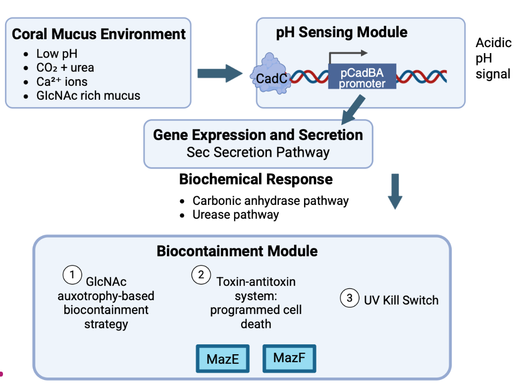 |
|---|
Device level
Our chassis organism, P. rubra, is a marine microorganism that naturally inhabits the coral’s mucus layer. Previous research indicates that this bacterium possesses a mutualistic role by protecting corals from disease. Specifically, evidence suggests P. rubra plays a significant role in defending coral from Gram-positive pathogens (Shnit-Orland et al., 2012). This proposal hypothesizes that engineering a bacterium with symbiotic properties increases the likelihood of a seamless integration within the coral microbiome without causing ecological imbalances.
Our design contains four modules: an environmental sensing module (CadC/pCadBA system); a biochemical-response module (dual buffering pathways); a secretion module (pelB exporting system); and a biocontainment control module (glmS-dependent design).
Environmental sensing module: CadC/pCadBA
Our design aims to integrate a sensor device (CadC/pCadBA system) that detects low environmental pH by gauging high H+ concentration. Its role is to convert chemical stress signals from elevated proton levels into a gene-expression response. This sensing system is based on the Escherichia coli CadC/pCadBA acid-stress regulatory system, which will be adapted to create a pH-sensitive transcriptional switch in the bacteria (see Figure 2). CadC is a membrane-integrated transcriptional activator that detects extracellular proton concentration changes. The CadC/pCadBA system functions as a threshold biological switch. Under relatively neutral water conditions (pH ~8.1-8.2), CadC remains inactive. When environmental acidification increases extracellular H+ concentration, CadC changes conformation and activates a pH-responsive promoter, pCadBA (Haneburger et al., 2011). Subsequently, a downstream expression of genes is triggered.
| Figure 2: Schematic of the pH-responsive CadC/pCadBA regulatory module. Under conditions of low extracellular pH and lysine availability, the periplasmic pH-sensing domain of CadC is activated. In this image, the CadC/pCadBA module is used as a pH-responsive switch to control engineered genes. The native cadA and cadB functions are shown only for context and are not used (Schlundt et al., 2017). | 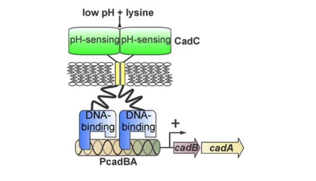 |
|---|
Biochemical response module
Under low extracellular pH and in the presence of lysine, the pH-sensing domain of CadC is activated. This allows CadC to dimerize and bind to the pH-responsive promoter pCadBA, turning on downstream gene expression. In our proposed system, the CadC/pCadBA module functions as a pH-responsive switch to control engineering genes. The native cadA and cadB genes are shown only for biological context and are not used in our design. (Schlundt et al., 2017). Once the CadC/pCadBA system is activated under acidic conditions, two complementary enzymatic pathways (a carbonic anhydrase pathway and a ureolytic pathway) are activated to chemically buffer the internal environment of the coral microbiome and restore chemical balance (see Figure 3).
| Figure 3: CadC/pCadBA-mediated dual buffering strategy in the coral microbiome under acidic stress. | 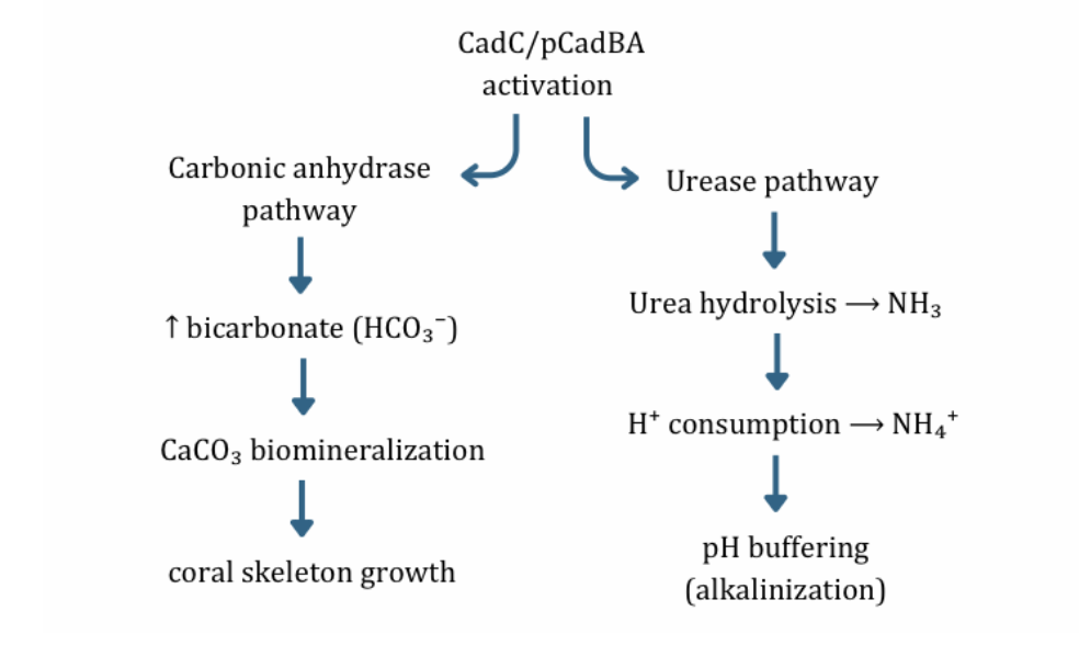 |
|---|
Following cadC/pCadBA activation, the carbonic anhydrase pathway will trigger the expression of large quantities of carbonic anhydrase (CA), which accelerates the reverse hydration of carbon dioxide: CO₂ + H₂O ⇌ H₂CO₃ ⇌ HCO₃⁻ + H⁺. While this reverse reaction does not completely remove protons from the cellular environment, it enhances local bicarbonate (HCO₃⁻) availability. This reaction provides essential substrates for calcium carbonate (CaCO₃) biomineralization: Ca²⁺ + HCO₃⁻ → CaCO₃ ↓ + H⁺. Thus, this pathway primarily accelerates the generation of calcium carbonate (CaCO₃), which is the most needed element for coral skeleton building (Hwang et al., 2021).
It also needs to be considered that cadC/pCadBA activated carbonic anhydrase activity is impacted by the Zinc availability of the environment. As in E.coli, Zinc acts as the essential catalytic cofactor that binds to the CA’s active site, required for the function of the enzyme(Cronk et al., 2001). However, the ocean has many other metallic ions dissolved within, including Mg2+, Ca2+, trace Cu2+, Ni2+, Co2+, Cd2+, Mn2+, Fe2+/ Fe3+ (Cheng & Gross, 2005). However, not all these elements compete effectively with each other nor with Zinc. Mg2+ and Ca2+ don’t substitute Zinc effectively as either their bond with the CA active site is too weak or wrong geometrically (Ohta & Hartwig, 1995; Strecker et al., 1987). Cu2+, Ni2+ and Cd2+are more realistic competitors for Zinc. Specifically, Ni-substituted CA can often become catalytically impaired, Cu-substituted CA can be strongly inhibited. (Yano et al., 2006; Kekki & Nikkilä, 1975; Lazar et al., 1970; Rossetti et al., 1984). In addition, Cadmium bonding with CA is rare because CA is evolved to preferentially bind to Zinc which has the optimal size, coordination geometry and catalytic properties for rapid Carbon dioxide hydration. Therefore, though it is true that there are cases where CA binds with Cd2+ and that results in CA becoming much less efficient, CA binding to Cd2+ only happens in some marine diatoms where Zinc is extremely scarce in sea water (Xue et al., 2008; Smith & Bryant, 1975). However, metal ions that either partially or totally impair CA activity are all trace elements, meaning they are dissolved in the ocean in relatively small amounts. Specifically, while Zinc has a molar concentration of 2.1 × 10⁻⁷ M, Cobalt has a concentration of 1.7 × 10⁻⁹ M, Nickel has 8.5 × 10⁻⁹ M and Cadmium has 5× 10⁻¹⁰– 1× 10⁻⁹ M (University of Washington, n.d.; Bruland & Lohan, 2003). Therefore, there is vastly a higher chance for the CA of our chassis to bond with Zinc and function rather than being impaired upon bonding with other competitively inhibiting metallic ions.
The ureolytic system (ureA/ureB/ureC) catalyzes the hydrolysis of urea (CH₄N₂O), producing ammonia (NH₃) and carbon dioxide (CO₂). Because CaCO3 precipitation releases H+, this NH3 production is necessary to offset the potential increase in local acidity. Therefore, for the ureolytic system to have a net positive effect on local pH, NH3 generation should be greater than or equal to twice of the CaCO3 precipitation rate. The resulting ammonia can further react with protons (H⁺) and water to form ammonium (NH₄⁺) and hydroxide ions (OH⁻). In this way, this pathway provides a direct protonconsuming and hydroxide-generating alkalinization mechanism, reducing the local hydrogen ion (H⁺) concentration. These chemical reactions consequently increase pH, helping to restore the coral’s microenvironment toward more favorable conditions as a partial mitigation strategy for acidification (Haneburger et al., 2011).
Carbonic anhydrase accelerates the reverse hydration of carbon dioxide using a zinc-bound hydroxyl ion generated by ionisation of the Zn²⁺-OH2 as the nucleophile. A common metal ion that exists in the coral mucus layer as a micropollutant is copper (Cu²⁺). Copper can act as a competitive inhibitor of carbonic anhydrase, inhibiting the reverse hydration of carbon dioxide. Increasing the concentration of CA contained in the coral mucus layer can greatly increase the rate of hydration even with the inhibition, without having to incorporate another enzyme that may alter the function of the buffer system (Johnsen et al., 2023).
Together, the carbonic anhydrase and urease pathway formulate an integrated buffering strategy that facilitates chemical stabilization and biological support within the coral microenvironment; the carbonic anhydrase pathway supports coral skeleton rebuilding and growth, while the urease pathway provides rapid pH buffering, which is important because it creates a safe environment for corals where the pH value is habitable.
Secreting module: pelB exporting system
To direct enzyme buffering activities outside of the cytoplasm of the engineered P. rubra, we aim to design a secretion device with the specific function of enzyme export. This step is crucial for successful buffering of the acidic conditions contributing to coral skeleton weakening, as buffering must occur outside of the cell of the engineered chassis where acidity is present. The secretion device, consisting of a signal peptide component (pelB) exports enzymes to the coral tissue and surrounding mucus chemical environment (see Figure 4). The N-terminal pelB signal peptide is fused to each enzyme coding sequence. pelB directs nascent proteins to Sec translocon, enabling translocation across the inner membrane into the periplasm and subsequent extracellular localization. This ensures that enzymatic activities occur directly within the coral mucus microbiome rather than intracellularly, where it would be functionally irrelevant to seawater pH regulation (Kim & Kendall, 2000; Su et al., 2015).
| Figure 4: PelB‑fused enzymes are exported via the SecA‑dependent post‑translational pathway (left), involving SecB chaperoning and translocation through the SecYEG complex (Oswald et al., 2021). | 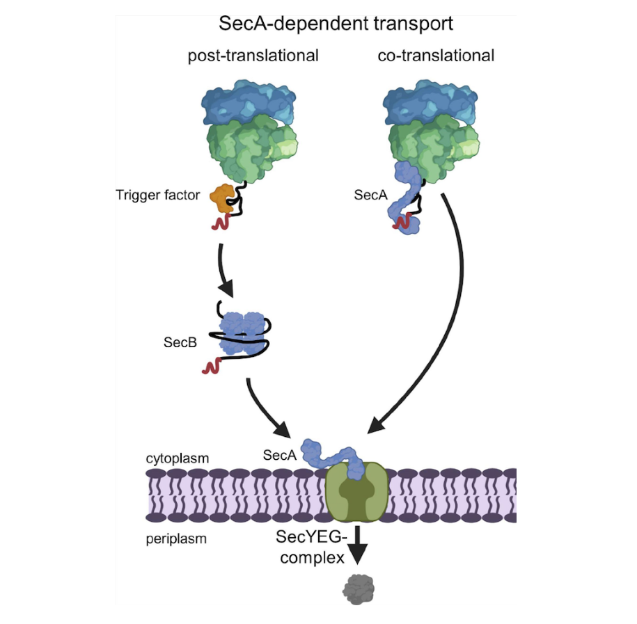 |
|---|
Biocontainment module
Finally, a biocontainment device will remove P. rubra’s ability to synthesize N-acetylglucosamine (GlcNAc), which is required for bacterial wall synthesis. The glmS gene encodes the glucosamine-6-phosphate synthase, an essential enzyme for the biosynthesis of UDP-N-acetylglucosamine (UDP-GlcNAc), a key precursor for peptidoglycan cell wall formation (see Figure 5). In the gene strain, glmS is knocked out, so the survival of P. rubra depends on external availability of GlcNAc. The coral mucus layer contains glycoprotein-rich mucins that provide sufficient GlcNAc derivatives, enabling survival in the target niche. In contrast, the open ocean environment lacks sufficient GlcNAc concentrations, resulting in loss of availability outside the coral-associated habitat. This deficiency prevents proper synthesis of N-acetylglucosamine-containing peptidoglycan precursors, impairing cross-linking of the bacterial cell wall. Consequently, the structural integrity of the peptidoglycan layer is compromised, ultimately leading to bacterial cell lysis. This system projects passive containment, ensuring that the engineered bacterium survives only in the coral mucus layer; it would die naturally if it leaked into open marine environments where GlcNAc is scarce (Konopka, 2012).
| Figure 5: GlcNAc auxotrophy‑based biocontainment strategy. Knockout of glmS disrupts UDP‑GlcNAc biosynthesis required for peptidoglycan formation, restricting survival of engineered P. rubra to GlcNAc‑rich coral mucus environments (Konopka, 2012). | 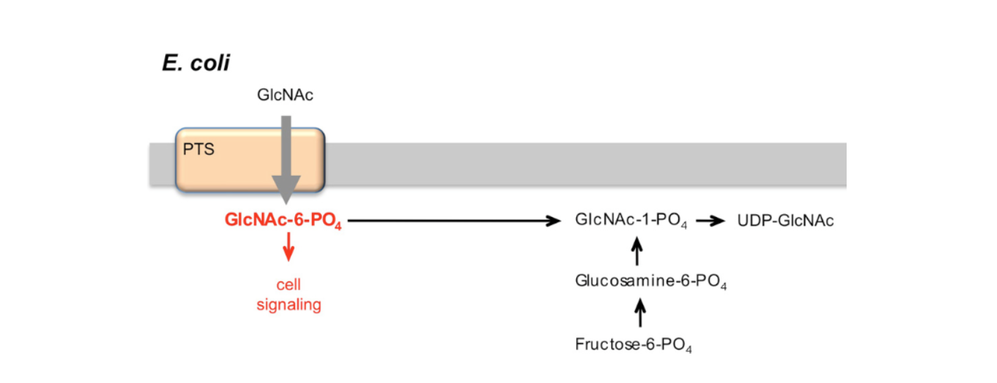 |
|---|
Parts level
pH sensor
The pCadBA promoter is a pH-sensitive ‘switch’ activated by the membrane-bound transcription factor CadC, which helps E. coli combat acidic stress. CadC is a transcriptional activator bound to the inner membrane with a disulfide bond which consists of a periplasmic domain that senses a decrease in pH and a cytoplasmic DNA-binding domain. When CadC gets exposed to a pH below 6.0, it undergoes a conformational change where its disulfide bonds are unable to form and coupled with the encounter with lysine from lysine permease, fully activates CadC (Tetsch et al., 2011). Then CadC binds to Cad1 and Cad2 in the CadBA promoter resulting in the transcription of the CadBA gene, and after going through translation, the pCadBA gene secretes proteins CadA and CadB. The enzyme CadA utilises protons and lysine to create cadaverine, and the antiporter CadB facilitates the intake of lysine in exchange for the export of cadaverine. This provides a constant supply of lysine for CadA and removes cadaverine, preventing intracellular toxicity. This mechanism acts as a pH-dependent switch to restore homeostasis and neutralize acidic pH (Haneburger et al., 2011).
Carbonic anhydrase pathway
The ureolytic pathway, which is mentioned below, can contribute to high excess levels of ammonia which can be toxic to the coral surface. The gene that encodes for carbonic anhydrase, the taCa gene, is controlled by the T7lac promoter, meaning that another molecule that prevents the repressor from binding to the T7lac promoter is necessary for taCa to be transcribed and to create carbonic anhydrase (“T7 Promoter System”). Carbonic anhydrase allows the cell to turn CO2 and water into HCO3– and H+ (acid). It uses a zinc ion (Zn2+), which activates a water molecule to form a hydroxide ion. This ion then has a nucleophilic attack on CO2 which creates bicarbonate (HCO3–). Additionally, although hydroxide production is accompanied by proton (H+) generation, these protons are rapidly neutralized by ammonia from the ureolytic process, reducing the acidity while simultaneously converting ammonia into its less toxic ammonium form (Kim et al., 2021).
Ureolytic pathway
ureA, ureB, and ureC are the urease genes, with accessory genes ureD, ureE, ureF, and ureG, which are responsible for the ureolytic part of the process that breaks down urea into carbon dioxide and ammonia. ureC codes for the active site, which holds 2 nickel ions (Ni2+), and has protein ‘flaps’ that allow in urea before closing to isolate the reaction area. The ureA gene creates the protein UreA, which holds the enzyme together, while ureB creates the protein UreB, which forms the docking sites for UreD, UreF, and UreG to attach to the enzyme and deliver the 2 nickel ions supplied by UreE (Konieczna et al., 2012).
pelB sequence
The pelB leader sequence acts as a signal peptide that directs the synthesized proteins (such as carbonic anhydrase) to the periplasmic space. By localizing these enzymes near the outer membrane, the bicarbonate produced can more efficiently exit the cell and react with environmental calcium to form calcium carbonate that precipitates on the coral surface, which allows corals to reinforce their skeletons (Lei et al., 1987).
Kill switch
There is a kill switch to ensure that this bacteria does not escape the coral and cause damage outside its intended environment. By getting rid of its ability to make N-acetyl glucosamine (GlcNAc) by knocking out the glmS gene, we ensure that the engineered P. rubra cannot leave the N-acetyl glucosamine-rich coral mucus by making it an auxotroph dependant on its intended environment, the coral. Without this sugar, the bacteria can’t build a cell wall which leads to its lysis and ultimate cell death (Lee et al., 2016).
In addition to nutrient dependency, the system incorporates a toxin-antitoxin regulatory module based on the mazF/mazE system. The toxin gene mazF is constitutively expressed, while the antitoxin gene mazE is expressed only under conditions specific to the coral mucus layer. If bacteria leave the mucus layer environment, mazE expression stops while mazF continues to be expressed, resulting in programmed cell death. This system provides an additional layer of security for environmental containment that is dependent on nutrient availability.
A third containment layer involves a UV-responsive kill switch. Outside the layers of the mucus layer, the bacteria are exposed to heightened levels of UV radiation. Employing UV-sensitive promoters can be used to activate toxin expression under external environmental conditions.
Together, these containment mechanisms are designed to reduce the likelihood of bacterial survival beyond its extended niche.
Operon structure
The operon structure will include an inducible promoter and structural operon. The structural operon is controlled by a single inducible promoter, pCadBA, which allows for the transcription of the structural operon once CadC attaches to it. The predicted benefit of implementing an inducible structural operon is to control the expression of carbonic anhydrase and urease genes, such that expression occurs only when environmental pH drops below a threshold sufficient to activate CadC. As environmental pH returns toward homeostasis, CadC activity decreases and pCadBA-driven transcription is passively downregulated, preventing excess alkalinization of the surrounding microenvironment (Tetsch et al., 2011).
Safety
While this study focuses on aiding corals in their production of calcium carbonate, through the chassis organism P. rubra, safety remains a central consideration throughout the future design and testing phases.
Thus, there are a few environmental risks that must be considered. Firstly, the genetic material of the engineered bacteria could potentially spread to other naturally occurring bacteria in an environment through horizontal gene transfer, which could unintentionally disrupt natural ecological systems. To reduce this risk, the plasmid could be designed without an oriT, or origin of transfer, a plasmid sequence which is required to initiate DNA transfer between bacteria (Zrimec & Lapanje, 2018). As a safety measure already incorporated, the plasmid uses a non-mobilizable backbone. Additional containment features can be added once real-time data from microcosm trials reveal the true rates of gene transfer when in the environment of a coral reef. Another way to reduce this risk is using a narrow-host-range oriT, which could potentially restrict replication of plasmids if it were transferred between bacteria. Another potential environmental risk is the disruption of natural ecosystems, since the coral mucus layer has many different microbial communities that are important for coral immunity, and disease resistance. Potentially, this plasmid could outcompete native bacteria and inadvertently weaken natural defenses of the coral. It can also have adverse effects on the non-coral organisms, if the plasmid were to spread past the coral environment. One way this can be managed is through feedback inhibition, where an excess of the product inhibits further production.
One way to contain the engineered bacteria so it only stays within the coral mucus layer, is to knockout glmS encodes, an enzyme that is responsible for synthesizing UDP-N-acetylglucosamine (fructose-6-phosphate and glutamine) into N-acetylglucosamine (Kim et al., 2013). By getting rid of the glmS gene in the bacteria, they depend on the coral mucus layer for N-acetylglucosamine (GlcNAc) and will not be able to function away from it, for example if they were in another environment. By getting rid of glmS in the bacteria, they depend on the coral mucus layer for glucosamine and will not be able to function away from it, for example if they were in another environment. This essentially operates as a kill switch if the bacteria leaves the coral mucus layer. To further monitor containment, fluorescent markers should also be incorporated into the bacteria to trace the movement and persistence of the bacteria in the coral biome.
In addition to this, there are biological risks that must be considered. In this study, there is a risk of the overproduction of ammonia, which is a product of the urease pathway. The urease pathway hydrolyzes urea into ammonia. Even though it is used to buffer acidification, excess ammonia is toxic to marine organisms and life. If the urease activity is unregulated, bacterial density is too high, or any other malfunction in the bacteria occurs, ammonia levels could skyrocket, harming coral tissue and life around the coral. To prevent this, urease expression could be tightly coupled to a promoter system (pCadBA ph-Sensitive), that way it is only active when the pH drops (Pflock et al., 2005).
Beyond the plasmid itself, some broader concerns exist around the introduction of an engineered bacteria into the coral microbiome. Through a process known as microbiome shifting, beneficial microbes are meant to shift the coral microbiome, to bring it to a healthier state when under stress from high water temperatures. However, engineered strains may compete with native microbes for resources, potentially disrupting the coral’s natural adaptive capacity to climate change. Ethically, some critics believe that deliberately altering a marine ecosystem already under stress may be unhelpful and the long-term consequences of introducing foreign bacteria have not been thoroughly studied (Zhang et al., 2022).
Nevertheless, as our research continues, further studies will be required to ensure the engineered bacteria does not leave the coral mucus layer, nor does it negatively affect the coral itself, and that it is suitable for use in the ocean ecosystem.
Discussions
Ocean acidification is a serious ecological threat caused by excess human‑emitted CO₂ dissolving in seawater, forming carbonic acid that releases hydrogen ions and lowers ocean pH (Mollica et al., 2018). This proposal aims to address the adverse effects of ocean acidification on the structural stability of coral skeletons by locally reducing acidity in the coral mucus layer. We will engineer P. rubra with an integrated four-module genetic system consisting of: an environmental sensing module (CadC/pCadBA system); a biochemical-response module (dual buffering pathways); a secretion module (pelB exporting system); and a biocontainment control module (glmS-dependent design).
The successful implementation of this strategy will provide a sustainable and feasible method to enhance coral’s resilience against ocean acidification. Unlike other solutions that are experiencing limits in keeping pace with the rate of global reef collapse (Erickson-Davis, 2025), our strategy is executed by a living chassis which provides a more effective way to combat the degradation of corals which are a keystone species in these communities and an environment that fosters a plethora of marine biodiversity.
While this study outlines an approach to improve coral mitigate pH stress in the coral microenvironment while maintaining ecological safety, several limitations must be acknowledged.
The scope of this study is ambitious and attempts to incorporate several genetic modules that have been studied independently. Consequently, the four systems integrated in our chassis organism may behave differently under varying environmental factors, such as temperature fluctuations, UV radiation, changes in salinity, and chemical shifts in ocean water conditions. Uneven performance across individual modules may weaken the sequential design of the four-module genetic system, as untested environmental variables may delay or disable intended design functions. Elevated temperatures, especially those exceeding typical reef conditions, can stress bacterial cells, disrupt protein folding, and reduce the efficiency of key enzymes involved in the buffering and calcium carbonate production pathways (van Woesik et al., 2021). Similarly, increased UV exposure can damage DNA and cellular structures, potentially impairing the function of the engineered genetic modules (Kciuk et al., 2020). Fluctuations in salinity can also affect osmotic balance within the bacterial cells, leading to reduced viability or metabolic activity (Gregory & Boyd, 2021). Together, these stressors may decrease the survival, consistency, and overall effectiveness of the engineered P. rubra within the coral mucus layer. Addressing these environmental challenges is essential to ensure that the system remains functional and reliable under the dynamic conditions of natural reef ecosystems.
The last step of the urease pathway involves the formation of carbon dioxide and ammonia. In the absence of similar levels of carbonic anhydrase to neutralize the toxicity of ammonia created by the urease pathway, increasing levels of ammonia may cause concern within the intracellular space of P. rubra. If excess ammonia is able to build up it could create the conditions to spark an algal bloom in the coral reef (Karcher et al., 2020).
Because our genetic system relies on the inducible expression of complex enzymatic pathways, its operation may pose a significant metabolic cost. This potential energetic trade-off highlights the need for potential optimization of expression levels and regulatory thresholds (Snoeck et al., 2024).
Next steps
If the experimental data supports the study’s hypothesis—that a combined approach via the bioengineering of P. rubra chassis with a sensor, response, secretion and biocontainment device is an effective solution to combating the negative effects of ocean acidification on coral skeleton strength—a novel synthetic biology approach to enhancing marine ecosystem resilience could be explored.
Individual validation of genetic modules
We will first validate the functionality of the environmental sensing module (CadC/pCadBA system) to ensure that it can reliably detect acidified conditions. The pCadBA promoter will be cloned upstream of the reporter gene and expressed in E. coli to evaluate pH responsiveness. Engineered strains will be cultured under two conditions: acidic (pH 5.0-5.8) and near-neutral (pH 7.4-7.6) both in the presence of lysine. High lysine levels allow the change in shape of the LysP protein which in neutral pH, would bind to the CadC and inhibit its activation. In this case, the high pH medium will be the control factor as the CadC regulator will only be activated in low pH levels. The two engineered strains will be left to grow. Analyzing the results, the medium with the low pH should show more enzymatic activity than in the high pH medium. Examining the pH response test of the pCadBA promoter, as acidity of the oceans increases, the CadC will activate the pCadBA promoter and the CadBA operon.
To validate the effectiveness of the integrated buffering module, we will assess the activity of urease and Carbonic anhydrase under different conditions of pH. Urease activity will be evaluated through pH-sensitive strips that detect ammonia production, where a color change from yellow to red indicates increased pH due to hydrolysis. Carbonic anhydrase activity will be measured using CO2 sensitive indicators, with a shift from blue to yellow reflecting changes in acidity. Following known assay protocols, we will use the pH indicator strips to validate each enzyme function reliably under relevant conditions (Medina Ferrer et al., 2020). Following this confirmation, the reporter gene will be replaced with functional enzymes from the biochemical response module, including Carbonic anhydrase and urease, to evaluate whether pH-dependent activation leads to measurable changes in local chemical conditions such as pH and carbonate availability. Once both the sensing and response modules are validated, they will be integrated into a single system to enable coordinated detection and buffering of acidified environments.
To validate the secretion module (pelB), we will test the secretion system of the pelB in E. coli, using a reporter gene to ensure that it will be able to export the taCa for effective pH increasing of the coral's mucus, creating a buffer system. In this case, we will measure the amount of cutinase in a culture medium with a strain of E. coli. with a defective type II secretion pathway. Type II secretion pathways guide the secretion of the cutinase, a fusion protein, so with this pathway being defective we can test the level of efficiency for pelB (Su et al., 2015).
To validate the biocontainment control module, the E. coli will be engineered with a knockout of the glmS gene which produces N-acetyl glucosamine, a necessary sugar to the bacteria commonly found in coral mucus. When it leaves the coral mucus area, the bacteria will be unable to get the N-acetyl glucosamine, and will die. This safeguard prevents uncontrolled spread and ensures that the system remains localized and ecologically contained. To gauge effectiveness of our biocontainment control module, we will test the survival of P. rubra under the absence of the glmS gene to ensure that the bacteria will not be able to thrive under the conditions outside the localized coral mucus layer, preventing any excess spread. We will prepare two cultures, both with the presence of the E. coli bacteria, one with the glmS gene which produces N-acetyl glucosamine and the other with the knockout of the glmS gene. N-acetyl glucosamine is a sugar that is a necessary nutrient for the bacteria’s survival, indicating that if the culture with the knockout of the glmS shows no population of the bacteria, our design that contains bacteria within the coral mucus layer will be validated. Our next step would be to test the bacteria without the glmS gene in a controlled simulation of the coral environment.
Optimization
Following initial validation of the individual and combined systems, we will focus on refining regulatory control and functional output. Because the pCadBA promoter is regulated by the CadC transcriptional system, promoter strength and basal expression levels can be tuned by modifying accessibility to the pCadBA promoter through gene expression techniques. As a result, we will aim to more precisely control gene expression in response to acidic conditions.
Parallel optimization of the buffering system will require adjusting bacterial density to determine the minimum effective bacterial population needed to achieve successful stabilization and increase of pH without disrupting the native coral microbiome.
Transformation
Following successful validation of the system in E. coli, we will assess whether the engineered plasmid can be introduced in P. rubra and support stable gene expression. This process would involve temporarily increasing permeability of the bacterial cell membrane for transformation, and involves chemical treatment to reduce electrostatic repulsion between negatively charged DNA and the bacterial cell surface. To ensure enhanced membrane permeability and the uptake of the plasmid, the bacteria cells will be incubated in CaCl2, and 10 picograms-100 nanograms of plasmid DNA will be added to the competent cells. After mixing, the DNA cell competent mixture will be heat shocked by placing it in water at a temperature of 42°C for about 45 seconds, then placing it in ice for 2 minutes. After the heatshock, we will allow the bacterial cells to grow overnight. The P. rubra will pick up the plasmid originated from the E. coli, and grow in its population, allowing transformation of the E. coli plasmid to the P. rubra
.
Microbiome analysis
To further validate our design, we can test the system in a simulated coral mucus environment, incorporating the engineered P. rubra into a living coral that is isolated and held in a stable laboratory environment. By testing the coral and P. rubra system in isolated laboratory conditions, we aim to troubleshoot safety concerns and limit the unwanted, adverse effects on other marine organisms within the same ecological niche. The laboratory conditions found to be most effective in reducing coral acidification and protecting coral from skeletal breakdown from individual testing will likely be used for experimentation with more complex laboratory conditions, such as multiple corals living in a simulated marine ecosystem. Factors such as ideal temperature conditions, nutrient availability, strain of coral, location (latitude and longitude), and density and number of P. rubra will all be factors simulated and considered in laboratory analysis.
Only once the engineered system has been rigorously tested in a controlled setting—with all relevant safety and ecological parameters that will be determined by coral type, ocean conditions, location, microbiome, and guidelines of local water safety laws—will P. rubra be incorporated within a more complex ecological environment with other organisms and corals.
By doing so, we will be able to ensure that the design will be functional for the localized microenvironment which could be used as a solution to the coral survival in seas with rising acidity levels without hindering the delicate coral microbiome.
Author contributions
All authors made contributions to the conceptualization, design, and execution of this project. Each author was involved in writing the various sections of the manuscript.
Acknowledgements
We would like to thank Mr. Hobbs for his continuous support throughout the process, assisting us in our project, keeping us on track, and teaching us the foundational concepts in biology and scientific communication we needed to succeed.
References
Chiriac, E. R., Chiţescu, C. L., Borda, D., Lupoae, M., Gird, C. E., Geană, E.-I., Blaga, G.-V., & Boscencu, R. (2020). Comparison of the Polyphenolic Profile of Medicago sativa L. and Trifolium pratense L. Sprouts in Different Germination Stages Using the UHPLC-Q Exactive Hybrid Quadrupole Orbitrap High-Resolution Mass Spectrometry. Molecules, 25(10), 2321. https://doi.org/10.3390/molecules25102321
Doney, S. C., Fabry, V. J., Feely, R. A., & Kleypas, J. A. (2009). Ocean acidification: The other CO2 problem. Annual Review of Marine Science, 1(1), 169–192. https://doi.org/10.1146/annurev.marine.010908.163834
Erickson-Davis, M. (2025, April 29). Scientists warn coral restoration can’t keep pace with global reef collapse. Mongabay Environmental News. https://news.mongabay.com/2025/04/scientists-warn-coral-restoration-cant-keep-pace-with-global-reef-collapse/
Escherichia coli K-12 substr. MG1655 cadC. (2026). Biocyc.org. https://biocyc.org/gene?orgid=ECOLI&id=EG10133
Fleurier, S., & Matic, I. (2025). MazF endoribonuclease promotes resolution of transcription-replication conflicts at ribosomal RNA genes in Escherichia coli. Nucleic acids research, 53(19), gkaf1034. https://doi.org/10.1093/nar/gkaf1034
Gregory, G. J., & Boyd, E. F. (2021). Stressed out: Bacterial response to high salinity using compatible solute biosynthesis and uptake systems, lessons from Vibrionaceae. Computational and structural biotechnology journal, 19, 1014–1027. https://doi.org/10.1016/j.csbj.2021.01.030
Haneburger, I., Eichinger, A., Skerra, A., & Jung, K. (2011). New Insights into the Signaling Mechanism of the pH-responsive, Membrane-integrated Transcriptional Activator CadC ofEscherichia coli. Journal of Biological Chemistry, 286(12), 10681–10689. https://doi.org/10.1074/jbc.m110.196923
In Seong Hwang, Joo Hyeon Kim, & Byung Hoon Jo. (2021). Enhanced Production of a Thermostable Carbonic Anhydrase in Escherichia coli by Using a Modified NEXT Tag. 26(19), 5830–5830. https://doi.org/10.3390/molecules26195830
Johnsen, L. (2023). Zinc–Lewis Acid and Gene Regulator. 10.1016/b978-0-12-819509-3.00010-9. https://www.sciencedirect.com/topics/neuroscience/carbonic-anhydrase.
Karcher, D. B., Roth, F., Carvalho, S., El-Khaled, Y. C., Tilstra, A., Kürten, B., Struck, U., Jones, B. H., & Wild, C. (2020). Nitrogen eutrophication particularly promotes turf algae in coral reefs of the central Red Sea. PeerJ, 8, e8737. https://doi.org/10.7717/peerj.8737
Kciuk, M., Marciniak, B., Mojzych, M., & Kontek, R. (2020). Focus on UV-Induced DNA Damage and Repair-Disease Relevance and Protective Strategies. International journal of molecular sciences, 21(19), 7264. https://doi.org/10.3390/ijms21197264
Kim, J., & Kendall, D. A. (2000). Sec-dependent protein export and the involvement of the molecular chaperone SecB. Cell Stress & Chaperones, 5(4), 267. https://doi.org/10.1379/1466-1268(2000)005%3C0267:sdpeat%3E2.0.co;2
Kim, K., Jeong, J. H., Lim, D., Hong, Y., Yun, M., Min, J.-J., Kwak, S.-J., & Choy, H. E. (2013). A Novel Balanced-Lethal Host-Vector System Based on glmS. PLoS ONE, 8(3), e60511. https://doi.org/10.1371/journal.pone.0060511
Konieczna, I., Zarnowiec, P., Kwinkowski, M., Kolesinska, B., Fraczyk, J., Kaminski, Z., & Kaca, W. (2012). Bacterial Urease and its Role in Long-Lasting Human Diseases. Current Protein and Peptide Science, 13(8), 789–806. https://doi.org/10.2174/138920312804871094
Konopka, J. B. (2012). N-Acetylglucosamine Functions in Cell Signaling. Scientifica, 2012, 1–15. https://doi.org/10.6064/2012/489208
Lee, S. T. M., Davy, S. K., Tang, S.-L., & Kench, P. S. (2016). Mucus Sugar Content Shapes the Bacterial Community Structure in Thermally Stressed Acropora muricata. Frontiers in Microbiology, 7. https://doi.org/10.3389/fmicb.2016.00371
Lei, S. P., Lin, H. C., Wang, S. S., Callaway, J., & Wilcox, G. (1987). Characterization of the Erwinia carotovora pelB gene and its product pectate lyase. Journal of Bacteriology, 169(9), 4379–4383. https://doi.org/10.1128/jb.169.9.4379-4383.1987
Li, J., Yang, Q., Dong, J., Sweet, M., Zhang, Y., Liu, C., Zhang, Y., Tang, X., Zhang, W., & Zhang, S. (2022). Microbiome Engineering: A Promising Approach to Improve Coral Health. Engineering. https://doi.org/10.1016/j.eng.2022.07.010
Liu, C., Xiao, Y., Xiao, Y., & Li, Z. (2020). Marine urease with higher thermostability, pH and salinity tolerance from marine sponge-derived Penicillium steckii S4-4. Marine life science & technology, 3(1), 77–84. https://doi.org/10.1007/s42995-020-00076-6
Medina Ferrer, F., Hobart, K., & Bailey, J. V. (2020). Field detection of urease and carbonic anhydrase activity using rapid and economical tests to assess microbially induced carbonate precipitation. Microbial Biotechnology, 13(6), 1877–1888. https://doi.org/10.1111/1751-7915.13630
Mollica, N. R., Guo, W., Cohen, A. L., Huang, K.-F., Foster, G. L., Donald, H. K., & Solow, A. R. (2018). Ocean acidification affects coral growth by reducing skeletal density. Proceedings of the National Academy of Sciences, 115(8), 1754–1759. https://doi.org/10.1073/pnas.1712806115
Ocean Portal Team. (2018, December 18). Ocean Acidification. Smithsonian Ocean. https://ocean.si.edu/ocean-life/invertebrates/ocean-acidification
Oswald, J., Njenga, R., Natriashvili, A., Sarmah, P., & Koch, H.-G. (2021). The Dynamic SecYEG Translocon. Frontiers in Molecular Biosciences, 8, 664241. https://doi.org/10.3389/fmolb.2021.664241
Pflock, M., Kennard, S., Delany, I., Scarlato, V., & Beier, D. (2005). Acid-Induced Activation of the Urease Promoters Is Mediated Directly by the ArsRS Two-Component System of Helicobacter pylori. Infection and Immunity, 73(10), 6437–6445. https://doi.org/10.1128/iai.73.10.6437-6445.2005
Phartade Vilas Bhagwat, Ravindran, C., & Lawrance Irudayarajan. (2024). Beneficial properties of mucus in coral adaptations and ecological interactions. Marine Biology, 171(2). https://doi.org/10.1007/s00227-023-04372-4
Schlundt, A., Buchner, S., Janowski, R., Heydenreich, T., Heermann, R., Lassak, J., Geerlof, A., Stehle, R., Niessing, D., Jung, K., & Sattler, M. (2017). Structure-function analysis of the DNA-binding domain of a transmembrane transcriptional activator. Scientific Reports, 7(1). https://doi.org/10.1038/s41598-017-01031-9
Shnit-Orland, M., Sivan, A., & Kushmaro, A. (2012). Antibacterial Activity of Pseudoalteromonas in the Coral Holobiont. Microbial Ecology, 64(4), 851–859. https://doi.org/10.1007/s00248-012-0086-y
Snoeck, S., Guidi, C., & De Mey, M. (2024). “Metabolic burden” explained: stress symptoms and its related responses induced by (over)expression of (heterologous) proteins in Escherichia coli. Microbial Cell Factories, 23(1), 96. https://doi.org/10.1186/s12934-024-02370-9
Song, X., Tian, C., Wang, P., Wang, J., Ma, J., Chen, L., & Zhang, W. (2025). Thermal kill-switches based biocontainment for genetically engineered cyanobacteria. Chemical Engineering Journal, Article 171691. https://doi.org/10.1016/j.cej.2025.171691
Su, L., Yu, L., Xu, C., & Wu, J. (2015). Extracellular expression of Thermobifida fusca cutinase with pelB signal peptide depends on more than type II secretion pathway in Escherichia coli. Journal of Biotechnology, 204, 47–52. https://doi.org/10.1016/j.jbiotec.2015.03.029
T7 Promoter System. (2016). Sigmaaldrich.com. https://www.sigmaaldrich.com/US/en/technical-documents/technical-article/genomics/cloning-and-expression/t7-promoter-system?srsltid=AfmBOorV72B2K3SGKLFIgT0Xe-fzYAlGnQzanqi29VFTC2ep3DAd9Drd.
van Woesik, R., & Cacciapaglia, C. W. (2021). Thermal stress jeopardizes carbonate production of coral reefs across the western and central Pacific Ocean. PloS one, 16(4), e0249008. https://doi.org/10.1371/journal.pone.0249008
Vardi, T., Rankin, T., Oliver, T., Moulding, A., Parrish, F., Moore, T., Enochs, I., Viehman, T., & Koss, J. (2020). NOAA Action Plan on Coral Interventions. https://cdn.coastalscience.noaa.gov/cdhc/docs/Vardi-2020-NOAA-Action-Plan-Coral-Interventions.pdf
Wang, X., Wang, M., Jia, Z., Qiu, L., Wang, L., Zhang, A., & Song, L. (2017). A Carbonic Anhydrase Serves as an Important Acid-Base Regulator in Pacific Oyster Crassostrea gigas Exposed to Elevated CO2: Implication for Physiological Responses of Mollusk to Ocean Acidification. Marine biotechnology (New York, N.Y.), 19(1), 22–35. https://doi.org/10.1007/s10126-017-9734-z
Wright, R. M., Strader, M. E., Genuise, H. M., & Matz, M. (2019). Effects of thermal stress on amount, composition, and antibacterial properties of coral mucus. PeerJ, 7, e6849. https://doi.org/10.7717/peerj.6849
Zrimec, J., & Lapanje, A. (2018). DNA structure at the plasmid origin-of-transfer indicates its potential transfer range. Scientific Reports, 8(1). https://doi.org/10.1038/s41598-018-20157-y
Cheng, H., & Gross, R. (Eds.). (2005). Polymer Biocatalysis and Biomaterials. American Chemical Society.
Cronk, J. D., Endrizzi, J. A., Cronk, M. R., O’neill, J. W., & Zhang, K. Y. J. (2001). Crystal structure of E. coli β-carbonic anhydrase, an enzyme with an unusual pH-dependent activity. Protein Science, 10(5), 911–922. https://doi.org/10.1110/ps.46301
Kekki, M., & Nikkilä, E. A. (1975). Turnover of plasma total and very low density lipoprotein triglyceride in man. Scandinavian Journal of Clinical and Laboratory Investigation, 35(2), 171–179. https://pubmed.ncbi.nlm.nih.gov/167428/
Lazar, E. C., Swango, L. J., & Binn, L. N. (1970). Serologic and Infectivity Studies of Canine SV-5 Virus. Experimental Biology and Medicine, 135(1), 173–176. https://doi.org/10.3181/00379727-135-35012
Ohta, Y., & Hartwig, J. H. (1995). Actin Filament Crosslinking by Chicken Gizzard Filamin Is Regulated by Phosphorylation in Vitro. Biochemistry, 34(20), 6745–6754. https://doi.org/10.1021/bi00020a020
Rossetti, O. L., Altman, R., & Young, R. (1984). Kinetics of Tn5 transposition. Gene, 32(1-2), 91–98. https://doi.org/10.1016/0378-1119(84)90036-2
Smith, R. J., & Bryant, R. G. (1975). Metal substitutions in carbonic anhydrase: a halide ion probe study. Biochemical and Biophysical Research Communications, 66(4), 1281–1286. https://doi.org/10.1016/0006-291x(75)90498-2
Strecker, W. B., Wood, M. B., & Schroeder, A. R. (1987). Stasis-induced thrombosis of rat microvascular anastomosis. Journal of Reconstructive Microsurgery, 4(1), 69–73. https://doi.org/10.1055/s-2007-1006966
Xu, Y., Feng, L., Jeffrey, P. D., Shi, Y., & Morel, F. M. M. (2008). Structure and metal exchange in the cadmium carbonic anhydrase of marine diatoms. Nature, 452(7183), 56–61. https://doi.org/10.1038/nature06636
Yano, Y., Ogura, M., & Matsuzaki, K. (2006). Measurement of Thermodynamic Parameters for Hydrophobic Mismatch 2: Intermembrane Transfer of a Transmembrane Helix. Biochemistry, 45(10), 3379–3385. https://doi.org/10.1021/bi052286w
University of Washington, (n.d.). Mineral makeup of seawater. Sea Urchin
Embryology. https://depts.washington.edu/embryology/index.php?id=mineral
Bruland, K., & Lohan, M. (2003). 6.02 Controls of Trace Metals in Seawater. https://www.whoi.edu/cms/files/6.02_32043.pdf