
Ella Jin, Lucia Sostak, Megan Wong, and Katrina Yeung, Concordia International School Shanghai, Shanghai, China
Reviewed on 9 May 2026; Accepted on 22 June 2026; Published on 26 October 2026
With help from the 2026 BioTreks Production Team.
Hepatitis C is a viral disease caused by the Hepatitis C Virus (HCV) that, in its chronic form, can lead to severe liver damage, liver cancer, and in some cases, death. The infection rate of HCV in China remains high, especially in rural and low-income communities, primarily among drug users and blood transfusion recipients. Current detection methods for HCV are often inefficient and expensive, in some cases taking up to 17 days for test results to become available. Due to these issues, as well as the often asymptomatic nature of HCV, the virus often goes undetected until damage to the liver is irreversible. Our objective is to use synthetic biology to create a cost-effective, at-home HCV detection system using HCV RNA biomarkers present in the saliva of those infected with chronic HCV. By creating an at-home detection system using a molecular beacon that binds to the target HCV RNA sequence, living in high-risk areas would easily be able to test themselves for HCV, even in its early stages, helping prevent HCV-caused liver diseases and deaths.
Keywords: DNA probe, HCV RNA, Hepatitis C Virus, molecular beacon, diagnosis
Authors are listed in alphabetical order. David Doyle mentored the group. Please direct all correspondence to mailto:david.doyle@concordiashanghai.org.
Background
Hepatitis C significance
Hepatitis C is caused by the Hepatitis C Virus (HCV), leading to liver inflammation. HCV is a bloodborne virus most commonly transmitted through unsafe injections and procedures during medical care and the sharing of needles and syringes during drug use, as well as sexual contact leading to the exposure of blood. The World Health Organization (WHO) estimates 50 million people living with chronic HCV, with around 1 million new infections each year. Many remain undiagnosed, slowing the progress towards the elimination of the virus (World Health Organization, 2024).
In the first 6 months of infection, HCV is considered to be acute. In some instances, the body will naturally rid itself of an acute infection, meaning that the HCV never progresses into the chronic stage. After six months of infection, HCV is considered to be chronic. If left untreated, chronic HCV is oftentimes a lifelong infection (CDC, 2024b.) Over half of those infected with HCV will develop a chronic infection, with some studies bringing that number up to 80% (Liang et al., 2000).
In China, chronic HCV is a major cause of liver disease, liver cirrhosis, and hepatocellular carcinoma (mainly liver cancer). The infection rate of HCV in China has been increasing yearly (Qin et al., 2013). There are around 360,000 liver cancer deaths every year and HCV infections account for 37.48% (Mei & Lu, 2021). A meta-analysis by Mei & Lu, 2021 estimated a 0.91% HCV antibody presence in China, with a higher prevalence in older populations.
| Figure 1. A diagram that shows the progression of chronic Hepatitis C infection in the liver. After initial infection, the liver progressively deteriorates, leading to severe consequences like cirrhosis and ultimately, liver failure. | 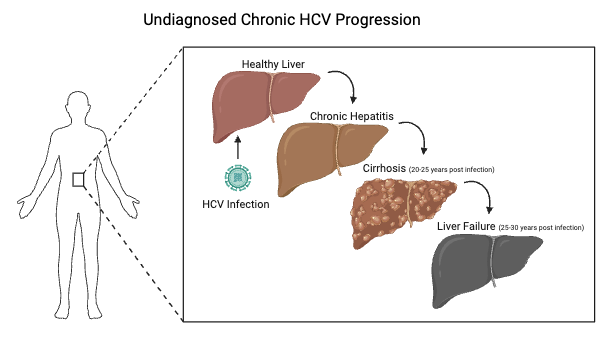 |
|---|
In most people, HCV infection becomes chronic. In these cases, CD8+ T cells become exhausted, losing cytokine production and killing abilities, and CD4⁺ T cells decline, which ultimately weakens the entire immune response. Antibodies appear later and are often isolate-specific, not broadly neutralizing, which allows HCV to mutate away from them (Green et al., 2010).
Current detection methods for Hepatitis C
Current Hepatitis C detection methods are oftentimes costly and take multiple days for results to become available. The current process includes two tests. First, there is an antibody test that determines prior HCV infection. Those who test positive for HCV antibodies then take a RNA NAAT test, which determines if the infection is active in the body. Both of these tests require blood to test for the presence of HCV antibodies and HCV RNA and combined take between 3 to 17 days (CDC, 2024a).
Due to the asymptomatic nature of chronic HCV, many of those infected do not get tested, especially because current testing occurs at hospitals and clinics and includes blood work at a lab or hospital, or the patient sending it to a lab to be tested (CDC, 2024a). There are no current widely used home-based testing methods for HCV RNA in saliva (CDC, 2024a). This is likely due to the fact that HCV RNA has a significantly lower concentration in saliva compared to blood, making detection more difficult and requiring highly sensitive technology only present in the lab (Cameron et al., 1999). Saliva also naturally contains enzymes and other inhibitors that may interfere with the molecular processes (such as PCR) used to amplify and detect viral RNA (Hooshmand et al., 2018).
The problem
HCV predominantly affects low-income populations in rural areas, often due to drug use and unsafe blood transfusions (Kermode, 2004), meaning that many of those infected with chronic HCV do not have access to the resources to be tested before the infection inflicts permanent damage to the liver and other bodily functions. Studies completed by the Chinese Society of Hepatology have indicated that the detection rates of HCV in Mainland China are around 2.1%, meaning that many infected go undiagnosed and therefore untreated (“Guidelines for the Prevention and Treatment of Hepatitis C.,” 2019).
Access to an efficient, low-cost, and at-home testing method for HCV is essential to increasing the rates of HCV detection in rural areas without easy access to hospitals and labs, therefore helping increase the rates of treatment for chronic HCV.
Proposed solution
Our plan is to genetically engineer a product that detects the presence of Hepatitis C RNA (HCV RNA) in saliva in a non-invasive and cost-effective manner for rural areas. Even though HCV RNA has not been clinically approved as an HCV biomarker, many research studies have been conducted. One study tested for HCV RNA in 23 subjects with chronic HCV infection. Results showed that 72% of 474 saliva samples were positive for HCV RNA. Thus, HCV RNA stands as a potential indicator for HCV infection (Wang et al., 2006).
Currently, saliva is gaining attention as a non-invasive and cost-effective alternative to traditional diagnostic methods, such as blood or tissue samples. Though saliva is composed of mainly water, it also contains proteins, enzymes, hormones, nucleic acids (DNA, RNA, miRNA), and metabolites, which can reveal a lot about the body’s overall health. These molecules found in saliva can be used as biomarkers for detection of a variety of diseases, including HCV. Notably, saliva collection is easier, patient-friendly, and does not require specialized medical expertise, making it especially applicable for at-home diagnostic purposes (Surdu et al., 2025).
We plan to engineer a plasmid to produce a strand of HCV RNA that, once binded to HCV RNA, will produce fluorescence. Nucleic acid hybridization exploits the inherent specificity of Watson-Crick base pairing, allowing complementary DNA or RNA strands to selectively bind to target nucleic acid sequences. For RNA viruses such as HCV, hybridization-based detection offers a direct means of sensing viral genomes without requiring enzymatic amplification (Shrivastav, 2025). This specificity makes hybridization probes preferred for distinguishing HCV RNA from host RNA and other viral pathogens.
To detect whether HCV RNA binds to its complementary strand, we will develop a fluorescent hybridization probe. We plan to develop a molecular beacon fluorescent hybridization probe, which consists of single-stranded oligonucleotides forming a stem-loop structure labeled with a fluorophore and a quencher. In the absence of the HCV RNA, fluorescence is quenched, but upon hybridization with complementary RNA, the probe will undergo a conformational change that separates the fluorophore and quencher, resulting in fluorescence emission (Tyagi & Kramer, 1996).
Our proposed solution aims to design a plasmid that will produce HCV RNA, simulating HCV infection in patients. After the HCV RNA is transcribed, it will bind to the loop on the molecular beacon, which will unquench the fluorophore and release fluorescence. The fluorescence will then be detected using a plate reader.
| Figure 2. Our proposed molecular beacon design system. Our designed plasmid aims to simulate the production of HCV RNA in patients, generating an accessible portion of HCV RNA. The partial HCV RNA will then bind with our designed molecular beacon. This will unquench the molecular beacon, resulting in fluorescence. | 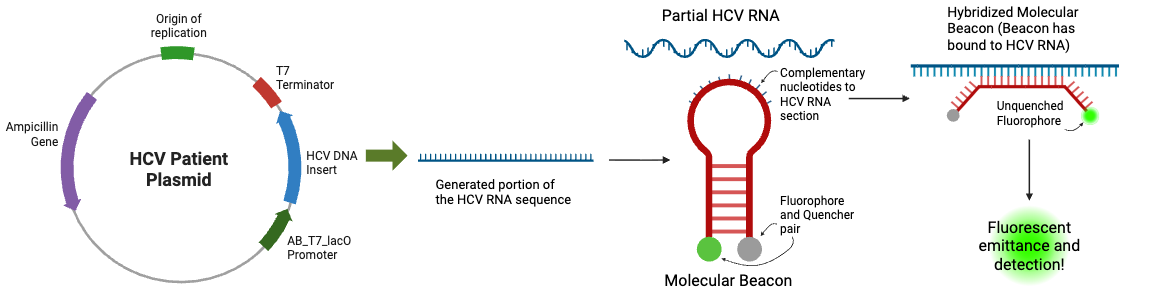 |
|---|
Systems level
Our system as a whole aims to engineer a molecular beacon for the detection of HCV RNA, proving the presence of an active acute or chronic infection. We plan to engineer E. coli strain HT115 (DE3) for the production of a non-viral portion of HCV RNA to act as a placeholder for an HCV patient with active HCV RNA in their saliva. Using Kernel software, we have designed a plasmid which will, once transformed into the E. coli cell and incubated at 37°C overnight, allow for our chosen portion of HCV RNA to be generated within the E. coli cell. Due to our project generating free RNA instead of a protein, E. coli strain HT115 (DE3) was chosen specifically because it has a suppressed ribonuclease III, allowing RNA accumulation in the cell cytoplasm (da Rosa et al., 2024). Once the chosen portion of the HCV RNA, specifically nucleotides 1-300 5’UTR, has been generated by our plasmid within the E. coli cell, cells will be lysed using a bead mill to preserve RNA integrity. The plasmid will also be expressed using a T7 plasmid expression kit, designed for cell-free expression of T7 plasmids, and RNA will be purified using a silica spin column and treated with DNase I to remove contaminant DNA. Using two expression systems, our construct will be tested in both cell-free and cell-based platforms. Once the generated portion of HCV RNA has been extracted and purified, it will be tested with our molecular beacon, which is specific to a portion of the HCV RNA present on the generated strand, nucleotides 117-145 5’UTR. This portion of the HCV RNA was chosen due to its relative accessibility, specificity to RNA, and lack of folding. When the purified HCV RNA is combined with the molecular beacon, the beacon should unfold and bind, unpairing the quencher-fluorophore pair, allowing the beacon to fluoresce in a way that is detectable by fluorescent plate readers. If fluorescence is present, it verifies the presence of the HCV RNA, offering proof of our concept of utilizing molecular beacons for disease diagnosis.
Device level
Our system includes two specifically designed devices: the HCV patient plasmid and the specific molecular beacon designed for diagnosis. All plasmid components were chosen for their relative stability and compatibility within our project targets.
Our chosen chassis is the E. coli strain HT115(DE3) as it is an RNase III-deficient strain of E. coli, meaning that our final product will not be quickly degraded by the cell. As our system is not producing a protein but instead is producing an RNA strand to replicate a portion of HCV RNA, the presence of a Ribosome Binding Site is not needed. Our chosen plasmid vector is the AE_lacZ pDest plasmid (BioBrick Number: BBa_J435320), which has the ampicillin gene for antibiotic resistance, induced by lactose, the ColE1 origin of replication, and the tonB terminator present in the vector. This vector was chosen for its high copy number, which will increase production of the RNA strand that replicates HCV RNA to increase detection sensitivity. Our chosen promoter is the AB_T7_lacO promoter (BioBrick Number: BBa_J435350), which is also induced by the presence of lactose. An inducible promoter was specifically chosen so that RNA production can be regulated by the plasmid user and only generated when lactose is added. This helps to simulate different concentrations of HCV RNA, allowing us to test the sensitivity of our molecular beacon.
The HCV genome sequence was inserted into the plasmid to create a replicated portion of the HCV RNA. Our selected terminator is the T7 terminator (BioBrick Number: BBa_K731721) which was chosen for its compatibility with other T7 system components and high success in cell-free T7 expression kits. Our chosen detection system is a molecular beacon, which will bind with the HCV RNA generated by our plasmid, specifically 5’UTR nucleotide 117 to nucleotide 145, and emit fluorescence. The molecular beacon will have the complementary strand to the HCV RNA within the loop to create a double stranded RNA and activate the molecular beacon’s fluorescence. Our chosen fluorophore for our molecular beacon system is the 6-Carboxyfluorescein fluorophore (6-FAM), which has an excitation peak at 493 nm and an emission peak at 517 nm. The quencher we chose is the DABCYL quencher and is often used with 6-FAM with high rates of success. Binding between the molecular beacon and HCV RNA replicate strand will offer proof of concept, as it will show that our molecular beacon binds with HCV RNA, effectively diagnosing Hepatitis C by the presence of HCV RNA. Any level of fluorescence above our control/base level will indicate success, as fluorescence should only occur in the event of binding between the RNA molecules.
Parts level
Our chosen promoter, HCV sequence, and terminator insert sequence will be inserted into our plasmid backbone using restriction enzymes EcoRI and BamHI. Our insert portion was designed with the necessary sticky ends for binding. These two restriction enzymes were chosen for their compatibility with our plasmid backbone and for their compatibility with each other in double enzyme digests. Transformation of E. coli with the recombinant plasmid and a T7 expression system will allow for our system to remain in biosafety level 1 while also allowing us to test the success of our molecular beacon at detecting HCV RNA. Our plasmid will be inserted into the E. coli using the heat shock method and additional lactose will also be added into the system to insure antibiotic resistance and the desired levels of gene expression and generated HCV RNA. The cell will be incubated overnight to allow for optimal plasmid expression of HCV RNA prior to RNA extraction and purification. Similarly, the T7 plasmid expression kit will be induced with lactose and the proper amount of time will be allowed for plasmid expression prior to purification and use. Once the RNA has been generated and purified, each system’s results will be tested for RNA concentration so the optimal system can be identified. Once RNA has been purified, it can then be tested with our molecular beacon and fluorescence can be measured.
Safety
Our project uses a genetically engineered E. coli strain HT115 (DE3), an RNase III-deficient laboratory strain commonly used for RNA production. Our strain is nonpathogenic and typically classified as a Biosafety Level 1 (BSL-1) organism, which would minimize risks under standard lab practices. All bacterial transformation, plasmid assembly, and RNA production experiments will be conducted in a controlled laboratory environment, following the biosafety protocols, including the use of personal protective equipment, such as gloves, lab coats, and goggles. Our workspace will be disinfected, and all biological waste will go through an approved disposal method.
Our system uses synthetic HCV RNA sequences from untranslated regions for simulation. These RNA fragments are noninfectious and incapable of replication or disease transmission because they lack a viral genome. Furthermore, our engineered plasmid system is designed only for laboratory proof-of-concept use; further research is needed for clinical testing.
This project prioritizes equal healthcare access for rural and low-income populations while ensuring privacy for at-home testing. False negatives would still remain a potential diagnostic risk, so clinical testing would still be recommended.
Discussions
This project proposes a synthetic-biology-based detection system for the early detection of Hepatitis C Virus (HCV) using a fluorescent molecular beacon. An advantage of this approach is its potential to serve as a rapid, low-cost, and non-invasive alternative to current HCV diagnostic methods, which commonly depend on blood sampling, expensive laboratory procedures, and extended result waiting times. As noted by the WHO, early detection of HCV is critical for reducing long-term complications such as liver cirrhosis and hepatocellular carcinoma, particularly in populations with limited access to healthcare (World Health Organization, 2024).
One of the major strengths of our system is the use of the molecular beacon–based hybridization for detection. Hybridization relies on highly specific Watson-Crick base pairing, allowing the molecular beacon to selectively bind to complementary HCV RNA sequences and produce a fluorescence signal upon activation. This provides high sequence specificity compared to antibody-based methods, reducing the likelihood of reaction with non-target RNA. Additionally, the nature of the molecular beacon design may allow for potential adaptation to detect other RNA-based viruses, making the system concept applicable beyond HCV diagnostics.
However, a challenge is the relatively low concentration of HCV RNA in saliva compared to blood, which may reduce detection sensitivity and increase the risk of false negatives. Saliva also contains natural enzymes and inhibitory molecules that may interfere with RNA stability and hybridization efficiency, which can further affect overall assay performance.
For future improvements, the target RNA sequence could be optimized by identifying a more accessible or stable region of the HCV genome to improve binding and detection efficiency. In addition, since saliva only contains detectable HCV RNA in about 80% of infected cases, the system may not be fully reliable for all patients. Therefore, future research can explore alternative non-invasive sampling methods that yield a higher percentage of detectable RNA to improve detection accuracy and reduce false negatives.
Next steps
To further develop and validate the project, our team plans to conduct the following laboratory experiments. First, we will use the restriction enzymes EcoRI and BamHI in a restriction digest reaction to isolate a fragment from the AE_lacZ pDest vector. Following digestion, gel electrophoresis and gel purification will be performed to isolate the DNA fragment from the plasmid vector. The purified vector backbone will then be used in a ligation reaction, where T4 DNA ligase will ligate the DNA insert into the vector.
Following ligation, the engineered plasmid will be used as a template in a T7 in vitro transcription system to generate HCV RNA transcripts. The engineered plasmid will also be transformed into E. coli HT115(DE3) cells, which will allow bacterial replication and therefore plasmid amplification, while also allowing us to later test whether the target gene is being expressed within the cells. After transcription and transformation, the samples will undergo hybridization with the molecular beacon, where complementary base pairing between the beacon and the produced target HCV RNA will trigger fluorescence activation. Finally, samples will be analyzed using a fluorescent plate reader to measure fluorescence intensity and assess successful target detection. Fluorescence levels between the cell-based samples and non-cell-based samples will also be compared to determine whether transformation of the plasmid into bacterial cells has a significant effect on RNA yield and overall fluorescence output.
Author contributions
EJ contributed as the main researcher for background information and necessary references to successfully complete this project. LS contributed as the main researcher for biological parts, plasmid design, and molecular beacon design. KY contributed as the main researcher for parts assembly and compatibility and future experiment methodology. MW contributed by researching future steps and experimental procedures, helping plan for where and how our project can be taken further.
Acknowledgements
We would like to thank Concordia International School Shanghai for providing us with the laboratory space and funding that made our research possible. We are especially grateful to Mr. David Doyle for his mentorship throughout the entire process and for his support in the lab.
We would also like to thank PhD candidate Ilkay Cisil Koksaldi from BSSE, ETH Zurich for her guidance and expertise during the early phases of our project, which helped establish the foundational knowledge needed to move forward.
References
Baber, A. S., Suganthan, B., & Ramasamy, R. P. (2024). Current advances in Hepatitis C diagnostics. Journal of Biological Engineering, 18(1). Journal of Biological Engineering. https://doi.org/10.1186/s13036-024-00443-2
Bao, Y., Larney, S., Peacock, A., Colledge, S., Grebely, J., Hickman, M., Degenhardt, L., & Leung, J. (2019). Prevalence of HIV, HCV and HBV infection and sociodemographic characteristics of people who inject drugs in China: A systematic review and meta-analysis. International Journal of Drug Policy, 70, 87–93. https://doi.org/10.1016/j.drugpo.2019.05.005
Bose, P. (2025, January 15). News-Medical. News-Medical. https://www.news-medical.net/health/At-Home-Test-Kits-How-They-Work-and-What-They-Can-Detect.aspx
Cameron, S. O., Wilson, K. S., Good, T., McMenamin, J., McCarron, B., Pithie, A., & Fox, R. (1999). Detection of antibodies against hepatitis C virus in saliva: a marker of viral replication. Journal of Viral Hepatitis, 6(2), 141–144. https://doi.org/10.1046/j.1365-2893.1999.00142.x
CDC. (2024a, April 18). Testing for Hepatitis C. CDC; Center for Disease Control and Prevention. https://www.cdc.gov/hepatitis-c/testing/index.html
Corstjens, P. L. A. M., Abrams, W. R., & Malamud, D. (2016). Saliva and viral infections. Periodontology 2000, 70(1), 93–110. https://doi.org/10.1111/prd.12112
Du, X., Mi, J., Cheng, H., Song, Y., Li, Y., Sun, J., Chan, P., Chen, Z., & Luo, S. (2023). Uptake of hepatitis C direct-acting antiviral treatment in China: a retrospective study from 2017 to 2021. Infectious Diseases of Poverty, 12(1). https://doi.org/10.1186/s40249-023-01081-4
Furuhata, Y., Kobayashi, M., Maruyama, R., Sato, Y., Makino, K., Michiue, T., Yui, H., Nishizawa, S., & Yoshimoto, K. (2019). Programmable RNA detection with a fluorescent RNA aptamer using optimized three-way junction formation. RNA, 25(5), 590–599. https://doi.org/10.1261/rna.069062.118
Gao, Y., Yang, J., Sun, F., Zhan, S., Fang, Z., Liu, X., & Zhuang, H. (2019). Prevalence of Anti-HCV Antibody Among the General Population in Mainland China Between 1991 and 2015: A Systematic Review and Meta-analysis. Open Forum Infectious Diseases, 6(3). https://doi.org/10.1093/ofid/ofz040
Guidelines for the prevention and treatment of hepatitis C. (2019). Chinese Society of Hepatology, 27(12), 962–979. National Library of Medicine. https://doi.org/10.3760/cma.j.issn.1007-3418.2019.12.008
Guo, J., Ju, J., & Turro, N. J. (2011). Fluorescent hybridization probes for nucleic acid detection. Analytical and Bioanalytical Chemistry, 402(10), 3115–3125. https://doi.org/10.1007/s00216-011-5526-x
Hooshmand, B., Alavian, S. M., Kouhestani, F., Firouzmandi, M., & Motamedian, S. R. (2018). Detection of Hepatitis C Virus RNA in Blood and Saliva of Transfusion-Dependent Thalassemia Patients Diagnosed with Hepatitis C. Contemporary Clinical Dentistry, 9(1), 5–9. https://doi.org/10.4103/ccd.ccd_297_17
Kermode, M. (2004). Unsafe injections in low-income country health settings: need for injection safety promotion to prevent the spread of blood-borne viruses. Health Promotion International, 19(1), 95–103. Oxford Academic. https://doi.org/10.1093/heapro/dah110
Liang, T. J., Rehermann, B., Seeff, L. B., & Hoofnagle, J. H. (2000). Pathogenesis, Natural History, Treatment, and Prevention of Hepatitis C. Annals of Internal Medicine, 132(4), 296. National Library of Medicine. https://doi.org/10.7326/0003-4819-132-4-200002150-00008
Mei, X., & Lu, H. (2021). Prevalence, diagnosis, and treatment of hepatitis C in Mainland China. Global Health & Medicine, 3(5), 270–275. https://doi.org/10.35772/ghm.2021.01080
Qian, H.-Z., Vermund, S. H., Kaslow, R. A., Coffey, C. S., Chamot, E., Yang, Z., Qiao, X., Zhang, Y., Shi, X., Jiang, Y., Shao, Y., & Wang, N. (2006). Co-infection with HIV and hepatitis C virus in former plasma/blood donors: challenge for patient care in rural China. AIDS, 20(10), 1429–1435. https://doi.org/10.1097/01.aids.0000233577.33973.fa
Qin, Q., Guo, W., Wang, L., Yan, R., Ge, L., Wang, L., & Cui, Y. (2013). Epidemiological characteristics of hepatitis C in China, 1997-2011. Zhonghua Liu Xing Bing Xue Za Zhi = Zhonghua Liuxingbingxue Zazhi, 34(6), 548–551. https://pubmed.ncbi.nlm.nih.gov/24125600/
Rosa, J. da, Carvalho, J., Rafael, F., Koltun, A., Mertz-Henning, L. M., Regina, S., Rech, E. L., & Nepomuceno, A. L. (2024). Optimizing dsRNA engineering strategies and production in E. coli HT115 (DE3). Journal of Industrial Microbiology & Biotechnology, 51. https://doi.org/10.1093/jimb/kuae028
Santangelo, P., Nitin Nitin, LaConte, L., Woolums, A., & Bao, G. (2006). Live-Cell Characterization and Analysis of a Clinical Isolate of Bovine Respiratory Syncytial Virus, Using Molecular Beacons. Journal of Virology, 80(2), 682–688. https://doi.org/10.1128/jvi.80.2.682-688.2006
Sherrill-Mix, S., Hwang, Y., Roche, A. M., Glascock, A., Weiss, S. R., Li, Y., Haddad, L., Deraska, P., Monahan, C., Kromer, A., Graham-Wooten, J., Taylor, L. J., Abella, B. S., Ganguly, A., Collman, R. G., Van Duyne, G. D., & Bushman, F. D. (2021). Detection of SARS-CoV-2 RNA using RT-LAMP and molecular beacons. Genome Biology, 22(1). https://doi.org/10.1186/s13059-021-02387-y
Shrivastav, S. P. (2025). Nucleic Acid Hybridization. https://www.ramauniversity.ac.in/online-study-material/agriculture/msc/gpb/isemester/principlesofbiotechnology/lec%207.pdf
Surdu, A., Foia, L. G., Luchian, I., Trifan, D., Tatarciuc, M. S., Scutariu, M. M., Ciupilan, C., & Budala, D. G. (2025). Saliva as a Diagnostic Tool for Systemic Diseases-A Narrative Review. Medicina (Kaunas, Lithuania), 61(2), 243. https://doi.org/10.3390/medicina61020243
Toehold Switches for Synthetic Biology. (2018, August 28). Wyss Institute. https://wyss.harvard.edu/technology/toehold-switches-for-synthetic-biology/
Tyagi, S., & Kramer, F. R. (1996). Molecular Beacons: Probes that Fluoresce upon Hybridization. Nature Biotechnology, 14(3), 303–308. https://doi.org/10.1038/nbt0396-303
Wang, C., Morishima, C., Chung, M., Engelberg, R., Krantz, E., Krows, M., Sullivan, D., Gretch, D., & Corey, L. (2006). High Serum Hepatitis C Virus (HCV) RNA Load Predicts the Presence of HCV RNA in Saliva from Individuals with Chronic and Acute HCV Infection. The Journal of Infectious Diseases, 193(5), 672–676. https://doi.org/10.1086/499602
Warkad, S., Nimse, S., Song, K.-S., & Kim, T. (2018). HCV Detection, Discrimination, and Genotyping Technologies. Sensors, 18(10), 3423. https://doi.org/10.3390/s18103423
World Health Organization. (2024, April 9). Hepatitis C. World Health Organization; World Health Organization: WHO. https://www.who.int/news-room/fact-sheets/detail/hepatitis-c