Yewon Kim, Riya Patel, Ruihan Qin, Maya Vaden, Jungin Yoon, Western Reserve Academy, Hudson, Ohio, United States
Reviewed on 3 May 2025; Accepted on 9 June 2025; Published on 27 October 2025
With help from the 2025 BioTreks Production Team.
Heavy metal use in cosmetics poses severe health risks. In the United States, 41% of consumers aged 30 to 59 use makeup daily, while more than 90% of cosmetics contain mercury (Hg), lead (Pb), and cadmium (Cd). As cosmetics are directly applied to the skin, metal ions easily enter the body through hair follicles and sweat pores, penetrating blood capillaries. Through this, metals bind to proteins or enzymes, disturbing the normal function, and accumulate in different organs. Therefore, the presence of heavy metals must be closely monitored, as their accumulation causes serious health problems ranging from dizziness and insomnia to neurological damage and miscarriage. Current heavy metal testing often requires a laboratory setting, specialized education, and complex detection processes. Existing biosensors are typically designed to address environmental issues and are incapable of simultaneous detection in cosmetics. This project aims to develop a lyophilized, in vitro test solution that detects mercury, lead, and cadmium concentrations in cosmetic products. The circuit employs metal-responsive transcription factors that activate fluorescent reporter gene expression in the presence of their corresponding ions. By enabling rapid, on-site screening of heavy-metal contamination, our platform significantly enhances transparency for both producers and consumers in the cosmetics industry. Once validated, it can be adapted to other fields to monitor heavy-metal contamination in household items, industrial materials, and environmental samples.
Keywords: Heavy metals, cosmetic contamination, biosensor, fluorescent protein detection, in vitro toxicology
Authors are listed in alphabetical order. Beth Pethel and Kosuke Seki mentored the group. Please direct all correspondence to pethelb@wra.net.
Background
Over the past few years, there has been a rising concern about heavy metal contamination in the environment and the health risks it poses to the population, as about 800,000 tons of lead have been released into the environment around the world (Zhao et al., 2022). The global cosmetics market was valued at 374.18 billion US dollars in 2023 and is estimated to reach 417.24 billion US dollars by 2030 (Cosmetics market size, share & industry analysis, n.d.). In the United States, 41% of consumers aged 30 to 59 use makeup daily, and 25% use it several times a week (Djordjevic, n.d.). However, this rapidly growing industry involving heavy metal components is responsible for exposing humans to heavy metals that can harm our health.
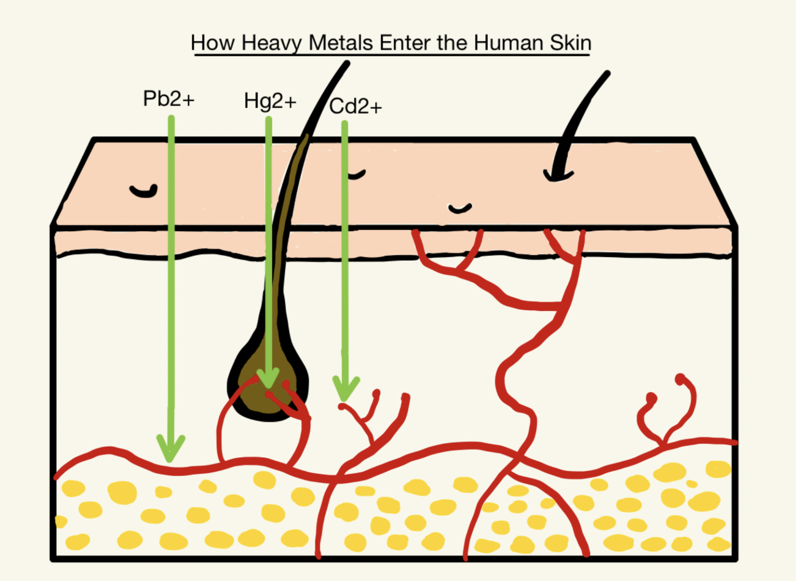
Common substances involved in cosmetics are mercury (Hg), lead (Pb), and cadmium (Cd), and even with strict controls of the manufacturing process and good practices, it is not possible to remove all such compounds or to ensure they are safe to use (Kicińska & Kowalczyk, 2025). Cosmetics are normally composed of small molecules, oil-soluble ingredients, and other chemicals that have similar properties to the skin. Since cosmetics are directly applied to the skin, heavy metals can enter the body through hair follicles and sweat pores, ultimately reaching the blood capillaries (Nnaji et al., 2015; Raza-Naqvi et al., 2022), as illustrated in Figure 1.
| Figure 1. Penetration of heavy metals through human skin. |
|
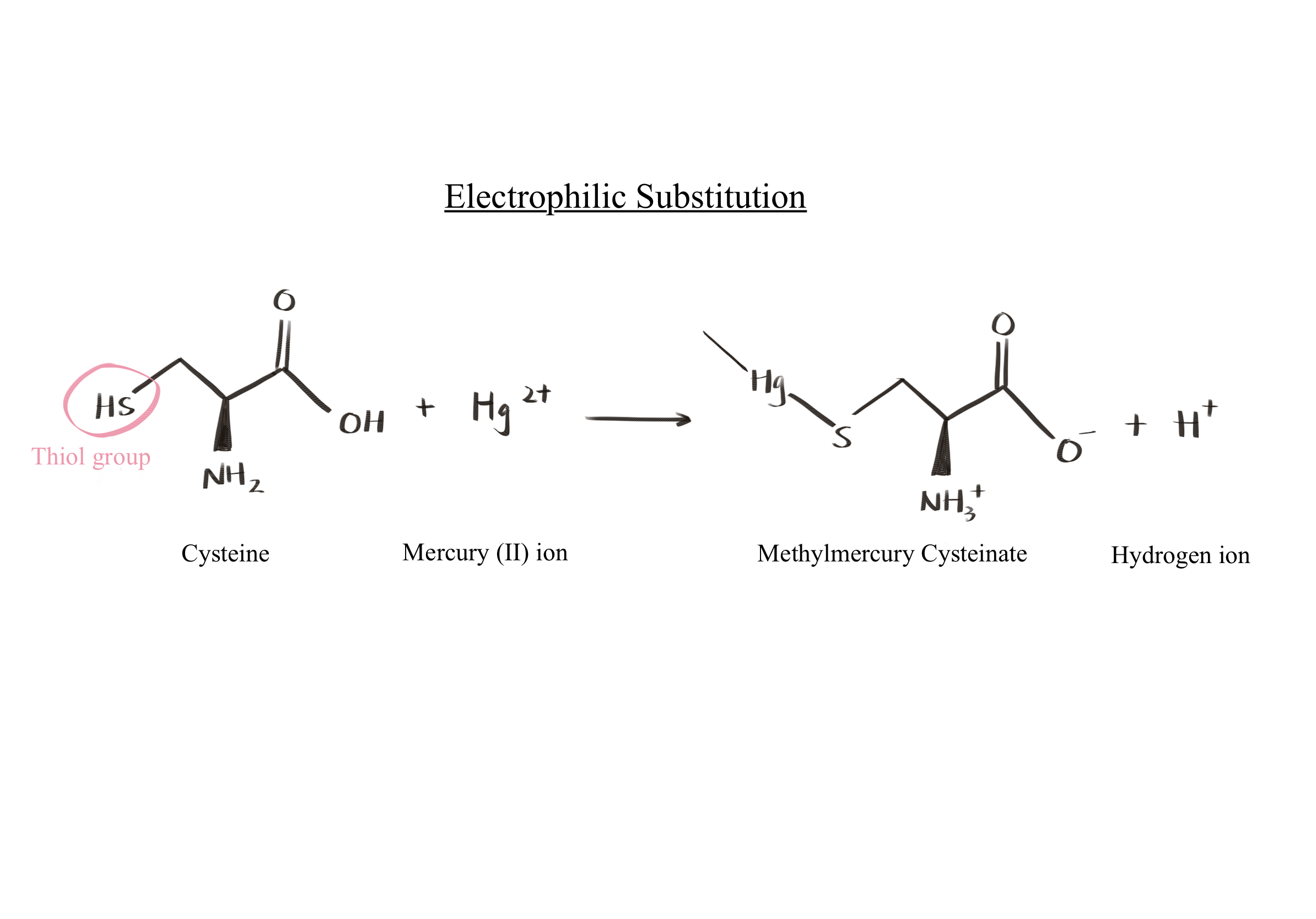
Over time, these metals accumulate in the body and interfere with biological processes by displacing normal functional groups through electron competition (Raza-Naqvi et al., 2022). Specifically, as illustrated in Figure 2, mercury has a high affinity for thiol (-SH) groups. Thus, it attacks the thiol group of the amino acid cysteine in proteins, distorting the tertiary structure of proteins, which leads to the loss of biological functions depending on the type of protein impacted. Meanwhile, lead attacks the hydroxyl (-OH) group. Since the body cannot metabolize or eliminate these heavy metals, prolonged exposure may lead to skin irritation, neurotoxicity, organ damage, and, in severe cases, cancer (Dinake et al., 2023; Coradduzza et al., 2024). Therefore, monitoring the concentration of heavy metals in cosmetic products is essential.
| Figure 2. Electrophilic substitution of mercury at the thiol group of cysteine. |
|
Unfortunately, many consumers are unaware of the risks associated with the cosmetics they use. Research on unregulated cosmetic products, such as whitening creams, reveals that these products frequently contain chromium, copper, and lead in amounts that exceed permissible limits (Arputhanantham et al., 2024). The FDA does not require cosmetic companies to report excess concentrations of heavy metals in their products, except for color additives (FDA, n.d.). Identifying the presence of heavy metals in cosmetics poses additional challenges for consumers due to the complexity of detection methods. Standard detection techniques such as microwave digestion, plasma-optical emission spectroscopy (ICP-OES), inductively coupled plasma mass spectroscopy (ICP-MS), and atomic absorption spectroscopy (AAS) are techniques currently used for sensing heavy metals; however, they require professional laboratory settings and are not easily accessible to individuals (Heavy metal test in cosmetic products, n.d.).
Therefore, this project aims to create a biosensor that detects heavy metal concentrations in various products, and in turn leading to more transparency regarding cosmetics components and safety risks. This project seeks to address three main heavy metals: mercury, lead, and cadmium.
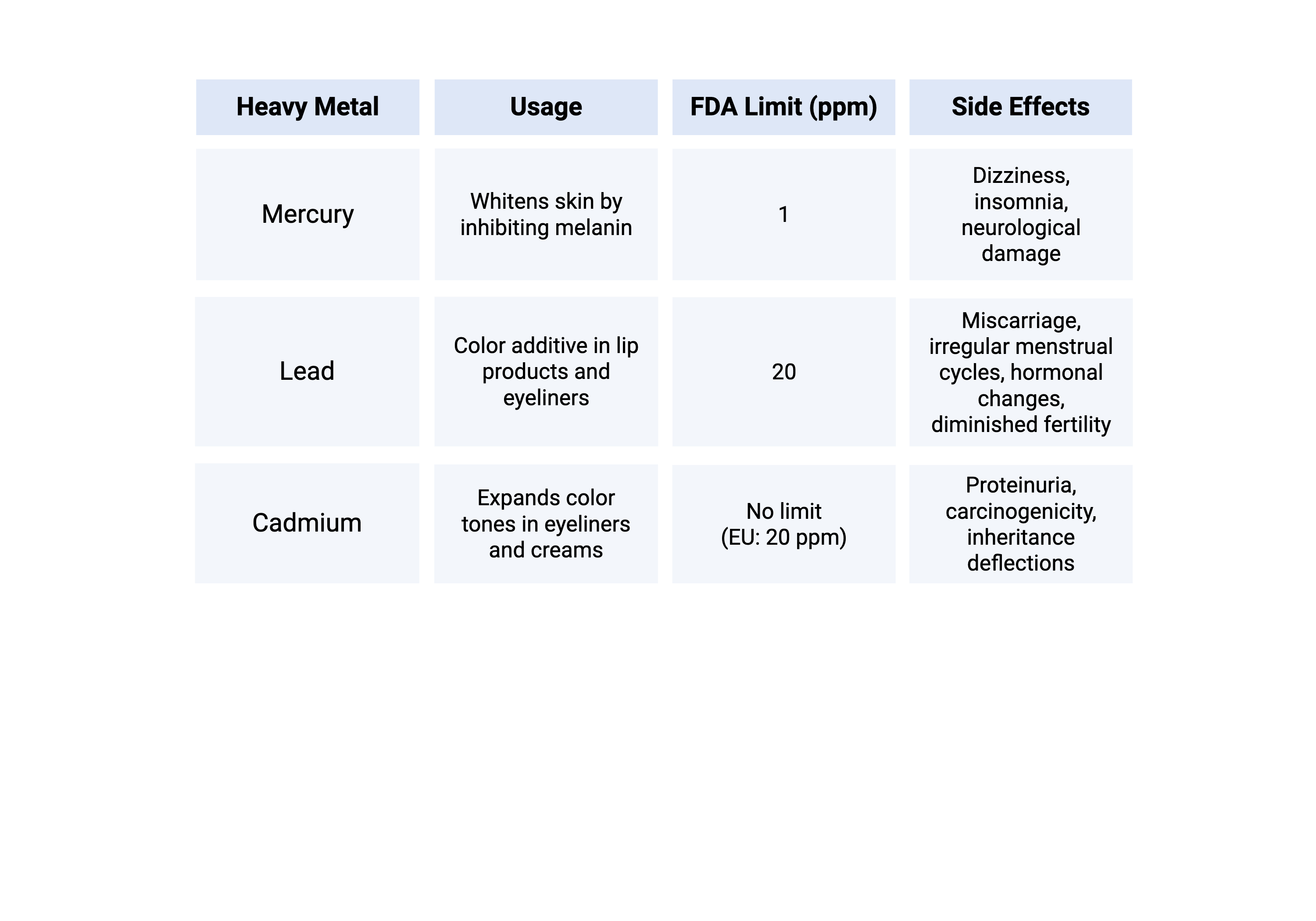
Mercury whitens the skin by inhibiting melanin production, a substance responsible for skin pigmentation. The FDA-recommended concentration is under one part per million (ppm), but many manufacturers use above 1000 ppm of mercury to maximize their whitening effect (FDA, n.d.; Sun et al., 2017). Side effects of mercury intoxication include dizziness, insomnia, and neurological damage (Fernandes Azevedo et al., 2012).
Lead is a color additive ingredient in lip products and eyeliners, which enables cosmetics to feature a variety of colors (Raza-Naqvi et al., 2022). However, blood lead concentrations of 3.5 µg/dL or higher might lead to harmful effects such as miscarriage, irregular menstrual cycles, changes in hormones, and diminished fertility in both genders (World Health Organization, 2024). The FDA recommends that lead levels in cosmetics not exceed 10 ppm (FDA, n.d.). While most products adhere to these guidelines, some intentionally exceed the limit to enhance certain effects such as increased whitening. For example, the Center for Disease Control and Prevention (CDC) documented cases of elevated blood lead levels for adults and children exposed to Surma, a type of eye cosmetic that acts as lead exposure (Hore et al., 2024). These findings highlight the ongoing need for not only stricter restrictions but also the development of accessible methods for customers to test products at home.
Cadmium sulfide is a yellow pigment that achieves specific colors. It is overused in many products to produce more intense or longer-lasting hues. Cadmium expands color tones in eyeliners, lip gloss, and beauty creams (Omenka & Adeyi, 2016). Excessive intake of 0.005 mg/kg/day for more than two weeks (U.S. Environmental Protection Agency, n.d.) can lead to kidney disorders, proteinuria (high levels of protein in urine), potential carcinogenic exposure , and genetic disorders (Raza-Naqvi et al., 2022). While there is currently no FDA limit to cadmium usage, the European Union’s limit is 20 ppm (Abed et al., 2024).
| Figure 3. Comparison of mercury, lead, and cadmium. |
|
Given the similar uses and the variety of purposes of the above heavy metals in cosmetics, two or more metals often coexist in a single product (Fernandes Azevedo et al., 2012). Moreover, since consumers often use multiple cosmetic products simultaneously, it would be ideal to develop a combined mechanism to test for various heavy metals at once. Current testing equipments, such as Inductively Coupled Plasma Mass Spectrometry (ICP-MS) and Atomic Absorption Spectrometry (AAS), as well as X-ray fluorescence (XRF) are complicated and require a laboratory setting, where samples need to be collected, washed, prepared before quantifying for the results (Arshad et al., 2020). Existing heavy metal biosensors lack a combination method that can test multiple heavy metals at the same time.
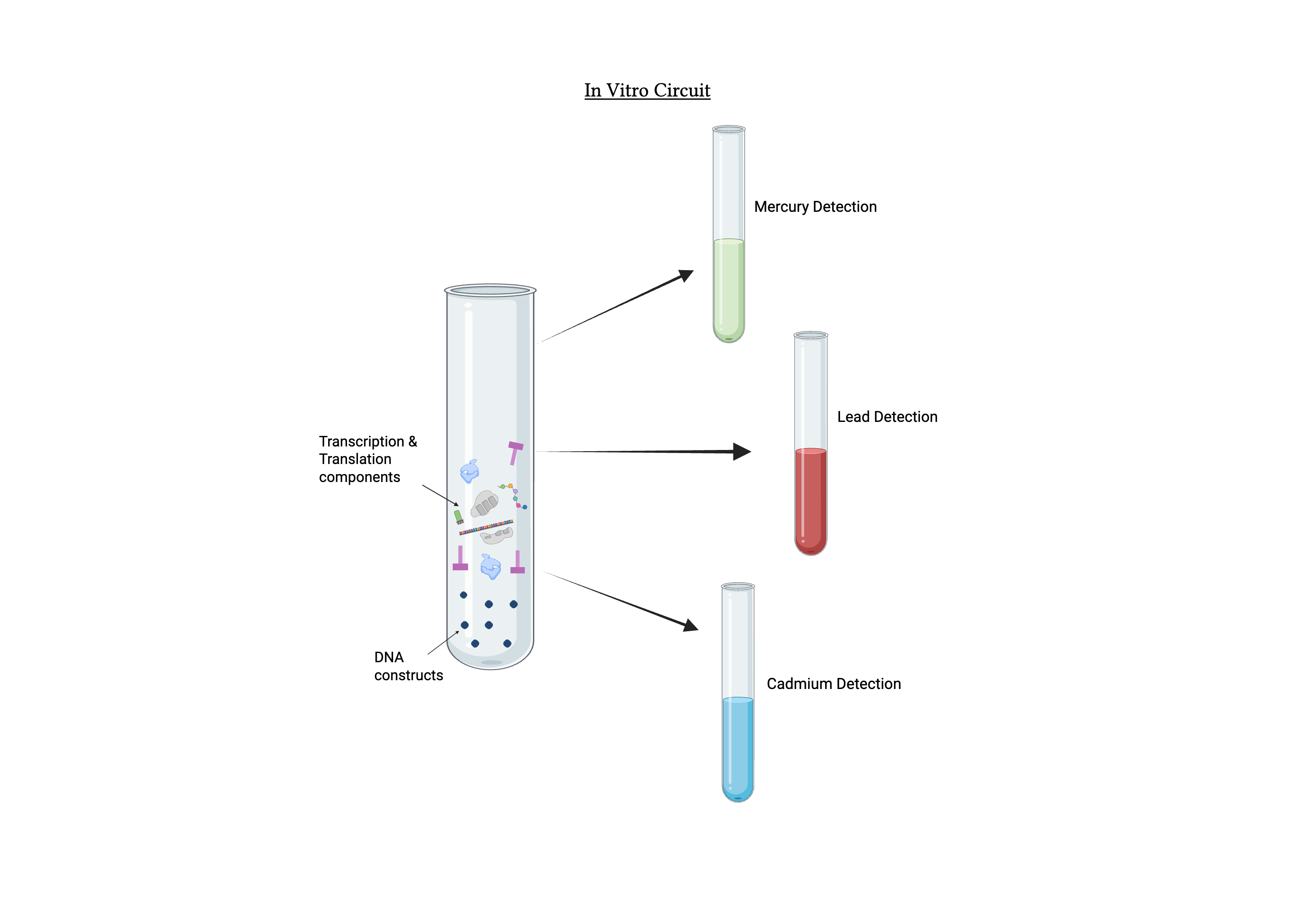
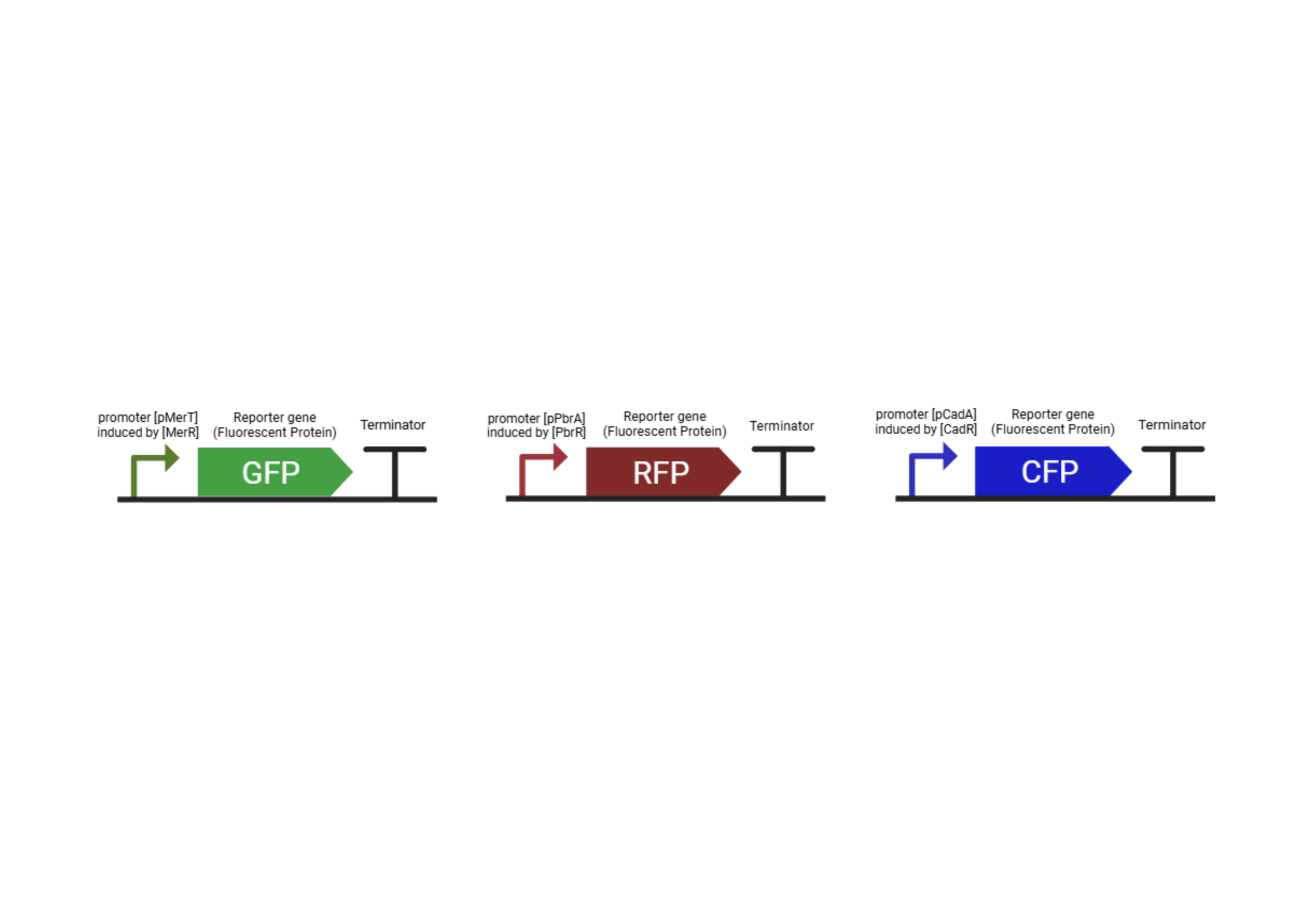
Each metal triggers a distinct color change for easy identification. MerR, a mercury-binding transcriptional regulator, is activated in the presence of Hg ions and initiates transcription of promoter pMerT down the gene, coding for Green Fluorescent Protein (GFP). Similarly, the lead-responsive transcription factor PbrR activates the pPbrA promoter in the presence of lead ions, then initiates the code for Red Fluorescent Protein (RFP) that indicates the presence of lead. Finally, the cadmium-binding transcription factor CadR activates the promoter pCadA and codes for Cyan Fluorescent Protein (CFP) to indicate cadmium ions.
| Figure 4. In vitro genetic circuit. |
|
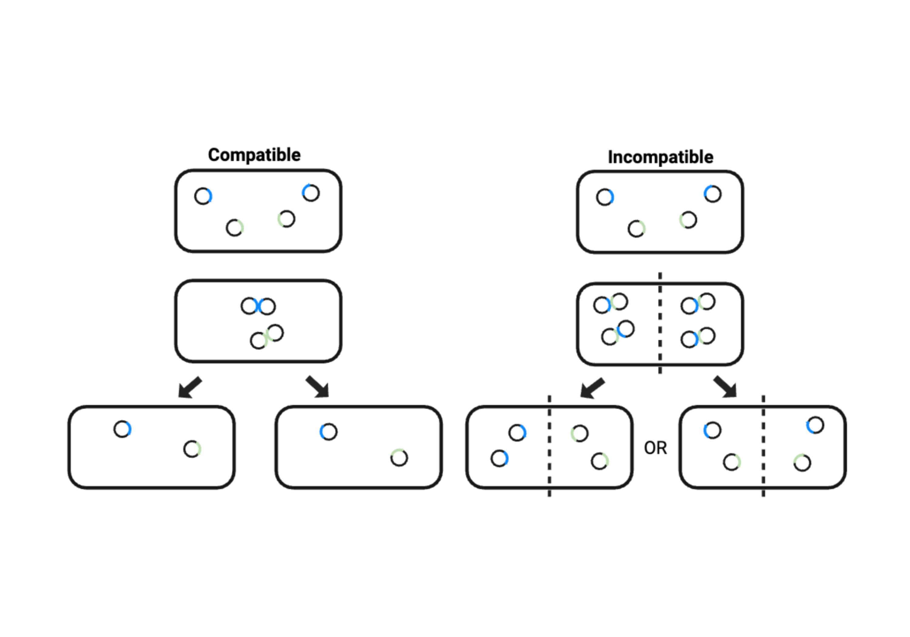
We opted for in vitro detection because it does not require a host, thus increasing success probabilities, as host organisms can easily die in heavy metal environments with which this project works (Wu et al., 2008). Additionally, combining three circuits simultaneously, whether by inserting them as three different plasmids or creating a super plasmid, can result in plasmid incompatibility, burden, reduced stability, and competition in cloning sites. In vitro circuits, on the other hand, mitigate these issues. Finally, our final product, a portable testing chamber, is easily accessible for consumers in a household setting. Since it does not rely on microbes, this biosensor does not require maintenance to keep it alive, ensuring safe and hygienic use. This enables the manufacturers to evaluate the contents of the components they use in cosmetics,allowing them to evaluate the risks of their own products.
Systems Level
This project aims to engineer an in vitro, lyophilized biosensor to detect contamination by heavy metal mercury, lead, and cadmium in industrial products that pose health and environmental risks. The biosensor, upon rehydration, contains genetic circuits that specifically bind each target metal ion and produces a visible fluorescent signal indicating such presence.
We aimed for the cell-free circuit design to address two concerns: toxicity and plasmid competition. The biosensor requires continuous detection of high concentrations of heavy metals, and since these chemicals inhibit the growth and impair the performance of many common host bacteria—such as Escherichia coli and Pseudomonas putida—the use of living organisms is unsuitable for this system. Integrating multiple genetic circuits into one host is also challenging due to plasmid incompatibility: when plasmids share the same replicon, the antisense-RNA replication control halts replication at high plasmid numbers indiscriminately, leading to insufficient plasmid number for each circuit (Schwiesow, 2020). Competition over centromere-binding proteins (CBPs) that controls partitioning and NTPase that serve as energy for cellular functions may lead to uneven distribution of genetic materials in daughter cells, as illustrated in Figure 5 (Schwiesow, 2020; Schumacher, 2012).
| Figure 5. Plasmid Incompatibility. |
|
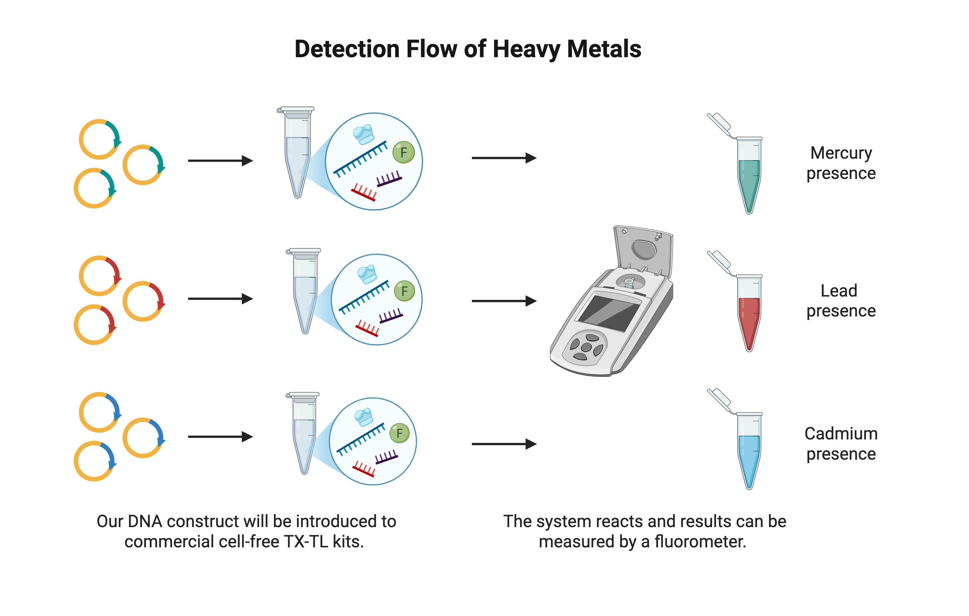
Low-viscosity fluids or small solid particles can be added directly to the rehydrated reaction mixture at room temperature. All reagents are pre-prepared in 1.5 mL microcentrifuge tubes containing a commercial cell-free expression kit purchased through manufacturers such as Arbor Biosciences or Thermo Fisher, where our custom DNA constructs are added. In the presence of a target ion, a metal-responsive transcriptional regulator binds the ion and activates its promoter, driving expression of a fluorescent reporter protein. Each metal is paired with a unique fluorescent reporter color, where green indicates mercury, red indicates lead, and cyan indicates cadmium. Users could interpret the results through a visible color change or by measuring the quantitative fluorescent data with a fluorometer.
The reaction operates optimally at pH 7.5 at room temperature, as the transcription-translation system is optimal at these conditions (Stephen and Mishanina, 2022). HEPES buffer with a pKa of 7.5 is chosen for system dehydration and operation for its strong stability (Sigma-Aldrich, n.d.). The freeze-dried systems can be stored at –50°C to –20°C or refrigerated at 4°C or below (Gregorio et al., 2019). The biosensor is useful for on-site quality control in manufacturing, environmental testing of water and soil, and rapid screening of household consumer products. This assay requires minimal training.
Device Level
The biosensor combines a 1.5 mL microcentrifuge tube containing a commercial freeze-dried cell-free protein expression kit and our own designed DNA construct. This master mix provides all the machinery required for reactions and can produce enzymes, transcription factors, toxic proteins, and other soluble proteins, with typical yields of 5 μg to > 500 μg in a reaction volume of 5 μL to > 250 μL (Arbor Biosciences). The system is compatible with any T7 or E. coli promoter, and accepts both plasmid and linear DNA at 10–40 ng/μL. Reactions are incubated at 27°C for 1–24 hours (Arbor Biosciences.). The cell-free system is lyophilized and can be stored frozen and rehydrated with a buffer immediately before use.
Each sensing module for the heavy metals is composed of a sensor, a promoter, and a reporter. Regulatory proteins are selectively activated by binding to their corresponding ion substrates. Upon activation, the expression of GFP, RFP, and CFP produces green, red, and cyan fluorescent signals, respectively, and specific values can be detected using a fluorometer. To enable quantitative interpretation, calibration curves are generated by testing the biosensor against a series of known heavy-metal concentrations (Andriani & Kubo, 2021). Starting from a 100 ppm stock solution, serial dilutions at 10 ppm, 1 ppm, and 0.1 ppm are prepared. Each test sample is placed in an enclosed black box, and its fluorescence is measured with a light-dependent resistor (LDR), which converts light intensity into an output voltage. By plotting known concentrations against the corresponding LDR voltages and fitting a regression, the user will have an easier time interpreting the outputs from not only what exists but also its concentration.
| Figure 6. Detection flowchart for heavy metal identification using a cell-free biosensor. |
|
Parts Level
In the mercury detection system, when mercury ions are present, they bind to the regulatory protein MerR, inducing a conformational change. This change activates the promoter PmerT, initiating the transcription of the reporter gene gfp, and encoding for the GFP (Doulix, n.d.; Lopreside et al., 2021). Consequently, the appearance of green fluorescence indicates the presence of mercury under UV light or visible light. Similarly, lead ions bind to the pbrR regulatory protein in the lead detection system, activating promoter PpbrA, transcribing the reporter gene rfp, and producing RFP (iGEM Registry, n.d.; Hui et al., 2023). In the cadmium detection system, cadmium ions bind to the CadR regulatory protein, activating promoter PcadA, which transcribes the cfp reporter gene and ultimately produces CFP, is then stopped by a terminator (Schulz et al., 2021).
| Figure 7. Genetic circuits for mercury, lead, and cadmium detection. |
|
The ColE1 origin of replication is used because of its high yield in DNA output (Morgan, 2020). A B0015 rho-independent terminator, known for its strong and protein-independent termination efficiency, ensures clean transcriptional ends (iGEM Registry, n.d.). Additionally, to improve signal strength and stability, superfolder variants of fluorescent proteins, such as sfGFP, are considered as they provide enhanced physical properties and protein expression (Pedelacq & Cabantous, 2019). All genetic parts are synthesized and assembled through Thermo Fisher or similar providers.
Safety
According to the Food and Drug Administration (FDA), mercury is permitted in cosmetics only as a preservative in eye products and must be present at a concentration of less than 1 ppm (FDA., 2020). It is not allowed in other cosmetic products or color additives. The FDA sets a limit of 10 ppm for lead in cosmetics and 20 ppm for lead in color additives. While there are no strict federal regulations in the U.S. regarding cadmium in cosmetics, Health Canada sets a limit of 3 ppm, above which it is considered an impurity (Canada, 2012). Any concentration of these heavy metals exceeding the specified limits is considered contamination and poses a risk to human health.
Since mercury, lead, and cadmium are toxic, proper safety protocols in laboratory environments are essential. Standard laboratory safety equipment, such as nitrile gloves, lab coats, and eye protection goggles, are used at all times. The required heavy metals are typically supplied as liquid solutions in sealed containers, so serial dilutions should be performed in a fume hood using pipettes to ensure accuracy. Deionized water is used during dilution to prevent unintended reactions. Waste is collected in a designated container for heavy metals and disposed of following the institution’s hazardous waste procedures.
As a heavy metal sensing circuit, regulations regarding biosafety levels (BSL) are not applicable, since heavy metals are not microbes or biological agents; however, biosafety and biosecurity must be secured. Biosensor testing chambers should be stored in the refrigerator until use and then disposed of under heavy metal waste standards after testing. Stock solutions of heavy metals should be stored in tightly sealed containers within secondary containment trays and kept in locked chemical cabinets designated for toxic substances.
Discussions
This project aims to create a biosensor that detects the presence of mercury, lead, and cadmium by generating a color change. Compared to traditional methods such as chromatography and capillary electrophoresis, this bioengineered construct provides a faster, portable, and simple lab procedure by utilizing lyophilized reaction kits (Wang et al., 2024). Without the need to maintain host cell viability, the DNA construct will be more stable when facing environmental stressors. By incorporating separate genetic pathways, the system produces distinct fluorescence signals that not only indicate the presence of specific heavy metals but can also be quantified against standard curves to determine exact concentrations, thereby empowering consumers and producers to verify safety claims and enabling sellers to understand the composition of their products better.
A key challenge in implementing this biosensor is obtaining governmental approval to work with heavy metals in laboratory settings. Experiment and disposal procedures will also strictly follow the guidelines set by the Environmental Protection Agency (U.S. Environmental Protection Agency, n.d.). In terms of functionality, the biosensor does not account for metal speciation; the color change only indicates the presence of a metal and gives no information on its type or role in the household or industrialized product. The protein transcription factors included in the DNA construct bind only to free metal ions (e.g., Hg²⁺, Pb²⁺, Cd²⁺) or loosely bonded metals, such as weak electrostatic or hydrogen binding, that can dissociate under the test conditions (e.g., Pb(OOCCH₃)₂, lead acetate) (Wang et al., 2024). On the other hand, they are unable to interact effectively with metals in complexed or nanoparticulate forms (e.g., Hg(SR)₂, CdSe/ZnS quantum dots), which often require chemical pretreatment or digestion to release free metal ions (Varun et al., 2018). Depending on the viscosity or color of the sample, the fluorescence-based color change may not be visible to the naked eye; in such cases, a fluorometer is recommended. Some products may contain only trace amounts of heavy metals, and the sensor’s effectiveness at these concentrations is unknown. The construct’s lower and upper limits will need to be determined experimentally.
Circuit components will continue to be optimized to match target detection thresholds and environmental concentrations. Together, these advancements underscore the potential for a safer, more efficient, and more accessible solution to addressing high heavy metal concentrations in the field of cosmetics.
If the biosensor successfully fulfills the need to detect heavy metals, future iterations may extend its application to other industrial areas, such as the detection of environmental toxins and contamination in household and industrial products. The system also has the potential to be integrated into a lateral flow device (test strips) rather than a kit for possible future consumer accessibility.
Next Steps
This project features a proof of concept. Target gene sequences will be customized from commercial gene synthesis suppliers such as Thermo Fisher and Biosupplies, and codon-optimized for use in cell-free systems. They will then be integrated with the TX-TL kits and lyophilized for storage. Experiments will be performed to validate the design and refine the system until it meets performance criteria.
To confirm successful DNA input into the kit, a positive control template that produces fluorescence will be used as a comparison. DNA will be pre-quantified using a spectrophotometer. Pathway efficiency will be measured through fluorescence intensity and standard curves, and comparing circuit responses will evaluate promoter strength and reaction time. The reaction does not produce byproducts; however, unreacted metals and waste may accumulate in the microtubes after detection, so the product should be disposed of as chemical waste.
The biosensor will then proceed to be tested with industrial or household contaminated products. With government approval, both pure heavy metals and cosmetics containing heavy metals will be tested at equivalent metal concentrations to evaluate the biosensor’s response and determine whether its ideal efficiency matches its actual efficiency. All experiments will be conducted in controlled laboratory environments first. To ensure consistency and accuracy, multiple tests will be performed on a range of heavy metal concentrations to determine detection thresholds. Safety precautions will be strictly enforced at all times. If the results prove successful, further steps towards product implementation may be considered.
Author Contributions
S.Q. came up with the original idea and initiated introductory research. Y.K., R.P., S.Q., M.V., and J.Y. conducted the early research process. Y.K., R.P., S.Q., M.V., and J.Y. contributed to the writing and proofreading of the paper. M.V. and J.Y. created the video. Y.K., S.Q., and J.Y. designed the images and graphics for this project.
Acknowledgements
We sincerely thank Western Reserve Academy for providing the resources and support that made this project possible. We are especially grateful to Dr. Pethel and Dr. Kosuke Seki for their invaluable guidance and encouragement throughout the challenges of this work. Their insight and mentorship have been instrumental to the success of this design.
References
Abed, M. S., Moosa, A. A., & Alzuhairi, M. A. (2024). Heavy metals in cosmetics and tattoos: A review of historical background, health impact, and regulatory limits. Journal of Hazardous Materials Advances, 13. 100390.
Andriani, R. T., & Kubo, Y. (2021). Voltage-clamp fluorometry analysis of structural rearrangements of ATP-gated channel P2X2 upon hyperpolarization. eLife, 10. https://doi.org/10.7554/elife.65822
Arputhanantham, S. S., Al-Thani, G. S. S., Al Amri, R. M. S., & Hossain, M. A. (2024). Comparative analysis of heavy metal content of unregulated facial whitening creams and branded facial whitening creams. Toxicology Reports, 26, 326-330. https://doi.org/10.1016/j.toxrep.2023.10.011
Heath Canada (2012). Guidance on Heavy Metal Impurities in Cosmetics.. https://www.canada.ca/en/health-canada/services/consumer-product-safety/reports-publications/industry-professionals/guidance-heavy-metal-impurities-cosmetics.html#a323
Cardinale, S., Joachimiak, M., & Arkin, A. (2013). Effects of genetic variation on the e. coli host-circuit interface. Cell Reports, 4(2), 231-237. https://doi.org/10.1016/j.celrep.2013.06.024
CAS. (n.d.). Phosphoenolpyruvic acid. CAS Common Chemistry. https://commonchemistry.cas.org/detail?cas_rn=138-08-9
Coradduzza, D., Congiargiu, A., Azara, E., Abulkahar, M., Rosaria, M., Zinellu, A., Carru, C., & Medici, S. (2024). Heavy metals in biological samples of cancer patients: a systematic literature review. BioMetals, 37:803-817. https://doi.org/10.1007/s10534-024-00583-4
Cosmetics market size, share & industry analysis. (n.d.). Fortune Business Insights. https://www.fortunebusinessinsights.com/cosmetics-market-102614
Deutscher, J., Francke, C., & Postma, P. W. (2006). How phosphotransferase system-related protein phosphorylation regulates carbohydrate metabolism in bacteria. Microbiol Mol Biol Rev. 70(4):939-1031. https://doi.org/10.1128/mmbr.00024-06
Dinake, P., Motswetla, O., Kereeditse, T., & Kelebemang, R. (2023). Assessment of level of heavy metals in cosmetics. Toxicology Research and Application. 7. https://doi.org/10.1177/23978473231156620
Djordjevic, M. (n.d.). 15 fascinating beauty industry statistics you should know in 2024. SaveMyCent. https://savemycent.com/beauty-industry-statistics/
Doulix. (n.d.). MerR regulator CDS (QR5CDW8). https://doulix.com/biomodules/QR5CDW8/
FDA. (n.d.). FDA’s Testing of Cosmetics for Arsenic, Cadmium, Chromium, Cobalt, Lead, Mercury, and Nickel Content. https://www.fda.gov/cosmetics/potential-contaminants-cosmetics/fdas-testing-cosmetics-arsenic-cadmium-chromium-cobalt-lead-mercury-and-nickel-content
Fernandes Azevedo, B., Barros Furieri, L., Peçanha, F. M., Wiggers, G. A., Frizera Vassallo, P., Ronacher Simões, M., Fiorim, J., Rossi de Batista, P., Fioresi, M., Rossoni, L., Stefanon, I., Alonso, M. J., Salaices, M., & Valentim Vassallo, D. (2012). Toxic Effects of Mercury on the Cardiovascular and Central Nervous Systems. Journal of Biomedicine and Biotechnology, 2012:949048. https://doi.org/10.1155/2012/949048
Futra, D., Heng, L., Ahmad, A., Surif, S., & Ling, T. (2015). An Optical Biosensor from Green Fluorescent Escherichia coli for the Evaluation of Single and Combined Heavy Metal Toxicities. Sensors, 15(6), 12668–12681. https://doi.org/10.3390/s150612668
Gonzalez, M. H., Souza, G. B., Oliveira, R. V., Forato, L. A., Nóbrega, J. A., & Nogueira, A. R. A. (2009). Microwave-assisted digestion procedures for biological samples with diluted nitric acid: Identification of reaction products. Talanta, 79(2), 396–401. https://doi.org/10.1016/j.talanta.2009.04.001
Gregorio, N. E., Levine, M. Z., & Oza, J. P. (2019). A User’s Guide to Cell-Free Protein Synthesis. Methods and Protocols, 2(1). https://doi.org/10.3390/mps2010024
Heavy metal test in cosmetic products. (n.d.). https://www.chemleg.com/articles/heavy-metal-test.
Hore, P., Sedlar, S., & Ehrlich, J. (2024). Lead poisoning in a mother and her four children using a traditional eye cosmetic — New York City, 2012–2023. Morbidity and Mortality Weekly Report, 73(30). 661-676. https://www.cdc.gov/mmwr/volumes/73/wr/mm7330a2.htm
Hui, C., Ma, B., Wang, Y., Yang, X., & Cai, J. (2023). Designed bacteria based on natural pbr operons for detecting and detoxifying environmental lead: A mini-review. Ecotoxicology and Environmental Safety, 267, 115662–115662. https://doi.org/10.1016/j.ecoenv.2023.115662
iGEM Registry. (n.d.). Part:BBa_K346054 – MerR. https://parts.igem.org/Part:BBa_K346054
Karthik, V., Koruna, B., Senthil Kumar, P., Saravanan, A., & Hemavathy, R.V. (2022). Development of lab-on-chip biosensor for the detection of toxic heavy metals: A review. Chemosphere, 299:134427.. https://doi.org/10.1016/j.chemosphere.2022.134427
Kicińska, A., & Kowalczyk, M. (2025). Health risks from heavy metals in cosmetic products available in the online consumer market. Scientific Reports, 15(1). https://doi.org/10.1038/s41598-024-83477-2
Lead poisoning. (2024, September 27). World Health Organization. https://www.who.int/news-room/fact-sheets/detail/lead-poisoning-and-health
Lopreside, A., Montali, L., Wang, B., Tassoni, A., Ferri, M., Calabretta, M. M., & Michelini, E. (2021). Orthogonal paper biosensor for mercury(II) combining bioluminescence and colorimetric smartphone detection. Biosensors & Bioelectronics, 194, 113569. https://doi.org/10.1016/j.bios.2021.113569
Morgan, K. (2020, November 11). Plasmids 101: Origin of Replication. Addgene Blog.
Nnaji, N. D., Anyanwu, C. U., Eke, U. E., Okeke, O. K., & Nwankwo, C. M. (2015). Possible health implications associated with cosmetics: A review. Science Journal of Public Health, 3(5), 58-63. https://doi.org/10.11648/j.sjph.s.2015030501.21
Nnaji, N. D., Anyanwu, C. U., Miri, T., & Onyeaka, H. (2024). Mechanisms of Heavy Metal Tolerance in Bacteria: A Review. Sustainability, 16(24), 11124. https://doi.org/10.3390/su162411124
FDA. (2020). FDA’s Testing of Cosmetics for Arsenic, Cadmium, Chromium, Cobalt, Lead, Mercury, and Nickel Content. https://www.fda.gov/cosmetics/potential-contaminants-cosmetics/fdas-testing-cosmetics-arsenic-cadmium-chromium-cobalt-lead-mercury-and-nickel-content
Omenka, S. S., & Adeyi, A. A. (2016). Heavy metal content of selected personal care products (PCPs) available in Ibadan, Nigeria and their toxic effects. Toxicology Reports, 3, 628-635.
Pedelacq, J.-D., & Cabantous, S. (2019). Development and applications of superfolder and split fluorescent protein detection systems in biology. International Journal of Molecular Sciences, 20(14), 3479. https://doi.org/10.3390/ijms20143479
Raza-Naqvi, S. A., Idrees, F., Sherazi, T. A., Anjum-Shahzad, S., Ul-Hassan, S., & Ashraf, N. (2022). Toxicology of heavy metals used in cosmetics. Journal of the Chilean Chemical Society, 67(3). 5616-5622. http://dx.doi.org/10.4067/S0717-97072022000305615
Schulz, V., Schmidt-Vogler, C., Strohmeyer, P., Weber, S., Kleemann, D., Nies, D. H., & Herzberg, M. (2021). Behind the Shield of Czc: ZntR Controls Expression of the Gene for the Zinc-Exporting P-Type ATPase ZntA in Cupriavidus metallidurans. Journal of Bacteriology, 203(11). https://doi.org/10.1128/jb.00052-21
Schumacher, M. A. (2012). Bacterial plasmid partition machinery: a minimalist approach to survival. Current Opinion in Structural Biology, 22(1), 72–79. https://doi.org/10.1016/j.sbi.2011.11.001
Schwiesow, L. (2020, March 31). Plasmids 101: Plasmid Incompatibility. Addgene Blog. https://blog.addgene.org/plasmids-101-plasmid-incompatibility
Sigma Aldrich. (2025). NMR Chemical Shifts of Impurities. Merck, 1(1). https://www.sigmaaldrich.com/MX/en/technical-documents/technical-article/genomics/cloning-and-expression/blue-white-screening
Stephen, C., & Mishanina, T. V. (2022). Alkaline pH has an unexpected effect on transcriptional pausing during synthesis of the Escherichia coli pH-responsive riboswitch. Journal of Biological Chemistry, 298(9), 102302. https://doi.org/10.1016/j.jbc.2022.102302
Sun, G.-F., Hu, W.-T., Yuan, Z.-H., Zhang, B.-A., & Lu, H. (2017). Characteristics of Mercury Intoxication Induced by Skin-lightening Products. Chinese Medical Journal, 130(24), 3003–3004. https://doi.org/10.4103/0366-6999.220312
Tchounwou, P. B., Yedjou, C. G., Patlolla, A. K., & Sutton, D. J. (2014). Heavy Metal Toxicity and the Environment. Experientia Supplementum, 101(1), 133–164. https://doi.org/10.1007/978-3-7643-8340-4_6
U.S. Environmental Protection Agency. (n.d.). Cadmium. https://archive.epa.gov/epawaste/hazard/wastemin/web/pdf/cadmium.pdf
Varun, S., Daniel & S. C. G. K. (2018). Emerging nanosensing strategies for heavy metal detection. In A. K. Mishra & C. M. Hussain (Eds.), Nanotechnology for Sustainable Water Resources, (pp. 199–225). Scrivener Publishing LLC. https://doi.org/10.1002/9781119323655.ch7
Wang, M., Zhang, L., Hao, H., Yan, M., & Zhu, Z. (2024). Applications of engineered skin tissue for cosmetic component and toxicology detection. Cell Transplantation, 33. https://doi.org/10.1177/09636897241235464
Wu, Y., Zhou, Y., Song, J., Hu, X., Ding, Y., & Zhang, Z. (2008). Using Green and Red Fluorescent Proteins to Teach Protein Expression, Purification, and Crystallization. Biochemistry and Molecular Biology Education 36(1):43–54. https://doi.org/10.1002/bmb.117.
Zhang, L., Lin, X., Wang, T., Guo, W., & Lu, Y. (2021). Development and comparison of cell-free protein synthesis systems derived from typical bacterial chassis. Bioresources and Bioprocessing, 8, 58. https://doi.org/10.1186/s40643-021-00413-2
Zhao, H., Wu, Y., Lan, X., Yang, Y., Wu, X., & Du, L. (2022). Comprehensive assessment of harmful heavy metals in contaminated soil in order to score pollution level. Scientific Reports, 12, 3552. https://doi.org/10.1038/s41598-022-07602-9